Iron deficiency
Worms
Allergy
53075 10 August
IMPORTANT!
The information in this section cannot be used for self-diagnosis and self-treatment.
In case of pain or other exacerbation of the disease, diagnostic tests should be prescribed only by the attending physician. To make a diagnosis and properly prescribe treatment, you should contact your doctor. We remind you that independent interpretation of the results is unacceptable; the information below is for reference only.
Clinical blood test: indications for use, rules for preparing for the test, interpretation of results and normal indicators.
Why do you need a general blood test? Why is this analysis so important?
Blood is a special tissue that transports various substances between other tissues, organs and systems, while ensuring the unity and constancy of the internal environment of the body. Thus, most processes affecting the condition of various tissues and organs, one way or another, are reflected in the condition of the blood.
Blood consists of plasma (the liquid part of the blood) and formed elements - leukocytes, platelets, erythrocytes. Each type of formed element has its own functions: leukocytes are responsible for immune defense, platelets for blood clotting, red blood cells provide transport of oxygen and carbon dioxide.
In a healthy person, the composition of the blood is fairly constant, but in case of illness it changes. Therefore, with the help of a blood test, it is possible to determine that the disease exists. Sometimes a general blood test can detect the disease at an early stage, when the main symptoms of the disease have not yet appeared. That is why OAC is performed during any preventive examination. If symptoms are present, clinical analysis helps to understand the nature of the disease and determine the intensity of the inflammatory process. Clinical analysis is used to diagnose various inflammatory diseases, allergic conditions, and blood diseases. A repeat general blood test will give the doctor the opportunity to judge the effectiveness of the prescribed treatment, assess the trend towards recovery and, if necessary, adjust the course of treatment.
COAGULOGRAM. HEMOSTASIOGRAM
COAGULOGRAM
(blood test for hemostasis) is a necessary stage in the study of blood clotting during pregnancy, before operations, in the postoperative period, i.e. in situations where the patient expects some blood loss, as well as with varicose veins of the lower extremities, autoimmune diseases and liver diseases. Violation of blood clotting, especially its increase or hypercoagulation, can lead to dangerous consequences for the body, causing a heart attack, stroke, thrombosis.
During pregnancy, a coagulogram always shows increased blood clotting. If coagulation values are higher than normal, then blood clots can form in the vessels of the placenta, as a result of which the baby does not receive enough oxygen, which can lead to miscarriage, premature birth or the birth of a child with severe brain damage.
Blood hemostasis is maintained due to the balance of three systems:
A coagulation system that activates platelets, their adhesion to the vascular wall and gluing (main components: fibrinogen, platelets, calcium, vascular wall).
Anticoagulant system that controls blood clotting and prevents spontaneous thrombus formation (antithrombin III)
Fibrinolytic system that dissolves clots (plasmin).
Clinical blood test indicators
A general blood test must contain the following indicators:
- hemoglobin;
- red blood cells;
- color index;
- reticulocytes;
- platelets;
- leukocytes;
- neutrophils;
- eosinophils;
- basophils;
- lymphocytes;
- monocytes;
- ESR (erythrocyte sedimentation rate).
If necessary, the doctor may prescribe an extended clinical blood test. In this case, he will specifically indicate which indicators should be additionally included in the analysis.
Interpretation of general blood test indicators
Hemoglobin
Hemoglobin is a protein that makes up a part of the red blood cell. Hemoglobin binds with oxygen and carbon dioxide molecules, which allows oxygen to be delivered from the lungs to tissues throughout the body, and carbon dioxide back to the lungs. Hemoglobin contains iron. It is what gives the red color to erythrocytes (red blood cells), and those to the blood.
Blood saturation with hemoglobin is an extremely important indicator. If it falls, the body's tissues receive less oxygen, and oxygen is necessary for the life of every cell.
The hemoglobin norm for men is 130-160 g/l, for women – 120-140 g/l. In children, there is no dependence on gender, however, in a newly born child, the number of red blood cells (and, accordingly, the hemoglobin level) significantly exceeds the “adult” norm. And during the first 2-3 weeks this figure gradually decreases, which must be kept in mind when assessing the results of a general blood test.
When hemoglobin values are below normal, anemia is diagnosed. Also, a low hemoglobin level may indicate hyperhydration of the body (increased fluid consumption). Hemoglobin is higher than normal, therefore, can be observed with dehydration (blood thickening). Dehydration can be physiological (for example, due to increased physical activity), or it can be pathological. An increased level of hemoglobin is a typical sign of erythremia, a blood formation disorder in which an increased number of red blood cells is produced.
Red blood cells
Erythrocytes are red blood cells. There are significantly more of them than all other formed elements combined. This is why our blood is red. Red blood cells contain hemoglobin and thereby participate in the process of oxygen metabolism in the body.
The norm for red blood cells for men is 4-5*1012 per liter of blood, for women – 3.9-4.7*1012 per liter.
A decrease in the content of red blood cells in the blood is observed with anemia, bleeding, pregnancy, and hyperhydration. Exceeding the norm may indicate dehydration, erythremia, tumor formations, as well as diseases such as a kidney cyst or hydrocele of the renal pelvis.
Color index
The color index is calculated using a formula that correlates the level of hemoglobin and the number of red blood cells. Normally, the color index should be close to one (0.85-1.05). Deviation from the norm is observed with anemia, and with different types of anemia it manifests itself in different ways: a color indicator below the norm indicates iron deficiency anemia (the hemoglobin level is reduced to a greater extent than the number of red blood cells); a color index higher than normal is characteristic of other types of anemia (the number of red blood cells decreases to a greater extent than the hemoglobin level).
Reticulocytes
Reticulocytes are young, immature forms of red blood cells. The process of formation of red blood cells is continuous, so reticulocytes are always present in the blood. Normal: 2-10 reticulocytes per 1000 red blood cells (2-10 ppm (‰), or 0.2-1%). If there are more reticulocytes than normal, this indicates that the body feels the need to increase the number of red blood cells (for example, due to their rapid destruction or blood loss). A reduced level of reticulocytes is characteristic of anemia, radiation sickness, oncology (if metastases have affected the bone marrow), and some kidney diseases.
Platelets
The main function of platelets is to ensure hemostasis, that is, simply put, platelets are responsible for blood clotting. They also participate in the body’s immune response to infection. Norm: 180-320*109 per liter. A low platelet count may indicate severe inflammation or an autoimmune disease. An increased level is typical for conditions after significant blood loss (for example, after surgery), and is also observed in cancer or atrophy (decreased function) of the spleen.
Leukocytes
Leukocytes are white blood cells that perform a protective function, that is, they represent the immune system. Normally, the total number of leukocytes should be in the range of 4-9 * 109 per liter.
An increase in the number of leukocytes indicates the body’s immune response and is observed in infectious diseases (primarily caused by bacteria), inflammatory processes, and allergic reactions. A high level of white blood cells can also be a consequence of recent bleeding, stress, tumor processes and some other pathologies.
A low level of white blood cells indicates a depressed state of the immune system. Such results can be observed with viral infections (influenza, measles, rubella), severe toxicosis, sepsis, diseases of the hematopoietic organs, radiation sickness, autoimmune diseases, etc.
It is not only the overall assessment of the white blood cell count that is important. There are five types of leukocytes - neutrophils, eosinophils, basophils, lymphocytes and monocytes; they all have different functions, and therefore it is important to know in what proportion they are present in the blood. The ratio of different types of leukocytes in their total volume is called the leukocyte formula
.
Neutrophils
Neutrophils are cells of a nonspecific immune response. Their task is to bind the invading infectious agent (bacteria, fungi, intracellular parasites, etc.). When a focus of inflammation occurs in the body, neutrophils begin to move in its direction, their number increases. Neutrophils are produced by the bone marrow, where they undergo various stages of maturation: first a myelocyte is formed, then it becomes a metamyelocyte, then the band neutrophil stage occurs. A mature neutrophil is segmented. Normally, myelocytes and metamyelocytes should be absent in the blood; segmented neutrophils should be 47-72% of the total number of leukocytes; stab - 1-6%. If the immune system is stressed, the proportion of band cells increases (the body is forced to send not yet fully mature neutrophils to fight the infection). Similar analysis results are called rod shift
.
An increase in the number of neutrophils in the blood, therefore, indicates the presence of an infection (first of all, a bacterial infection should be suspected), an ongoing inflammatory process. It can also be the result of stress, intoxication, or cancer.
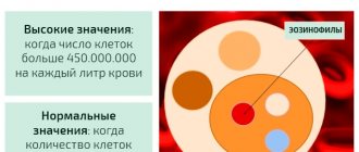
Eosinophils
Eosinophils primarily neutralize immune complexes resulting from the penetration of the allergen. Normally, eosinophils make up 1-5% of the total number of leukocytes. An increase in the level of eosinophils in the blood indicates an allergic reaction or a parasitic infection (primarily infection with worms).
Basophils
Normal: 0-1% of the total number of leukocytes.
Lymphocytes
Lymphocytes are the main cells of the immune system. They provide specific immunity, that is, they recognize an invading foreign agent and destroy it. With the help of lymphocytes, the body fights viruses. Normally, lymphocytes make up 19-37% of the total number of leukocytes. In children, the proportion of lymphocytes is higher. At the age of 1 month to two years, lymphocytes are the main type of leukocytes, and they make up the bulk of the observed mass. By 4-5 years, the number of leukocytes becomes comparable to the number of neutrophils. As the child grows older, the decline continues, but even at the age of 15, children have more lymphocytes than adults.
An increased level of lymphocytes in the blood indicates the penetration of a viral infection; It is also observed in toxoplasmosis, tuberculosis, and syphilis.
A low number of lymphocytes is a sign of a depressed immune system.
Monocytes
Monocytes remain in the blood for an average of about 30 hours, after which they leave the bloodstream and move into tissues, where they turn into macrophages. The purpose of macrophages is to completely destroy bacteria and dead body tissue, clearing the site of inflammation for subsequent regeneration (restoration of healthy tissue). The norm for monocytes is 3-11% of the total number of leukocytes.
An increased number of monocytes is characteristic of sluggish and long-term diseases; it is observed in tuberculosis, sarcoidosis, and syphilis. It is a specific sign of mononucleosis.
ESR – erythrocyte sedimentation rate
If a tube of blood is left in an upright position, red blood cells - as a heavier fraction of blood compared to plasma - will begin to settle to the bottom. Ultimately, the contents of the test tube will be divided into two parts: a thick and dark part at the bottom (these will be red blood cells) and a light part at the top (blood plasma). The erythrocyte sedimentation rate is measured in mm/hour. Normal: 2-10 mm/hour in men and 2-15 mm/hour in women. In children, pregnant women and the elderly, the range of normal values will be different (in children it varies greatly with age).
The erythrocyte sedimentation rate increases if the erythrocytes begin to stick together more strongly (at the same time, their combined mass increases, which means they settle faster). The acceleration of red blood cell adhesion depends on many factors. The most common cause is the existence of an inflammatory process in the body. In this case, as a rule, the stronger the inflammation, the higher the ESR. In addition, an increased ESR value may indicate:
- diseases of the liver and biliary tract;
- processes associated with tissue death (heart attack, stroke, tuberculosis, malignant tumors);
- blood diseases;
- endocrine diseases (thyrotoxicosis, diabetes mellitus, etc.);
- autoimmune diseases;
- and some others.
smear on flora
smear on flora
- microscopy of scrapings from the urethra, the contents of the posterior wall of the vagina and the cervix.
Decoding the smear:
- squamous epithelium
is a layer of cells lining the vagina and cervix. In a normal smear, epithelium should be present. If the smear does not contain epithelium, then the gynecologist has reason to assume a lack of estrogen, an excess of male sex hormones. The absence of squamous epithelium in the smear indicates atrophy of epithelial cells. - leukocytes
- the norm is up to 15 units in the field of view. A small number of leukocytes will be considered normal, since leukocytes perform a protective function and prevent infection from entering the woman’s genitals. Elevated leukocytes in the smear are observed during inflammation of the vagina (colpitis, vaginitis). The more leukocytes in the smear, the more acute the disease. - rods
make up the normal microflora of the vagina. Apart from rods, there should be no other microorganisms in the smear. - small sticks
are most often gardnerella - the causative agents of gardnerellosis or vaginal dysbacteriosis. - “key” cells
(atypical cells) are squamous epithelial cells glued to a small rod. As with Gardnerella, if the smears contain atypical cells, the doctor can diagnose vaginal dysbiosis. - fungus
is a sign of candidiasis (thrush). In latent (asymptomatic) stages of thrush, the fungus in the smear can be detected in the form of spores.
Even if the smear results show the presence of cocci, small rods and “clue” cells indicating bacterial vaginosis, the smear results alone may not be enough to make a diagnosis. It is necessary to conduct an additional examination: bacteriological culture and DNA diagnostics (PCR smear).
BAKPOSEV
The bacteriological method of examining a smear taken from the vagina or urethra is that this material is placed in a special nutrient medium that promotes the proliferation of certain bacteria. Bakposev allows you to differentiate nonspecific bacterial flora, determine the species and quantity of the pathogen. In addition, which is very important for subsequent treatment, bacterial culture makes it possible to determine sensitivity to antibacterial drugs
PCR DIAGNOSTICS
PCR – polymerase chain reaction. The main advantage of the DNA method is that it allows you to determine small quantities of the pathogen, as well as persistent forms of pathogens that are encountered in the treatment of latent and chronic infections. The sensitivity and specificity of the PCR method is high – 95%.
How to take a general blood test. Preparing for the UAC
Blood for a general blood test can be taken either from a finger or from a vein.
It is advisable to take the test on an empty stomach. If the test is taken during the day, then at least 4-5 hours should pass after the last meal. However, this requirement is not strict.
Be sure to avoid eating fatty foods the day before. You cannot take a test while there is alcohol in your blood, or after an X-ray examination, physical therapy, or sunbathing.
If you are taking any medications, be sure to tell your doctor: some medications can affect your blood composition.
For women undergoing routine examination, it is advisable to wait until the end of their period for analysis. If a general blood test is prescribed during the treatment of a disease, you can donate blood regardless of your period - the doctor will take this into account when interpreting the results.
smear for cytology
Cytology smear is a cytological examination of smears taken from the surface of the cervix and from the cervical canal. This test is performed annually on all women over 18 years of age who are sexually active. The procedure is absolutely painless. The examination is not carried out during menstruation and in the presence of an inflammatory process.
Normally, the smear reveals squamous and columnar epithelial cells without any features. The appearance of atypical cells in a smear is a signal of trouble. The cause may be inflammatory processes caused by urogenital infections (mycoplasma, gonococci, trichomonas, chlamydia, etc.), background diseases (erosion, ectopia, leukoplakia, polyps, etc.), as well as precancerous conditions (dysplasia) and malignant degeneration of cells.
Each pathology has its own cytological features, which will be described in the cytogram.
Further examinations depend on the results of cytology: colposcopy (examination of the cervix under magnification using a special device - a colposcope), PCR examination, PAP test, bacteriological examinations (cultures), biopsy followed by histology (taking a piece of tissue from suspicious areas and examination under a microscope).