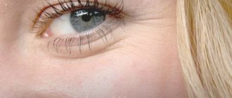
Benign epithelial tumors - senile warts, age-related keratomas (photos in the article show the appearance of the formations). In most cases, this type of skin lesions is discovered after 40–50 years. The disease occurs equally in men and women. The cause is not precisely established, however, genetics and sun exposure are considered to be the main factors.
Causes of keratomas
Keratomas most often affect exposed areas of the skin - face, neck, arms.
Less commonly, this disease affects the back or abdomen. This arrangement of growths gives reason to believe that keratosis is caused by excessive exposure to the sun.
These conclusions are confirmed by scientific research.
Aging skin is at greatest risk; it is the skin that is especially sensitive to excess ultraviolet radiation. People over 50 years of age often suffer from keratomas. This disease even received a separate name - “senile warts” .
It is worth emphasizing that this type of wart, unlike other varieties, is not caused by a virus and is not transmitted from person to person in any way.
However, there is a possibility of a genetic predisposition to this disease.
In addition to excess ultraviolet radiation, other possible causes of senile warts should be mentioned:
- Lack of vitamins in the body.
- The prevalence of animal fats in the diet and the lack of plant fats.
- Predisposition to seborrhea.
Patient reviews
To choose the right way to get rid of growths, you need to consult a doctor and also study reviews. Treatment of senile keratoma with traditional and folk remedies has both advantages and some disadvantages. For example, laser and radio wave removal, cryodestruction and electrocoagulation make it possible to get rid of growths in one go. In addition, all these procedures are completely painless. Unfortunately, according to users, in some cases the formation appears again. Most often this is observed after cryodestruction. After surgical excision, the risk of recurrence of the keratoma is reduced to zero.
Treatment with folk remedies is affordable and safe. But, judging by the reviews of patients, not everyone has the patience to complete it. In the case when the course of treatment is completed completely, nothing will remind you of the growth. The patient will receive clear, scar-free skin.
Kinds
Senial keratoma
This type of disease is characterized by the appearance of gray-white growths .
Such keratomas are prone to inflammation, they often peel off and bleed.
Such formations appear mainly in people over 30 years of age.
Affected areas are exposed skin: face, neck, forearms.
Seborrheic keratoma
The most dangerous type of keratoma is seborrheic.
During the course of this disease, a yellowish spot on the skin transforms into a dark brown growth.
Such formations are prone to constant itching, peeling, and cracks. Keratomas often fall off, which entails bleeding.
In such cases, there is a need to consult a doctor, since an infection can enter the body through the resulting wound.
Horny keratoma
The so-called cutaneous horn is particularly impressive in size.
These dark brown formations rise several millimeters above the surface of the skin.
The height of such keratomas can reach seven to eight millimeters. Often several such formations appear on the skin at the same time.
It is horny keratomas that most often develop into a malignant tumor . This type of growth requires maximum attention and, possibly, surgical intervention.
Solar keratoma
Ketactinic keratosis is considered a precancerous stage of the disease .
This type of disease is characterized by a large number of foci. The formations are covered with dry grayish scales.
This type of keratoma is especially common among men over forty years of age.
The disease affects areas of the skin most exposed to sunlight. This disease often occurs among summer residents.
This disease is very insidious, as it may not cause concern to the patient or even to the attending physician. Keratosis almost imperceptibly develops from a benign tumor into cancer.
The only way out in this situation is to be extremely attentive to new growths on the skin and timely medical care.
Follicular keratoma
This type of keratoma is quite rare.
It appears in the form of pinkish-brown nodules.
The dimensions of such formations range from one and a half centimeters.
Women are susceptible to keratosis pilaris. Keratomas appear on the head: under the hairline, on the upper lip.
Forms
Taking into account the cause of formation, as well as the formation, age-related keratomas are able to acquire a certain shape. At the moment, there are 5 types of senile keratomas.
Spotted
At this stage, a spot with increased pigmentation forms on the skin. It does not protrude above the skin, but has a somewhat flaky surface. The contours are round or unclear. It occurs in the form of a single growth or multiple neoplasms. Mainly found on the upper limbs, back and chest, and on the face.
Nodular
At this stage, the spot begins to protrude above the skin, its surface is smooth. Here individual scales can already be distinguished, but they fit very tightly to the lower layers. This stage is characterized by slow dynamics.
Plaque
Senile keratomas that take the form of a disc. They are flat neoplasms of irregular shape, but with outlined contours that are gray in color. Under the top dense crust, when scraped off, there is a lower layer. It may bleed.
Seborrheic
Individual plaques begin to merge together, their junctions resemble cracks. The top layer is very peeling. In some places the scales fall out and the formation bleeds.
At this stage, multiple keratomas are noted on the surface of the face and body. Such growths cause physical discomfort as they begin to itch and become inflamed. In such a situation, it is imperative to consult a doctor.
Cutaneous horn
The keratoma becomes keratinized. The scales protrude significantly above the skin and become dark brown or black.
This new growth resembles a mulberry or cauliflower. At this stage, the keratoma should be checked for benignity, as it can degenerate into a squamous cell form of cancer.
It is optimal to be observed by a specialist from the moment the first spot forms in order to control the situation.
Treatment
In order to avoid the development of a benign tumor into cancer, you should consult a doctor at the first symptoms of keratoma. Your doctor will make a diagnosis based on examination and biopsy.
For a benign course of the disease, the following treatment may be prescribed:
- Taking vitamin C in large doses . This method will prevent the formation of new growths, but will not be able to destroy existing ones. Please note that long-term use of ascorbic acid is harmful to the body. It can lead to both stomach diseases and the formation of kidney stones. The duration of taking any medication should be determined by the attending physician.
- Using hormonal ointments to lubricate spots . These drugs eliminate inflammation and slow down metabolic processes in the skin, which in turn reduces the growth of epidermal cells. Be carefull! The period for using these creams is also prescribed by the doctor. Long-term use of hormonal drugs can provoke the development of fungus.
- Elimination of tanning as the main factor in the occurrence of keratomas.
- Enrichment of the diet with vitamins and vegetable fats.
- Reducing stress . In particular, it is recommended to change the type of activity, place of work or position.
- Full sleep.
Cryodestruction
You can remove age-related formations using liquid nitrogen. During the procedure, no anesthesia is provided; the person may only feel a slight tingling sensation. After exposure to nitrogen, a crust forms, under which the tissue heals. After a week, it disappears and in its place remains an inconspicuous pink spot. After a month it fades and becomes invisible.
Unfortunately, during the procedure it is impossible to control the destruction of all keratoma cells. Because of this, relapses are possible in the future. We can say that cryodestruction is as effective as the treatment of senile keratomas with folk remedies. The main advantage of liquid nitrogen is the speed and painlessness of removing the growth.
Removal
In the vast majority of cases, removal of the formations is still recommended.
Indications for the procedure are the following factors:
- Predisposition to cancer. This is determined by the type of keratoma and the results of clinical tests. This information will be clarified by the attending physician.
- Constant peeling, bleeding, ulceration of keratomas.
- Regular mechanical damage to formations. Keratomas often cling to clothing and are damaged during sleep or water procedures.
- For cosmetic purposes. Often, patients themselves express a desire to get rid of keratomas located on the face or other open parts of the body.
- In case of a large number of tumors on the body.
The following methods are used to remove keratomas:
Laser removal of keratoma
This method is considered the most effective at the moment. Removal occurs under the influence of laser beams. This procedure has no contraindications and eliminates the possibility of relapse.
Keratoma removal with nitrogen
Cryodestruction is performed without the use of anesthesia. During the action of liquid nitrogen, the patient may feel a slight burning sensation. The keratoma disappears on its own within a week after the procedure. A small pink spot remains at the site of formation. It disappears within a month.
Radiosurgery
This is a non-contact method of removing keratoma using a special radio knife. This method is especially popular in cosmetology, as it does not damage adjacent tissues and does not leave any scars.
Removal of keratoma by surgical excision
The classic way to remove tumors. It is performed under anesthesia using a regular scalpel. After the operation, a cosmetic suture remains, which must be removed after a few weeks. This method of removing keratomas can lead to the formation of a scar on the skin.
Surgical excision
Surgical removal of tumors is still the most reliable method. It allows you to completely eliminate suspicious tissue and save it for histological examination. Unfortunately, after the operation, a scar may remain. This is a significant drawback of the method if the formation is on the face.
Surgical excision is chosen when treatment of senile keratoma with folk remedies or drug therapy is impossible. And also when there is a suspicion of its malignant degeneration. Since other methods cannot guarantee the removal of all cancer cells, which can provoke their increased growth.
Folk remedies for treating seborrheic warts
There are traditional medicine recipes, the purpose of which is to treat keratosis.
- It is recommended to treat formations on the face with vegetable oil . Sea buckthorn, fir or even sunflower will do. The oil must first be calcined. Carry out the procedure daily. These manipulations will reduce the stiffness of the keratomas.
- Combine baby cream and nut pericarps crushed in a coffee grinder in proportions of five to one . Apply this ointment to the affected areas of the skin.
- The nut is also used in the following recipe . Chopped slightly unripe fruits are poured with heated vegetable oil (temperature - 45°). The proportions are one to six. The mixture should be infused in a thermos for 24 hours. The product is then cooled and filtered. The resulting liquid mass should be rubbed into sore spots for two weeks.
- Bay leaves are used to reduce the growth of tumors and relieve pain . It is mixed with butter in a ratio of one to twelve. Add 12-15 drops of fir or lavender essential oil to one hundred grams of the resulting mass. This remedy is especially useful for older people.
- In precancerous situations, use the following recipe . Crushed dry leaves of celandine are mixed with rendered pork fat. This mixture can only be stored if 10 drops of carbolic acid are added to it.
- It is also recommended to drink various herbal infusions and teas . Plants such as burdock root, St. John's wort, rose hips, and celandine have proven themselves well.
If you use any folk recipes, do not forget that keratomas can develop into cancer! Even such treatment still needs to be agreed upon with a doctor.
Diagnostics
Based on the appearance of the growth, it becomes clear that this is a senile wart. However, before removing age-related keratomas, the growth should be examined. It must be distinguished from other types of keratomas, neoplasms caused by HPV, and melanoma.
The main diagnostic method is biochemical analysis. To carry it out, you need a biopsy - taking a fragment. To exclude HPV as the root cause of the formation of a neoplasm, sometimes PCR is needed - diagnostics that can exclude the DNA of the pathogen in keratoma cells or show its absence.
Prevention
In order to prevent the appearance of such neoplasms, certain rules should initially be followed. These conditions are similar to all keratosis treatment methods.
- You should not get carried away with tanning . It is necessary to use protective creams and give preference to light-colored clothing. You should not sunbathe at critical times - from 10 am to 4 pm.
- It is necessary to saturate the body with vitamins . Both medications and a varied diet will help here.
- Proper nutrition . Animal fats not only increase cholesterol levels, but also contribute to the development of keratomas.
- A healthy lifestyle will improve immunity, metabolism, and protective functions of the body. Which in turn will prevent the risk of getting sick.
- Reducing the amount of stress will also have an extremely positive effect on your health.
- Adequate sleep is one of the fundamental factors affecting human health.
| Take care of your health and the health of your loved ones. The older generation needs special care and attention. You should not be dismissive of such a seemingly common problem as spots on the skin of older people. Spots noticed and diagnosed in time will not have a chance to turn into cancer. Taking good care of your health will provide you with long and fulfilling years of life! | |
Why are plaques dangerous?
Plaques that are not caused by malignant processes do not pose a threat to the patient’s life. In some cases, the risk of relapse or the disease becoming chronic increases significantly. Of particular danger are cancers and diseases that provoke the appearance of seals on the skin. For example, with psoriasis on the genitals, problems with erection, psychological disorders, and disruptions in the functioning of the cardiovascular system may begin.
Shingles, which damages the nervous system, can lead to infection of the entire body, autoimmune diseases worsen the functioning of internal organs, and high cholesterol levels increase the risk of stroke and heart attack.
Important! The appearance of plaques on the skin often signals a disruption in the functioning of internal organs and systems. Therefore, it is necessary to contact a specialist and look for the root cause of the dermatological defect.
Can a mole fade?
There is no need to worry if the mole becomes lighter with age. A normal process, during age-related hormonal changes, hormonal protection decreases, especially in women after menopause. Sometimes the neoplasm completely disappears. It happens in the absence of stimulation of the sebaceous subcutaneous glands, the effect on melanocytes (melanin production is blocked).
With age, the skin loses adequate nutrition from blood vessels, becomes thinner, and lipid substances and sweat are not released. Involves susceptibility to dryness and desquamation, removal of the outer keratinized layer of the nevus with its subsequent fading. Large and dark spots become pale, hanging or convex spots may fall off and become less noticeable.
Harmless nevi:
- Galonevus
- Spindle cell virus (Spitz)
- Balloon cell nevus
- Fibroepithelial nevus
- Papillomatous nevus
- Verrucous nevus
- "Mongolian spot", so-called Halo-nevus (Setton's nevus)
All neoplasms must be monitored. If a mole suddenly changes color, begins to grow, the edges or outlines change, or itching appears, you need a dermatologist.
The presence of melanoma-dangerous nevi, as well as an inconvenient location of the nevus, making self-control inaccessible, permanent damage (for example, by clothing or jewelry) are indications for removal.
Classification and photo
1. A flat nevus or birthmark is a pigmented island of skin with clearly defined boundaries. It can take the form of lentigo - multiple brown or brownish formations in the upper layers of the epidermis.
2. Convex nevus or mole. It has a diameter of up to a centimeter, a smooth or lumpy surface and rises above the level of the skin. Its color varies from beige to black, and a hair is usually located in the center of such a formation.
3. Blue nevus or blue mole. It looks like a smooth hemisphere, slightly raised above the skin, sometimes reaching a size of 2 cm. The color of this benign formation ranges from blue to dark blue.
4. Giant nevus. It is a large spot on the body, gray, bright beige (sometimes brick), black or brown.
Possible complications
A dangerous complication of keratoma is the likelihood of it developing into a malignant form. Most often, the cause of the degeneration of a neoplasm into a cancerous tumor is the solar and horny varieties of keratoma.
In addition, the wound surfaces of senile keratomas are a favorable environment for herpes infection and human papillomavirus, through which they penetrate the body.
If the neoplasm is subjected to mechanical damage, fungal or bacterial infections can also penetrate through it, causing microbial eczema and pyoderma.
What to do if you find spots on the lower extremities
If the spots on the legs differ in size and shape, are white, yellowish, brown, pink, red, itchy, wet or swollen, this may be a skin pathology. It is recommended to consult a dermatovenerologist.
If the spots form clusters, or turn into blisters, ulcers, or become crusty, you should contact an infectious disease specialist, as this may be a manifestation of measles or rubella.
Severe swelling and itching may indicate an allergy, so contact an allergist for testing and clarification of the diagnosis.
If large dark or, conversely, whitish spots (chloasis, vitiligo, leucoderma) appear on the legs, you should consult an endocrinologist and oncologist. They indicate a malfunction of the adrenal glands, thyroid gland, liver or kidneys.
Precancerous conditions
Precancrosis is a pathological condition of any tissue of the body, which, with varying degrees of probability, can contribute to the occurrence of a malignant process.
The following diseases are considered precancer of the skin:
- Bowen's disease;
- Paget's disease, etc.
Bowen's disease is an intraepidermal cancer that tends to develop into squamous cell carcinoma. It is a chronic inflammatory disease that is associated with excessive proliferation of atypical keratinocytes. Occurs in older people.
The tumor has invasive growth, growing not only into the epidermis, but also into deeper tissues. It can be located on any part of the skin and mucous membranes, but more often on the torso.
The elements look like pink spots with fuzzy, rounded edges. Under them there is an infiltrate, due to which the formations are slightly elevated. They are rough to the touch and covered with scales. When the latter are peeled off, an erosive, bleeding surface opens.
Paget's disease is an adenocarcinoma prone to metastasis. The source of growth, as in Bouin's disease, is located intraepidermally. The typical location is the mammary glands, most precisely the area of the nipple and its areola.
Tumor growth is infiltrating (grows into underlying tissues). Clinically manifested as a unilateral itchy plaque with clear contours in women over 40 years of age. The surface is covered with scales and crusts. The element increases in size and begins to metastasize. The outcome is breast cancer.


