- home
- X-ray
- Fluorography
21.03.2019
Fluorography is one of the types of radiographic examination, which is used to diagnose pathology of the lungs, as well as other organs and structures located in the chest cavity. The main disease that can be recognized using this method is tuberculosis, but the list of identified pathologies, as shown by fluorography, includes a number of other diseases. Therefore, the technique is used not only as a screening examination, but also to clarify the diagnosis.
Unfortunately, many patients do not know that fluorography reveals diseases not associated with tuberculosis infection, and when ordering an examination they try to challenge its appropriateness. Experts are confident that a fluorogram can become no less a valuable source of information about the patient’s health condition than an x-ray.
Fluorography for pneumonia
Pneumonia (pneumonia) is an acute lesion of the pulmonary lobes that is infectious and inflammatory in nature. This inflammatory process involves the alveoli, lung tissue, and in case of improper treatment or severe form, other structural elements of the respiratory system. Diagnosis of pneumonia is carried out by auscultation, blood tests, and also using such common methods as x-rays or fluorography. With pneumonia, the clinical picture is characterized by the following symptoms:
- fever;
- cough (dry, wet, with mucus, paroxysmal or continuous);
- shortness of breath or shortness of breath;
- pain in the chest (most often in the right side);
- dizziness and headaches;
- general weakness and weakness;
- increased sweating;
- drowsiness and decreased appetite.
There are a number of factors that predispose to the development of pneumonia:
- old age or under 5 years of age;
- smoking and alcoholism;
- hypothermia;
- insufficient physical activity;
- unbalanced diet;
- diabetes;
- poor immune system function (including HIV);
- chronic heart failure;
- atherosclerosis;
- oncological diseases.
Today, pneumonia is one of the leading causes of death among adults and children. Most deaths occur precisely as a result of untimely treatment. It is very important to start treating pneumonia at the first stages of its development, as this reduces the risk of developing side pathologies several times.
How often should I be checked?
All adults, with a few exceptions, must undergo fluorography. In addition, there are certain categories of the population who, due to their work activities or life circumstances, need to undergo FLG at least once a year.
These include the following persons:
- Professionals whose activities involve the threat of contracting tuberculosis or infecting others. This group includes medical workers, as well as people working in specialized institutions such as kindergartens, schools, or those employed in trade and the food industry.
- Patients at medical risk. This includes people with serious illnesses that pose a danger to the patients themselves and/or to others. These are people suffering from diabetes mellitus, pulmonary pathologies, immunodeficiency status, including HIV, as well as serious diseases of the digestive system, such as hepatitis and colitis. Due to their weakened general condition, it is very easy for these patients to become infected with tuberculosis, which will rapidly develop in them.
- People who constitute a social risk group. These are persons who abuse alcohol, psychotropic or narcotic substances, lead an antisocial lifestyle, have no fixed place of residence, as well as former convicts and those in prison.
For other citizens, the rules established by the Ministry of Health state that they need to undergo FLG at least once every two years. It also states that if you have been in contact with a patient with tuberculosis for two years, you need to be observed by a TB doctor, undergoing an X-ray examination of the chest organs once every six months.
If a person in contact with a person suffering from tuberculosis has a question about how many times FLG can be done, and whether it will be harmful to the body, then he should explain how difficult and lengthy the treatment of the disease itself is.
Pneumonia: X-ray or fluorography?
It is generally accepted that fluorography of the lungs shows inflammation, and x-rays can only confirm this. In fact, fluorography is more of a preventive method than a method for examining a specific pathology. The answer to the question “Does fluorography show pneumonia and bronchitis?” - controversial, since the resulting picture depends entirely on both the severity of the pathology itself and the thickness of the tissue through which the radioactive rays penetrate. A preventive fluorography image will protect against chronic pneumonia or its atypical form and prevent the development of other pathologies, but it is the x-ray that will help determine the localization of the inflammatory process and its complexity. Moreover, the X-ray method is safer, since its radiation dose is not as high as the radiation dose from fluorography.
The essence and benefits of the study
It’s no longer a secret to anyone that fluorography is an x-ray method for studying the organs of the chest, and in particular the lungs and heart. In fact, the technique is very simple. The so-called photo is obtained as a result of exposure to x-ray radiation, which, passing through the human body, is reflected from a special screen.
Fluorography of the lungs differs from conventional X-rays by much lower radiation exposure. The radiation used in FLG also has a lower rigidity. The dose that those tested receive in this case is approximately equal to what people are exposed to who are outside for several days under the hot spring sun.
According to many scientists, not a single source of both foreign and domestic medical literature provides information about the cause-and-effect relationship between frequent FLG and the development of malignant neoplasms.
According to numerous studies, a person traveling on a transatlantic flight from the United States to Europe is exposed to 0.05 mSv of radiation, which clearly corresponds to the dose during a fluorographic examination. And at such moments no one thinks about the dangers of X-ray radiation.
In addition to the low dose, this type of procedure has other advantages compared to conventional x-rays. Firstly, FLG is done faster, secondly, the images used in such a study are much cheaper, and thirdly, the studied area is larger, which makes it possible to detect pathologies in several organs at a time.
X-ray of the lungs for pneumonia
An X-ray for pneumonia is performed if the doctor has recorded all visible signs of the disease and if laboratory tests (blood and urine tests) have confirmed this. There are no contraindications to x-rays, but it is recommended that women avoid this test during pregnancy. But in cases where the expected benefit to the mother’s health outweighs the harm to the child, x-rays are still used during pregnancy, while doing everything possible to minimize exposure to radioactive rays. There are two classifications of pneumonia, the differences of which are clearly visible on an x-ray: focal pneumonia (clinical manifestations include fever, wheezing during breathing, a slight increase in leukocytes in the blood. In the image, the initial stage of focal pneumonia may not be revealed, since of the obvious signs only the presence of a small infiltrate in the pleural cavity. However, a competent Yusupov hospital is able to determine the inflammatory process in the respiratory system by the following signs: by an increase in the size of the root of the lung due to effusions in the pleural cavity, by the presence of an uneven structure of the lungs, the level of the pleural cavity (pleurisy), dark spots in the lung area , which indicate a decrease in the airiness of the lung tissue) and lobar pneumonia (usually seen in the picture as a large, medium-intensive darkening. Can be localized in one or two pulmonary lobes. The causative agent of lobar pneumonia is most often Friendler's bacillus, which is dangerous to human life and can cause more severe consequences. Signs of lobar pneumonia on fluorography or x-ray include: total or subtotal darkening in the lung area, deformation of the domes of the diaphragm, total or subtotal deformation of the lung structure, pleural effusions, changes in the shape of the roots of the lungs).
Main objectives of the study
Often patients, upon receiving a referral, are indignant about why fluorography is needed or simply do not fully understand the importance of regular examinations. But this diagnosis is one of the simplest, inexpensive and, no less valuable, fastest ways to identify pneumonia, tuberculosis or neoplasms of various types.
We should not forget that pneumonia in almost all cases is accompanied by severe symptoms, such as cough, high temperature, etc. Therefore, this disease is not difficult to determine, and fluorography is necessary only to confirm the alleged diagnosis, which cannot be said about tuberculosis and cancer
Oncological processes and tuberculosis often do not manifest themselves for a long time, that is, they do not give doctors the opportunity to recognize them in the initial stages, when there is a high probability of a favorable prognosis with therapy. The only way out for patients with such diseases is to undergo fluorography, and as early as possible.
Fluorography: pneumonia, photo and diagnosis
A fluorogram is also an effective examination method, especially in the absence of other methods. There are a number of changes that fluorography can show:
- changes in the roots of the lungs and pulmonary contour;
- fibrosis;
- the focus of pneumonia and its localization;
- petrificates and calcifications;
- swollen lymph nodes;
- infiltration and foci behind the heart shadow.
X-ray images are quite informative, but they are not able to fully clarify the diagnosis. For example, it is difficult to see the thin linear shadows associated with pneumonia, as well as some types of pneumonia caused by certain infections.
What can be seen in the photo?
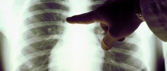
It is generally accepted that chest fluorography makes it possible to see the condition of only the lungs and heart. This is even evidenced by the constant medical stamp that is given to all patients without diseases of these organs - “Lungs and heart without visible pathology.” But for an experienced specialist, the x-ray photograph created during FLG will tell a lot.
This kind of image will show the lungs, the shadow of the heart muscle with the pericardium (pericardial sac), and the shadow of the spine. Sometimes the doctor can see the trachea, part of the esophagus, large bronchi and even the diaphragm on fluorography. But at the same time, of course, in relation to the lungs and heart, the picture is most informative.
While studying the image, the doctor checks the photographed organs for the presence or absence of changes caused by pathological processes, notes whether there are structural lesions of the lungs, and whether the heart muscle is enlarged in size. In addition, to an experienced specialist, such an examination can show neoplasms or atypical shadow areas, which are often a consequence of the development of certain diseases.
Fluorography is a quick screening method that makes it possible to assess the condition of the chest organs. It is considered very important that diagnostics reveals pathologies in the early stages, making treatment faster and more effective. Many people only after FLG learned about the presence of a disease that did not manifest itself with any symptoms.
Can fluorography show pneumonia?
Pneumonia can be seen both on fluorographic and x-ray examinations. The examination must be carried out by a qualified specialist, because the further treatment of the patient and, as a result, his well-being will depend on how he reads the photo of the lungs during pneumonia.
Experienced doctors at the Yusupov Hospital identify pneumonia and its varieties using both fluorographic and x-ray photographs. Pulmonologists, in turn, will be able to carefully and competently prescribe treatment, including high-quality drugs proven by many years of experience. You can make an appointment by phone or on our website by contacting our 24-hour online service.
What diseases can be diagnosed?
Most people think that FLG is only used to check the lungs for tuberculosis and do not know what diseases can be recognized with just one image. Indeed, the fluorographic method is capable of providing comprehensive information about such pathology, but this is not all of its capabilities. Then why else is this procedure needed?
Its results help diagnose other diseases of the lungs and mammary glands. Thus, based on the results of diagnostics, the following can be identified:
- neoplasms of both benign and malignant nature;
- areas of the inflammatory process (when it spreads to a large volume of tissue);
- pathologically formed cavities - cysts, abscesses, cavities, and it is also determined what they are filled with - gases or liquid;
- sclerotic changes (replacement of normal connective tissue);
- fibrosis (scarring and thickening of connective tissues).
If a person has had an incessant cough, shortness of breath, general weakness, or lethargy for a long time, then he or she should definitely undergo fluorography to rule out tuberculosis or pneumonia. According to fluorography, it is possible to determine the anatomical features of the cardiovascular system and respiratory organs, some diseases that can sometimes have a non-standard clinical picture.
For example, foreign objects in the respiratory tract are not always accompanied by typical symptoms, so doctors find it difficult to make a diagnosis. But timely FLG quickly helps to determine the causes of certain pathological manifestations. The procedure is indispensable for the diagnosis of malignant neoplasms and, in particular, central lung cancer.
It is typical for such a disease to develop latently for a long time and without affecting the patient’s condition in any way, since the lung tissues themselves are not subject to pain. And only when a person begins to feel certain symptoms, the disease may already be at a stage at which surgical intervention is not performed. Latently developing pathologies include sarcoidosis of the lungs and lymph nodes of the thoracic region.
What is it for?
When conducting a comprehensive examination of the patient, many specialists conclude that fluorography needs to be done. The operating principle of this technique is based on the ability of X-rays to penetrate tissue structures, which allows one to visualize the condition of the body.
The first mentions of the use of this technology appeared at the end of the 19th century, when scientists were looking for a more gentle alternative to the classic options for radiographic examination. Due to its high accuracy and accessibility, it was actively used for diagnosing organs of the respiratory system in order to determine all kinds of pathologies. Among them:
- Inflammatory processes.
- Oncological diseases.
- Infectious pathologies.
There are other problems that require fluorography. Thus, modern specialists prescribe it for intractable or chronic colds. The key object of examination is the chest area, which allows us to study the condition of the lungs, heart, mammary glands and bones. The technique is effective in the early stages of dangerous pathologies that have mild symptoms. These include:
- Tuberculosis.
- Pneumonia.
- Malignant formations.
As a preventive measure, fluorography of the lungs is performed for the following groups of people:
- Citizens who are over 16 years old. The optimal frequency of diagnostics is at least once every 2 years. If a person works in a hazardous enterprise and receives a high dose of radiation exposure, he is prescribed fluorography once a year.
- Patients of medical centers who came for an initial examination.
- Citizens living with a pregnant woman or newborn child.
- Diagnosed with HIV.
An unscheduled study of the lung is necessary if there are symptoms of the following pathologies and diseases:
- Various forms of pneumonia, pleurisy and inflammation in the lungs and other organs of the respiratory system.
- Tumor formations.
- Tuberculosis.
- Lung diseases.
In this X-ray examination, rays pass through the human body. An image of 256 shades of black and white appears on the screen, which reflect the condition of the chest and nearby organs.
Understanding the importance of fluorographic examination, you need to pay attention to the following features and advantages:
- Availability. Every medical facility has equipment for chest X-ray examination. To quickly detect lung cancer, bronchitis, tuberculosis and other dangerous pathologies, powerful installations are used that ensure high accuracy of the examination. On-site diagnostics are also practiced, which allows us to reach a larger number of people.
- High execution speed. The average duration of a lung study session for tuberculosis, bronchitis and other diseases is no more than 5-7 minutes. During this time, the doctor sets up the equipment and records the results. To make an accurate diagnosis, the patient needs to undress to the waist and stand in front of the matrix. The remaining actions are corrected by a specialist. After a couple of seconds, a picture of the lungs appears on the screen.
- Painless. Since the procedure is non-invasive, the patient does not feel pain or discomfort during it. This allows you to see any pathologies without negative effects on the skin.
- Information content. The technique provides accurate results and qualitatively displays the inflammatory focus.
Contraindications
Checking the respiratory system through fluorographic diagnostics is safe and harmless. This technique does not harm the human body, since the radiation dose remains minimal. In this case, the radiation quickly leaves the body, leaving no traces.
Under high loads, the likelihood of developing pathological processes at the cellular level, which are fraught with cancer, increases.
However, it is impossible to receive such radiation in a medical facility.
The use of this diagnostic technique is contraindicated for:
- Children under 15 years of age. To identify fibrosis of lung tissue or other manifestations of tuberculosis, it is enough to evaluate the body’s reaction to the Mantoux vaccination.
- Pregnant women and breastfeeding women.
- Bedridden patients who are unable to hold the body in an upright position.
- Patients with respiratory failure.
If there are such contraindications, other examination options are used, and pregnant women are allowed to undergo fluorography only after the birth of the child.
X-ray of the lungs
When performing a chest x-ray, no special preparation is required from the patient. Before starting the study, the patient undresses, removes metal jewelry and puts his long hair up. Then, specialists cover the abdomen with a special anti-radiation apron and place it in the gap between the radiation tube and the device for receiving X-ray data.
The procedure lasts only 1-2 seconds, during which the patient must hold his breath and remain absolutely still. In this case, there is a chance to get a very clear and informative picture.
When conducting targeted radiography, it is sometimes necessary for the patient to press at the angle required by the specialist to the radiation source. In general, a chest X-ray procedure may not take more than a few minutes.
It is important to understand that for preventive purposes, x-rays should be taken no more than 2 times a year, but if the doctor needs to monitor the progress of treatment or the progression of the disease, such an examination can be done several times a week.
Preparation and execution
Diagnostics does not require special or complex tedious preparation. The only thing the doctor will recommend on the eve of the procedure is abstaining from smoking for several hours and a light breakfast if the examination is scheduled for the next morning. The entire procedure, including undressing and dressing, will take no more than 5 minutes.
Step by step it will look something like this:
- the patient is invited to a special room intended for taking the image;
- he undresses to the waist, approaches the apparatus, and rises to a low step;
- the chin is located in a depression approximately corresponding to the average height of a person;
- the subject is warned not to hold his breath for a minute and not move;
- the nurse turns on the device, the picture is taken, the procedure is over.
In some situations, during fluorography, a protective apron is used, which protects the organs located in the lower part of the abdominal cavity from radiation. The results of the study are usually ready the next day.
Other forms of lung testing
When wondering how to check the lungs other than fluorography, you should pay attention to methods such as:
- Primary examination of the chest, during which not only the size, but also the shape of the sternum and the symmetry of its halves are determined.
- Assessment of external indicators of breathing, which include depth and rhythm.
- Using palpation, you can determine the shape and size of the organ.
- Thanks to percussion, you can analyze the sounds that are heard directly during tapping. They can be quite indicative of some diseases.
- Auscultation is possible, that is, an assessment of the sounds that are formed during the activity of the organ.
- In laboratory conditions, sputum can be examined.
- Carrying out linear or computed tomography.
- MRI.
Important!
If at least one of the symptoms appears, it is worth conducting a fluorographic examination.
Video: what can be determined using fluorography
Detection of tuberculosis
Tuberculosis is a disease that occurs quite often. First symptoms
, which should become an indicator for the survey, are the following:
- persistent cough that occurs throughout the day, at any time of the day;
- shortness of breath that occurs even without physical activity;
- wheezing in the lungs.
Tuberculosis on fluorography looks like focal shadows
, which are located in the upper part of the organ. If necessary, it is worth using other diagnostic methods, such as CT or MRI.
It is worth noting that early detected disease is easily treatable.
Therefore, it is very important to seek help from a specialist at the first alarming symptoms.
What are the contraindications?
The only contraindication is pregnancy. It is not recommended to perform a CT scan or x-ray with it. Pregnant women can undergo these tests only if vital signs of health are affected. For cancer patients, the elderly and young children, X-ray examinations are also not recommended. “And in these, as in other cases, the doctor must decide whether it is advisable to give the patient a load, whether this makes sense,” explains Sergei Anikeev.
For reference. X-ray, fluorography, computed tomography (CT) and magnetic resonance imaging (MRI) are hardware diagnostic methods. The foundations for the use of fluorography and radiography were laid immediately after the discovery of X-rays by Wilhelm Roentgen in 1895. CT and MRI are more modern diagnostic methods. The use of computed tomography was proposed in 1972 by engineer Godfrey Hounsfield and physicist Allan Cormack . The founding year of MRI is considered to be 1973, when chemistry professor Paul Lauterbur published an article about this technique in the journal Nature.
The essence of the procedure
Often, what diseases fluorography can detect depends on the correctness of the procedure. Anyone coming for an X-ray examination must remove all clothing from the waist down. For accuracy, remove the chain, earrings and anything “foreign” that may be reflected on the screen, and remove long hair up.
Pneumonia on X-ray
You need to press your chest as tightly as possible to the screen, lower your arms along your body, then follow the specialist’s instructions, take a deep breath and exhale. Now they additionally take a second picture by moving the person being examined with his right side towards the screen, while his arms should be raised up as much as possible, without bending.
Features when analyzing pulmonary fields
For the convenience of describing the localization of pathological shadows in the fields, they are usually divided into segments. In the description of the radiograph, the doctor indicates the serial number of the segment and the exact size of the formation.
Digital code
In the right lung it is customary to distinguish 10 segments, in the left, since its field is smaller due to the overlap with the cardiac shadow - 9. The principle of division into segments is based on the study of the branching of large bronchi. One segment is formed by one large bronchus.
Smoker survey
What else can a lung test show?
Any smoker has at least once thought about the question, is it clear on fluorography that a person smokes? It is worth saying here that healthy organs in the picture should look light gray and clearly defined.
Therefore, if smoking is the result of pathological changes, then fluorography of the smoker will not show anything.
But long-term, intense smoking will cause some changes to be visible in the picture.
The FG of a smoker with a good experience has its own characteristics and will indicate the presence of certain ailments.
- A smoker's organs have a denser pattern
compared to healthy organs. They may have a reticular structure, and the alveoli appear outlined black. - An enhancement of the vessel pattern can be observed in the peripheral parts of the organs. Dark dense structures alternate in the image, which shows non-functioning connective tissue.
- The density of the lung tissue changes
, which will certainly affect the results of the study. Lighter areas appear. - The roots of the organ also change, which will not be so clear in the picture. Additional shadows appear and the shape of the roots changes. In smokers, it is almost impossible to distinguish
the constituent parts of the lung root.
Fluorography of the lungs of a person with such a bad habit with these changes may indicate certain problems:
- Poor clarity indicates that fibrous tissue predominates in the organ and the lumen of blood vessels increases.
- Deformation of the roots indicates the course of some inflammatory processes.
- Increased density, easily noticeable in the image, indicates that the organ’s self-cleaning function is impaired, the lymph nodes are enlarged, and lymphatic fluid stagnates.
- In addition to changes in the structure of the lungs and improper functioning of the organ, diseases can occur, the appearance of which is provoked by nicotine.
Pregnancy and other contraindications
Among the contraindications to manipulation, there are two categories of prohibitions. The first is absolute, providing for pregnancy and childhood. Many mothers are puzzled by the question of at what age can babies have radio photography. It is believed that the minimum threshold is 15 years, since before this time it will be difficult for children to tolerate increased radiation exposure.
Among the relative contraindications, which are sometimes ignored in case of excess of benefit over harm, are:
- claustrophobia;
- dyspnea;
- general serious condition.
But sometimes even pregnancy, which is a contraindication for almost all types of diagnostics, does not become an obstacle to the procedure. It should be done only after preliminary consultation with your doctor. Control must be carried out by a specialist throughout the entire manipulation.
An important factor in the success of diagnosis and safety for the baby is scheduling an appointment no earlier than the fetus is 25 weeks old. During this period, he will have time to form basic systems. You also need to take care to find in advance a special lead apron that will cover your stomach from increased background radiation.
The reasons for “photographing” the respiratory system of pregnant women and women during the breastfeeding period should be as compelling as possible. As an argument, conventional prevention will not work.
Radiograph description protocol
Any therapist can decipher an X-ray of the lungs and see gross pathology, but a detailed conclusion is provided by a radiologist, based on a special protocol.
For convenience, the protocol contains a special analysis algorithm, which includes the following points:
- the exact name of the study (the doctor indicates the anatomical area of the image, projections: direct, lateral);
- the symmetry of the pulmonary fields is assessed;
- the presence of pathological shadows (focal, infiltrative, diffuse) or clearing in the area of the lung tissue is detected;
- strengthening of the pulmonary pattern: a violation indicates pathology of the pulmonary vessels;
- condition of the roots of the lungs - there is a violation of the structure of the lymph nodes, a study of the pathology of large bronchi, the lymphatic system (lymph nodes);
- description of the shadow of the mediastinal organs (important for diagnosing heart diseases): arch of the ventricles of the heart, aorta, pulmonary artery;
- the condition of the diaphragm and pulmonary-phrenic angles (sines): the symmetry of the position of the diaphragm, the angle of the sinus, its fullness (the presence of effusion in pleurisy) is assessed.
Encapsulated exudative pleurisy
Decoding the results
The need to undergo fluorography can be either forced or preventive. Therefore, not only sick people, but also healthy people who want to make sure of the presence or absence of pathologies of the chest organs are examined. To ensure the accuracy of the diagnosis, you should figure out how to independently decipher the results.
What the results look like for a healthy person
An experienced doctor will need seconds to confirm a fluorography image of a healthy person. To do this, it is enough to be guided by the following principles:
- The image of healthy lungs does not have dark spots and has a homogeneous structure. Any spot on the lungs during fluorography may indicate the development of an oncological process, infiltration, pneumonia or cysts.
- A photograph of a healthy person's lungs clearly shows the 5 separate sections and their actual dimensions. Traces of expansion indicate diseases of the respiratory system.
- Soft tissues have a uniform surface and clear dimensions.
The specialist also pays attention to the shadow of the heart. If there are abnormalities, you may need to be examined by a cardiologist.
What does tuberculosis look like in a photo?
Tuberculosis in a fluorography image is deciphered according to several signs. The first of these is a visible spot in the image, which has a heterogeneous structure and large size. Other arguments confirming the presence of tubercle bacilli in the body include:
- Patchy white or dark spots. To increase the accuracy of the results, the patient is prescribed additional diagnostics. If the spot is small, there may be no cause for concern. But if it constantly increases and becomes heterogeneous, the presence of an acute form of tuberculosis or an inflammatory process cannot be ruled out.
- Any darkening in the image for pulmonary tuberculosis with contours. In this case, additional therapeutic measures are not prescribed, since the body has overcome the infection on its own.
To make an accurate diagnosis, comprehensive fluorography should be performed.
General concept
Fluorography is a diagnostic procedure that arose at the end of the 20th century.
Important!
FG is an X-ray examination of the sternum using X-rays.
Such an examination has its advantages and disadvantages. Among the positive aspects
it is worth highlighting:
- low cost of examination due to the use of inexpensive large format film;
- the compactness of a modern fluorograph makes it possible to conduct research at the place of work,
using special mobile units; - It is possible to conduct several studies in a short period of time.
Recently, digital fluorography has become increasingly used, what does it show? Digital FG allows you to get a snapshot and identify lung pathologies. This simplifies the procedure and makes it even more economical.
The most significant disadvantage is the low resolution
the resulting image. If signs of disease are detected, specialists have to send the patient for x-rays.
Kinds
In modern medicine, 3 options for fluorographic examination are used:
- Film.
- Digital.
- Scanning digitally.
They differ in the type of equipment used to detect pathologies and the degree of effectiveness. Each option has the following properties:
- Film type. It does not guarantee high accuracy and operates by recording information on film through a special screen located behind the patient’s back. The advantage of this type is accessibility, because... Such equipment is installed in almost every medical institution.
- Digital. Representatives of this group are equipped with a matrix system that allows you to take photos and videos of pulmonary tuberculosis. Digital chest fluorography is characterized by a low degree of radiation, but increased accuracy. After recording the results, the specialist can print the image or send it to the patient by email. The option provides long-term storage of results.
- Scanning. Unlike the previous two types, this equipment reduces the intensity of radiation exposure, but does not guarantee the accuracy of the results detected.


