A cyst on the neck is a benign neoplasm, most often located on the front or side surfaces of the neck. This is a relatively rare disease of predominantly congenital nature. In the absence of complications - fistulas or infection, cysts grow slowly and do not cause noticeable discomfort for a long time. Varieties of cystic cavities require timely differential diagnosis from each other, from inflammation of the lymph nodes and a number of specific diseases. Treatment of cysts on the neck is surgical; the prognosis for recovery is favorable.
Development mechanism
Usually develops in babies during pregnancy.
Median neck cyst usually develops in babies during pregnancy. The embryos have gill grooves with a cavity that should grow with the baby. In some cases, this does not happen and the cavity does not heal, but remains.
In this place, a lateral or median cyst of the child’s neck is formed. The mechanism of occurrence of the neoplasm is not fully understood. Experts believe that congenital cysts and fistulas are formed in the presence of the following unfavorable factors:
- mechanical abdominal injuries;
- exposure to radioactive elements;
- intoxication of the body;
- uncontrolled use of medications prohibited during pregnancy;
- the expectant mother has serious chronic diseases;
- bad habits;
- genetic predisposition;
- nervous and exhausted state while carrying a child.
Lymphadenitis increases the risk of developing pathology.
A median neck cyst in adults (thyroglossal) develops with certain diseases and exposure to external factors. These include lymphadenitis, malignant tumors, infections, and mechanical injuries. In the vast majority of cases, purulent exudate is formed, which causes discomfort during swallowing. The tumor can rupture on its own, causing a fistula to appear.
Symptoms
Some forms of tumors on the neck in children or adults may be asymptomatic for a long time. As the hollow tumor grows, the following symptoms may be present:
- inability to fully flex the neck;
- when palpating the neoplasm, pain is felt;
- the tumor is mobile, the skin is unchanged, but redness is possible;
- the child cannot hold his head up;
- weakness, lethargy;
- low-grade body temperature, local fever is also possible;
- signs of general intoxication of the body - nausea, vomiting, general malaise.
If the process of suppuration has begun, the following clinical symptoms may be present:
- local redness of the skin, swelling;
- increased body temperature;
- weakness, dizziness;
- severe pain on palpation;
- purulent exudate flows out, less often into the oral cavity;
- the skin around the mouth may become crusty.
If such clinical signs are present, you should immediately seek medical help. The purulent process can lead to an abscess, phlegmon and other life-threatening diseases.
Symptoms of a neck cyst
It should be understood that the release of purulent exudate cannot be regarded as recovery and eliminating the need to see a doctor. The formed fistula never heals on its own, and the accumulation of fluid in the tumor almost always occurs again after some time. In addition, the risk of malignancy increases significantly.
Symptoms
Thyroglossal or branchiogenic cyst of the neck is detected after birth. There may be no symptoms until a certain age. As the child grows further, the lump increases in size and characteristic manifestations occur:
- pain when pressing on the affected area;
- inability to fully turn the neck;
- redness at the site of growth;
- decreased sensitivity of the facial nerves;
- The newborn cannot hold his head up on his own.
Inability to fully turn the neck.
If suppuration forms, the list of symptoms expands.
Swelling in the area of formation, increased body temperature, pain that increases with pressure, deterioration of well-being and weakness are noted. If the thickening opens spontaneously, a yellowish liquid flows from the site of the breakthrough.
Possible complications and consequences
- Cellulitis of the neck
- Branchiogenic cancer (rare)
Infected branchiogenic cyst. CT scan with contrast: a cyst at the level of the right angle of the lower jaw. There is thickening and strengthening of the cyst wall, as well as a decrease in density in the center of the formation. The sternocleidomastoid muscle is displaced posteriorly and laterally, the neurovascular bundle is displaced medially.
MRI, T2-weighted image without contrast: branchiogenic cyst in the left submandibular region. The signal intensity in the center of the cyst is significantly increased, the signal from the cyst wall is of intermediate intensity. The sternocleidomastoid muscle is displaced posteriorly and laterally, the neurovascular bundle is displaced medially.
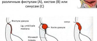
Classification
1 and 2 - median tumor, 3 - lateral cyst.
4 - median cleft, 5 - dermoid cyst, 6 - lateral fistula, 7 - cartilaginous remains. Based on their structure, there are several types of neoplasms:
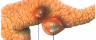
- dermoid cyst. Formed on soft tissues, most often on the surface. The capsule contains the contents of the sweat and sebaceous glands. Does not connect to the pharynx;
- cystic hygroma of the fetal neck is a soft lump that is located at the bottom of the cervical spine;
- the branchial is localized at the hyoid bone;
- venous is rare and is a thickening of a blue or brown hue;
- a lymph node cyst in the neck appears in the lymphatic vessels, the development of which is disrupted.
Based on the type of location, lateral and median neoplasms in the neck area are distinguished.
Lateral neck cyst
The lateral one is dangerous because it often turns into a malignant tumor.
This type of growth may not manifest itself until adolescence. During the period of rapid growth of the body, the lateral neck cyst also begins to increase in size. In some cases, pathology can be diagnosed after the birth of the child. This variety is dangerous because it often turns into a malignant tumor.
When suppuration occurs, the nerve endings are compressed and pathological processes develop. The patient has difficulty swallowing food. When spontaneously opened, a non-healing fistula forms in the neck.
Median neck cyst
The standard age for diagnosing this pathology is 5–6 years. It is detected when suppuration appears. The thickening is located under the tongue or at its root.
Located under the tongue.
There is difficulty speaking and swallowing, and the patient experiences a sensation of the presence of a foreign object in the mouth.
What are the advantages of Top Ichilov - clinic No. 1
Reviews about the treatment of head and neck cysts in Israel are only positive and confirm the undisputed reputation of Israeli clinics in the world of medicine, and in particular the Top Ichilov medical center, which is ensured by many factors:
- high level of qualifications of doctors and medical personnel;
- the latest diagnostic and treatment equipment;
- use of medicines only of the latest generation;
- affordable prices and comfortable payment terms - no prepayment and the ability to pay for each procedure separately;
- 24-hour support for the patient in his native language with resolution of all everyday and organizational issues.
- 5
- 4
- 3
- 2
- 1
(4 votes, average: 5 out of 5)
Diagnostics
A visit to a doctor begins with an external examination. The doctor palpates the affected area, feels the lymph nodes and neoplasm, then writes a referral for additional examination. A urine test, a blood test (general and for tumor markers), an ultrasound scan of the neck, and in some cases a computed tomography scan are prescribed.
Ultrasound of the neck.
Additionally, a puncture will be taken to examine the contents of the cyst. A needle will be inserted into the tumor formation, the existing fluid will be pumped out and sent for testing to the laboratory. Diagnostics will determine whether there are cancer cells in the contents. The examination includes fistulography. A special substance will be injected into the fistula canal, then an x-ray will be taken.
Diagnostic tests
Diagnosis begins with a visual examination and palpation of the cystic formation and lymph nodes. The doctor collects individual data and information about the course of the patient’s illness. To confirm or refute the diagnosis, laboratory and instrumental studies are required:
- histology – puncture of cystic contents is taken;
- blood sampling to determine the content of tumor markers;
- fistulography - x-ray examination to detect fistulas;
- Ultrasound of cervical cysts;
- Based on the results of the studies, the patient may be referred for a CT or MRI.
Ultrasound of a cervical cyst
Prescribing general urine and blood tests to eliminate the problem is not valuable, so they are prescribed directly during preparation for surgery to remove the pathology.
The main role is played by differential diagnosis - it is this that is decisive and sets the direction and procedure for surgical treatment.
A lymph node cyst in the cervical region must be differentiated from the following diseases: lymphadenitis - its development is provoked by the presence of infection in the lymph node, tuberculosis of the lymph nodes of the neck, hemangiomas, lymphomas, lymphogranulomatosis. The only possible solution is to remove the cystic cavity. The surgery is considered both easy and difficult at the same time. Any cystic tumor formed in the maxillofacial areas is completely removed, regardless of the results of differential diagnosis.
Carrying out diagnostic procedures in small patients is difficult, this is due to the anatomical features of the child’s body and the size of the respiratory organs.
Treatment
Treatment of a median cyst involves surgery. It cannot be eliminated using traditional medication methods. The drugs do not bring the desired effect and cannot reduce the size of the thickening. The only possible way to get rid of it is to remove the median cyst of the neck.
Treatment of the tumor involves surgery.
The operation is prescribed if the child is 3 years old. For newborns, surgical intervention is indicated only if the tumor makes breathing difficult, festers and can lead to death.
It is necessary to remove a growth on the neck as early as possible, since in some cases it can transform into a malignant formation. Spontaneous opening will also complicate the operation, as a non-healing fistula will form.
Surgical intervention has its own characteristics, which depend on the type of seal being removed:
- It is better to eliminate the median cyst after identification, since it can fester and infection can penetrate inside. If there is a fistula, its tissues turn blue. During the operation, all contents are completely removed to prevent relapse;
- It is more difficult to eliminate the lateral thickening, since there is a risk of touching the blood vessels. If a capsule is found, it is also removed.
The procedure is performed under local anesthesia. After completion, the patient is prescribed antibacterial drugs and anti-inflammatory drugs. For a long period it is necessary to treat the oral cavity with antiseptics. After the operation, the attending physician monitors how the patient swallows and whether there are any difficulties in speech.
A cosmetic suture remains at the site of excision, which is almost invisible.
A cosmetic suture remains at the site of excision, which is almost invisible. For scar resorption, special ointments are additionally selected. The recovery period takes no more than 2 weeks. A few months after surgery, a follow-up ultrasound will be required to rule out recurrence.
Prevention
Experts believe that it is impossible to prevent the development of the pathological process. During normal pregnancy, the risk can be slightly reduced if the following conditions are met:
- provide proper nutrition;
- avoid stress;
- to refuse from bad habits;
- minimize contact with toxic substances.
After the birth of the child, it is necessary to visit the pediatrician promptly. During a preventive examination, neoplasms are detected at the initial stage.
It is impossible to prevent the formation of a neck cyst. It is formed during the period of intrauterine growth, at the beginning of pregnancy. Often neither parents nor doctors notice the lump until it begins to actively grow and fester. The only treatment is complete removal. With timely surgery, the risk of possible complications is minimized.
Causes of pathology
What are the causes of this pathology dangerous to health? The culprit is a violation of intrauterine development at the embryonic stage, when the thyroid gland, hyoid bone and tongue are formed. According to doctors, there is a displacement of the thyroid rudiment to the middle part of the neck through the lingual-thyroid canal.
The resulting pathologies are congenital; their formation at the stage of gestation is facilitated by:
- heredity;
- stress load;
- smoking;
- alcohol abuse;
- production factors;
- uncontrolled use of medications contraindicated during pregnancy;
- mechanical injury to the expectant mother’s abdomen;
- use of medications that exhibit teratogenic effects.
The manifestation of the harmful effects occurs in the early stages of gestation.