Home > Encyclopedia of Oncology > Tumor marker CA 125: norm and interpretation of indicators
Thanks to innovative technologies, doctors have the opportunity to diagnose many diseases at the initial stage of development. Scientists have achieved particularly great success in cancer research, developing methods for identifying signs of pathology using blood tests. When identifying cancer in the ovaries, the most informative is the tumor marker Ca 125. An increase in this glycoprotein may indicate a cell mutation, but there are other diseases in which there is a deviation from the norm. You can understand what the analysis shows, how decoding is carried out and what is considered an increase in the norm by reading our article.
Important! The accuracy and reliability of the study largely depends on where and how the woman will donate blood for the Ca 125 tumor marker. Israeli clinics are considered the best, one of them is Assuta.
Some people believe that spending money on biochemical studies if a tumor is suspected is inappropriate, especially if there are no obvious symptoms. But in reality, a timely analysis with competent decoding can save your or a family member’s life. At the initial stage of cancer, it is much easier to cope with it, and the recovery period takes significantly less time. Having discovered that the indicators are elevated, the doctor will promptly prescribe additional tests and select an individual treatment program. Therefore, you should not delay contacting the diagnostic center.
Tumor markers and their significance
Tumor markers are specific proteins that are produced by malignant tumors or normal tissues in quantities exceeding the permissible limits due to the entry of cancer cells.
They cannot be used to make an accurate diagnosis, but detection of these substances in the blood and/or urine allows:
- suspect cancer and its location;
- distinguish a malignant tumor from a benign one;
- study the effectiveness of tumor therapy;
- detect relapse of the disease early;
- detect metastases before their clinical manifestation.
Reasons for increasing cancer antigen
The main malignant processes in the human body, in which the CA 724 tumor marker produces increased values:
- Oncological pathologies of the gastrointestinal tract:
- Colon cancer;
- Gastric adenocarcinoma;
- Malignant tumor of the esophagus;
- Tumor in the pancreas.
- Ovarian adenocarcinoma in women and testicular epithelial tumor in men;
- Small and large cell lung cancer;
- Adenocarcinoma of the mammary glands;
- Metastases in the liver.
Types of tumor markers
Currently, scientists have identified more than 200 types of tumor markers, because all neoplasms secrete their antigens.
The most commonly used tumor markers in diagnostics include:
- Alpha fetoprotein (AFP) is determined to detect liver carcinoma, the formation of metastases of oncological pathologies in other organs and monitor the effectiveness of therapy;
- Carcinoembryonic antigen (CEA) is a protein secreted by embryonic cells; its detection in an adult allows one to detect colorectal cancer with more than 50% accuracy, monitor postoperative relapse, and determine the stage of cancer;
- Human chorionic gonadotropin (hCG) is a marker that increases during pregnancy (it is the increase in the hormone that confirms its presence). If the indicator increases in men and non-pregnant women, testicular or ovarian cancer can be suspected, respectively;
- Prostate specific antigen (PSA ) is a polypeptide, a high level of which allows one to suspect a benign prostate tumor or prostate cancer in a patient;
- CA 15-3 (breast tumor marker) is a highly specific marker that allows you to diagnose breast cancer pathology in the initial stages, evaluate the effectiveness of therapy and identify relapses and metastases in the earliest period;
- CA 19-9 (pancreatic tumor marker ) is a glycoprotein that does not have sufficient specificity, suitable for studying the dynamics of tumor development and differential diagnosis with other formations of the pancreas;
- CA-125 is a specific high-molecular glycoprotein, a tumor marker used in the diagnosis of both ovarian cancer and its metastases;
- HE 4 (epididymal secretory protein) is a glycoprotein, increased production of HE4 has been detected in ovarian and endometrial cancer, rarely in lung adenocarcinoma; has greater sensitivity than CA-125 and is used together with it to confirm the presence of ovarian cancer.
CA 72-4 cancer marker
CA 72 4 - is a complex high-molecular glycoprotein (protein containing a carbohydrate component) of the mucin-like type, which is formed by synthesis on the surface of the epithelium. The tumor marker CA72-4 was first described in the study of breast adenocarcinoma. In most cases, the reason for the appearance of this tumor marker in the blood of men and women is the development of cancer cells in various organs of the human body:
- Gastrointestinal tract;
- Breast;
- Ovaries;
- Pancreas;
- Reproductive system.
But predominantly this tumor marker CA 72-4 is detected as a tumor marker of the gastric mucosa.
What is SA-125?
CA-125, or mucin-16, carbohydrate antigen 125 is an antigen located on the membranes of ovarian cancer cells.
Ovarian tumors represent a serious gynecological problem in all age groups of patients.
In no other human organ is there such a histological diversity of tumors as in the ovaries.
The CA-125 protein belongs to a specific type of epithelium and is normally found in the endometrial tissue of healthy women of reproductive age.
In this situation, changes in CA-125 depend on the phase of the menstrual cycle: a slight increase in its level is observed during menstruation (especially in the presence of endometriosis), as well as during normal pregnancy during the third trimester.
Physiological is the content of CA-125 in the uterine fluid, while it does not penetrate into the bloodstream. Minimal amounts of the glycoprotein can be found in the mesothelial tissues of the thoracic and abdominal organs. Reference (threshold) protein values in laboratory diagnostics are up to 35 U/ml.
MRI of ovarian cyst
On MRI, Graafian follicles appear as hyperintense formations with thin walls, which are surrounded by ovarian stroma, which gives a not very intense signal.
Hemorrhagic cysts are characterized by high signal intensity; a component with high signal characteristics (fat, blood, etc.) is visualized. With fat suppression, the signal intensity does not decrease, which confirms the presence of hemorrhagic fluid.
The contents of an endometrioid cyst on MRI are manifested by increased signal intensity. Remains hyperintense, unlike teratomas.
Ovarian dermoid cyst is a cystic formation with hyperintense signal containing septa. In fat-suppressing mode, a decrease in signal intensity is determined.
Who needs to get tested?
- First of all, this test must be taken by every woman who monitors her health. For screening purposes, the analysis is carried out for early detection and most effective treatment of cancer. The sooner an increase in tumor marker levels is detected, the greater the chances of successful treatment of the disease.
- It is important for those women whose relatives have been diagnosed with cancer to undergo the test. For this purpose, it is recommended to carry out the analysis once a year.
- If a woman has previously been diagnosed with benign neoplasms , such as leiomyoma, fibromyoma, functional ovarian cysts, neoplastic lesions, the doctor may prescribe a blood test for tumor markers to diagnose and differentiate tumors.
- It is mandatory for women who have symptoms of a malignant neoplasm to undergo the test. However, do not forget that a positive test for tumor markers is not specific and 100% confirmation of cancer, so additional instrumental research methods (ultrasound, tumor tissue biopsy, MRI) will be prescribed.
- After diagnosing a malignant tumor and carrying out conservative (chemotherapeutic, radiotherapeutic) and surgical (radical removal) treatments, the doctor prescribes repeated blood tests for CA-125 tumor markers. This is done to evaluate the effectiveness of the therapy.
- Subsequent analysis is carried out to identify metastases in distant organs, as well as for the early detection of tumor relapse. To do this, the test is taken monthly in the first year after treatment, then once every 2 months during the second year, and once every 3 months in the third year. In the absence of relapses and metastases, the test is performed 1-2 times a year until the end of the woman’s life.
What is ovarian cancer
Ovarian oncology is a malignant tumor located on the tissues of the ovary. Of the general statistics of mortality from cancer, 5% is occupied by oncology of the female prostate organs. The likelihood of the disease increases with age-related changes and hormonal processes in the body. Women aged 40 years and older are susceptible to ovarian disease.
The tumor is characterized by rapid growth, the cancer cell is actively dividing, spreading a malignant effect on the body. The causes of the disease are not fully understood. Experts suggest that oncology develops under the influence of the following factors:
- Late birth.
- Genetic predisposition.
- The result of abortion.
- Miscarriages.
- Chronic inflammation of the ovaries.
- Cysts and benign tumors of the female organs.
- The effect of hormonal contraceptives used orally.
- Lack of breastfeeding after childbirth.
Bad habits such as smoking and excessive alcohol consumption provoke ovarian cancer. Early onset of puberty and late menopause are also causes of the development of malignant formation of the female prostate organs. Timely diagnosis and examination make it possible to identify oncology at an early stage and begin timely treatment. Tumor markers are a way to determine oncology by collecting venous blood. The examination takes from one to ten days. The CA 125 test is used to diagnose a malignant ovarian tumor. Women who have reached adulthood are recommended to undergo preventive testing annually.
How to take a tumor marker test correctly?
To obtain the most accurate result of a tumor marker test, you need to prepare and adhere to simple but very important rules:
- Blood from a vein must be taken on an empty stomach ; the patient must not eat 8 hours before taking blood for analysis. Only water is allowed among drinks to prevent distortion of results.
- It is better to take the test in the morning , between 8 and 11 o'clock.
- A woman should give up alcohol and smoking at least three days before taking the test.
- Immediately before the test, you need to calm down , because... Nervous strain, along with nicotine and alcohol, can affect the final result.
- For a certain number of days before the analysis, you should not engage in intense physical activity..
- It is necessary to exclude medical procedures (physiotherapy, massages, ultrasound examinations) 3-4 days before the test.
- It is very important to monitor your diet during the week before the test: avoid fatty, fried and spicy foods.
- Consult your doctor about taking medications before the test , because... some of them may affect the result of the analysis.
- If a woman has any inflammatory diseases , the test should be postponed and taken after they are completely eliminated.
- The test cannot be performed during menstruation , because... how this period can be accompanied by a physiological increase in the level of tumor marker in the blood.
If all these rules are followed, the analysis will be clearly and correctly interpreted by the doctor. The result can be expected within 1-2 days after delivery.
Ovarian cyst or pregnancy
Most often, women confuse pregnancy with a follicular cyst or corpus luteum cyst. The signs of such cysts are very similar to the early symptoms of pregnancy. The hCG level increases with an ovarian cyst, as with pregnancy, so at the first sign you should consult a gynecologist.
The Oncology Clinic of the Yusupov Hospital offers a wide range of surgical interventions in the field of gynecology. Our gynecological oncologists are true professionals in their field, using in their practice only the latest treatment protocols that meet international standards.
Thanks to the qualified specialists of the Yusupov Hospital and modern European equipment, you will receive high-quality specialized advice. The medical staff guarantees an individual approach to each patient, as well as comfortable conditions of stay in the hospital. The clinic’s doctors will be happy to provide you with detailed information about ovarian cysts, treatment features, diagnostic ultrasound, as well as prices for services. You can make an appointment and consultation by phone.
Decoding the results of the CA-125 analysis
After blood is drawn, it is sent to a laboratory where the tumor marker level is determined. After receiving certain numbers, a very important and responsible stage begins - deciphering the results. It requires a high level of professionalism for accurate verification of the diagnosis, and, accordingly, correctly selected treatment for the patient.
Results:
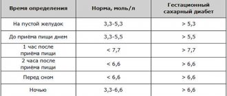
- Threshold values for protein in laboratory diagnostics are up to 35 U/ml.
- Under normal conditions, in the absence of pathology, the tumor marker level fluctuates between 10-15 U/ml.
- An increase in its level to 35 U/ml is observed in women during menstruation, as well as in the first trimester of pregnancy.
- If a woman’s screening examination revealed an increase in the level of the CA-125 tumor marker above 35 U/ml, you should not make hasty conclusions and think about the worst prognosis.
Reasons for increased antigen not of an oncological nature
In many cases, a slight increase in the cancer marker of the stomach can be detected in non-oncological diseases:
- Gastrointestinal diseases:
- Chronic stomach diseases (stomach ulcer, gastritis, multiple polyposis);
- Diseases in the pancreas (acute and chronic pancreatitis, cysts);
- Intestinal diseases (ulcerative colitis);
- Reflux diseases of the digestive system.
- Chronical bronchitis;
- Cirrhosis of the liver.
For example, with gastritis, the CA 72 4 level can be increased to 13-16 units/ml. Pregnancy may also be the cause of an increase in this tumor marker.
Would you like to receive an estimate for treatment?
*Only upon receipt of data on the patient’s disease, a representative of the clinic will be able to calculate an accurate estimate for treatment.
SA-125 analysis indicators
An increase in the CA-125 antigen in the blood to 100 U/ml can result in various non-tumor processes in a woman’s body:
- inflammatory changes in the abdominal cavity (chronic hepatitis and cirrhosis of the liver, chronic pancreatitis, peritonitis),
- pelvis (pelvioperitonitis),
- cystic ovarian abnormalities,
- endometriosis,
- adnexitis,
- other gynecological infections, pleurisy, autoimmune diseases.
Cystic ovarian abnormality
Ovarian cysts are not true tumors, since in their presence blastomatous (cellular) growth is not observed in the tissues.
They are formed as a result of retention or accumulation of various contents and gland secretions in the cavity. Cysts can form due to tissue softening due to hemorrhage and necrosis.
The presence of cysts in a woman can affect the level of tumor marker CA-125 in the blood, which begins to reach 60-70 U/ml (up to 100 U/ml).
Correct differentiation of ovarian cysts, timely detection, and examination of patients is very important. To accurately confirm the diagnosis, a bimanual examination and ultrasound of the female genital organs are performed.
After a correct diagnosis, the doctor chooses the most appropriate treatment tactics: from conservative and expectant (functional cysts can resolve on their own) to surgical.
Women who have reached the age of menopause require a careful approach. If an elevated level of the CA-125 tumor marker is detected, it is necessary to undergo regular tests to monitor the course of the disease and take the necessary measures.
Endometriosis
This disease is benign and is characterized by the presence of endometrial (tissue inside the uterus) glands and cells outside the uterus.
In 75% of cases, it is observed in women aged 25-50 years, regardless of its location. The incidence of endometriosis in women during the reproductive and later periods averages 10-15%.
The level of tumor marker CA-125 in the blood with this pathology can reach 100 U/ml, which is significantly higher than normal.
Considering the fact that the disease is widespread, it is necessary to carefully differentiate it from malignant neoplasms when an elevated level of CA-125 protein is detected.
A histological examination of the biopsy material, as well as ultrasound data of the female genital organs, can help with this.
It should be remembered that patients with endometriosis are not cured until menopause. Drug therapy is carried out to suppress and reduce endometrioid lesions. For this purpose, properly selected hormonal treatment is used. If indicated, various surgical interventions are performed.
Uterine fibroids
A benign tumor develops from the smooth muscle tissue of the uterus and ranks first in frequency among tumors of the female reproductive system.
According to statistics, 20% of women over the age of 30 have uterine fibroids of various sizes.
The tumor usually does not appear until puberty, develops only during reproductive age and regresses after menopause.
Scientists associate its development with hormonal disorders in a woman’s body.
After the development of this pathology, the level of tumor marker CA-125 in the blood can reach 90-110 U/ml.
To confirm the diagnosis, it is necessary to undergo a bimanual examination and ultrasound of the female genital organs. Only a thorough examination and high professionalism of the doctor will help identify the cause of the increase in tumor markers, as well as take the necessary measures to treat the pathology.
The first step in the management of patients with uterine fibroids is to clarify the shape and growth rate of the tumor. Depending on this, the patient’s treatment tactics are chosen. The main component of conservative treatment is hormonal therapy with progestogens.
In the presence of large tumor sizes and the development of complications from the uterus and adjacent organs, surgical treatment is indicated.
Tumor marker level during pregnancy
In some cases, it is possible to detect elevated levels of CA-125 protein during a normal pregnancy in the first trimester.
This is due to the fact that during this period the female body undergoes significant hormonal changes, general changes in the body, and emotional instability.
Therefore, an increase in the level of CA-125 antigen in the blood is a physiological change. In addition, fetal cells are capable of producing this antigen, as a result of which its level in the blood increases.
Due to the above reasons, the level of CA-125 protein can reach the threshold level of 35 U/ml in the blood and even slightly exceed it. However, this situation only requires careful monitoring of the antigen level and additional examination of the woman. Subsequently, it is mandatory to donate blood again for analysis.
Menopause (menopause)
After menopause (cessation of menstruation), a woman’s body becomes more vulnerable, which is associated with changes in hormonal metabolism.
Detection of an increased level of the CA-125 tumor marker in the blood during this period will no longer be characteristic of pregnancy, menstrual irregularities, endometriosis (the disease regresses) or functional cystic ovarian abnormalities.
Detection of deviations from normal values requires additional research: ultrasound of the female genital organs, bimanual examination, repeated testing for tumor markers in the blood.
If the level of the CA-125 antigen increases and there is no increase in it, the presence of a benign tumor can be assumed. A significant increase, together with symptoms characteristic of the menopausal period (bleeding from the genital tract), will confidently indicate in favor of a malignant neoplasm.
Significance of CA-125 in ovarian cancer
The main purpose of testing for CA-125 in the blood is to laboratory confirm or exclude the presence of a malignant neoplasm in a woman.
In the case of ovarian cancer, the CA-125 antigen level increases more than 5 times compared to the threshold level, thus reaching numbers of more than 100 U/ml. Do not forget that in ovarian cancer the level of the CA-125 antigen may be normal.
This, in turn, should not be regarded as a clear exclusion of cancer. The diagnosis can be established only after conducting a double analysis with increasing indicators over time.
To clarify the diagnosis, if there are controversial indicators of the CA-125 protein, it is useful to take a test for HE-4, which is more sensitive. A combined test with the calculation of a special index makes it possible to identify oncopathology at an early stage, as well as differentiate malignant pelvic tumors from benign ones.
In the early stages of cancer, the CA-125 indicator increases slightly or does not change. As the tumor grows and the stages of the disease progress, its level in the blood may exceed the norm. Testing for this antigen can be used to predict the course of the disease: if the level decreases after starting treatment, patients have a significantly increased survival rate.
It is very important to monitor the woman after treatment in the tumor remission stage. During this period, the level of CA-125 protein is reduced to zero. Increasing it even to a threshold value may mean a relapse even before its clinical manifestation. This condition requires careful examination.
If a constant antigen level is established after the start of treatment, one can judge a poor response to the therapy and continued growth of oncological pathology.
False-positive results when assessing CA-125
Diseases that do not have a tumor origin lead to false-positive results when assessing the level of the CA-125 antigen. They are named as such because the main purpose of the test is to confirm and show the presence of cancer.
These pathologies include:
- inflammatory diseases of the abdominal cavity (peritonitis, chronic hepatitis, chronic pancreatitis);
- pelvic inflammatory diseases (pelvioperitonitis);
- inflammatory diseases of the chest cavity (pleurisy);
- autoimmune diseases;
- infectious lesions of the female genital organs.
Additional examination methods help in differentiating these diseases and oncological pathologies. It is important to promptly confirm and treat, or exclude any damage to internal organs.
Preventive examination
Tumor marker CA 125 is a high molecular weight glycoprotein that every woman should know about. With age, hormonal and physiological changes occur in the body. The period of menopause and the onset of menopause is accompanied by complications, diseases of the ovaries and genitourinary system. The CA 125 test identifies emerging pathologies of the internal reproductive organs. Women over 40 years of age are recommended to undergo preventive examinations annually to rule out cancer.
A genetic predisposition to malignant tumors is also an indication for antigen testing. Environmental pollution, chemicals and unhealthy foods contribute to the development of cancer. The CA 125 tumor marker makes it possible to identify a dangerous disease at an early stage of development, which increases the chances of recovery. Missed treatment time leads to death. In the best case, stable remission is achieved and pain symptoms are eliminated.
The diagnosis must be made by a professional doctor who knows the subtleties of variations and deviations of indicators from the norm. It is important to remember that positive examination results do not exclude a cancer process. For tumors in the early stages, the indicators do not show noticeable deviations from the norm. It is important to undergo a comprehensive examination to confirm the correct diagnosis.
Preventive analysis is also carried out after removal of a malignant neoplasm. Cancer may return after a while. To avoid relapse, the body must be regularly examined for the concentration of antigens in the blood. Timely diagnosis will ensure life expectancy and material savings.
What else can the CA-125 tumor marker indicate?
When a high level of protein is initially detected, it is necessary to carry out a qualitative and detailed differential diagnosis with other oncological diseases.
The CA-125 protein is not strictly specific for ovarian cancer; it is also detected in a number of other oncopathologies:
- mammary cancer,
- uterus, endometrium,
- pancreas,
- lungs,
- liver and stomach.
If there are no signs of ovarian cancer on ultrasound, MRI, or histological examination of biopsy material, it is necessary to conduct additional diagnostics of the pathologies of other organs listed above.
Indications
Most often, a referral to test CA-125 levels is given by an oncologist or gynecologist.
An antigen test is recommended in the following cases:
- for suspected ovarian cancer
- assessment of the risk of recurrence or metastasis of serous ovarian cancer after therapy;
- control of endometriosis treatment;
- an additional marker in the diagnosis of adenocarcinoma of the pancreas, gastrointestinal tract and others (bronchi, kidneys, testes, mammary glands, etc.).
Most often, CA-125 is used to monitor the recurrence of an ovarian tumor: an increase in the level of CA-125 (even in the absence of clinical and radiological evidence of relapse) indicates a biochemical relapse of the tumor, which is ahead of the clinical one by 2-6 months.
In clinical studies, CA-125 has proven to be a reliable marker for monitoring patients with B-cell non-Hodgkin lymphoma.
Bottom line
When prescribing an analysis for a tumor marker, a woman must take preparations before it with complete seriousness, otherwise the result and further interpretation may be incorrect.
The following factors may affect the test, so it is worth preparing:
- eating before delivery;
- drinking alcohol and smoking several days in advance;
- drinking coffee, tea before delivery;
- use of medications;
- undergoing ultrasound and x-ray examinations;
- eating spicy, fatty and fried foods;
- stress;
- menstruation.
After eliminating these factors, it remains to correctly decipher the table of analysis results. This requires an excellent specialist and additional research methods. If necessary, the test is repeated.
In addition, an additional analysis for HE-4 may be prescribed. We must not forget that the absence or presence of an increased level of antigen cannot completely exclude or confirm the formation of a malignant tumor.
A timely diagnosis and correctly chosen treatment tactics are the key to an effective and speedy recovery for the patient!
Interaction with other markers
Due to the rather low specificity of analysis for the CA 72-4 tumor marker, there is a need to conduct additional studies of this antigen in combination with different tumor markers.
- CA 242 – general monitoring of malignant processes in the gastrointestinal tract;
- CA 199 – pancreatic tumor monitoring;
- CA 153 – monitoring of metastatic adenocarcinoma of the breast;
- CA 125 – monitoring of ovarian cancer;
- MSA (mucin-like cancer-associated antigen) – monitoring of breast adenocarcinoma;
- CEA (carcinoembryonic antigen).
Carrying out these combined studies of the antigenic properties of cells helps to establish a more accurate diagnosis. Let’s say that in case of tumor recurrence, the CA72-4 tumor marker gives a value within 78%, then in combination with CEA it increases to 87%.
For what purpose is analysis carried out on TAG 72:
- For the initial diagnosis of adenocarcinoma and ovarian cancer, clinical and instrumental research methods are used;
- To assess the result after surgery and further monitor the condition of a patient with stomach or ovarian cancer;
- For early detection of metastases and relapses before their active manifestation;
- To detect neoplasms of a malignant and benign nature in the reproductive system of women.
A blood test means a study of blood composition and comparison with standard indicators.
Usually, along with analysis for the CA72-4 tumor marker, patients are prescribed biochemical and general blood tests, clinical urine analysis, ultrasound and FGDS (fibrogastroduodenoscopy). It is also possible to use a relatively new method for diagnosing carcinoma using saliva to detect Ca2+ levels.