Absorption of nutrients occurs in the intestine; the important role of this organ in the formation of immunity has also been proven relatively recently. In this regard, intestinal problems affect the general condition of the body and require immediate treatment. One of the diagnostic methods that makes it possible to identify many diseases is intestinal irrigoscopy.
More information about the irrigoscopy research method
Irrigoscopy (irrigography) helps to conduct a qualitative and informative examination of the intestines. Irrigoscopy uses barium sulfate, a contrast agent, for examination. This method is one of the safest, and it can be used to identify the vast majority of diseases of the large intestine - polyposis, diverticulitis, Crohn's disease, fistulas, ulcerative colitis, various malignant neoplasms (cancer).
The principle of irrigoscopy is based on the ability of the intestine to transmit x-rays through itself. When a contrast agent is injected into an internal organ, the image taken will make it possible to see all the anomalies present in it, which is not possible when examined by other methods.
The procedure itself is quite easy to tolerate, lasts from 10 to 50 minutes, is not as traumatic as a colonoscopy, and requires less radiation exposure than an MRI.
The examination is carried out with a Bobrov apparatus, which is a container (volume from 1 to 3 l) for a contrast agent with 2 silicone tubes attached to it. At the end of one of the tubes there is a bulb for pumping air, the other tube ends with a tip for introducing a contrast agent into the rectum.
What is the difference between irrigoscopy and irrigography?
Irrigography and irrigoscopy are fundamentally different from each other in that in the first case, X-ray irradiation is carried out in small single doses only 4-5 times and that’s it (this examination is carried out mainly for children, to reduce radiation exposure). And in the second study, X-ray irradiation is carried out continuously in several portions, the radiation load is greater, and is used in adults. When performing fluoroscopy, images are always taken as documentary evidence of the pathology obtained.
When and to whom is irrigoscopy performed?
The procedure is prescribed to patients who cannot undergo colonoscopy or whose results are in doubt. As a result of irrigoscopy, information is obtained about the state of the intestinal system not only in straight sections, but also in bends.
In this way you can find out information about:
- the state of the intestinal mucosa, the functional state of its various parts - the appendix, the ascending colon, the small intestine, the descending rectum;
- size, diameter and location of the lumen of the large intestine;
- elasticity of intestinal walls;
- the functioning of the bauginian valve (intestinal valve), which is located in the place where the ileum passes into the colon (in normal condition it allows intestinal contents to pass in only one direction).
Using this method, you can evaluate the course of the disease over time. A study of this kind is prescribed to clarify the diagnosis if there are the following complaints from the patient:
- discomfort and pain in the anal area;
- bleeding from the rectum (suspicion of hemorrhoids);
- chronic constipation or diarrhea;
- abnormal discharge (pus, mucus) from the anus.
If there is a suspicion of the presence of a malignant tumor, then such a study is preferable, as it gives more accurate results.
An appointment for irrigoscopy is given by a coloproctologist, taking into account the general condition of the patient, possible contraindications and concomitant diseases.
Patient reviews
When patients are prescribed irrigoscopy, they first of all understand what it is. And the next step for many is to find reviews of irrigoscopy from those who have already done it.
Oksana, 40 years old: I went for irrigoscopy quite boldly, since I had already encountered sigmoidoscopy and it turned out that it was not at all fatal. An X-ray of the intestines with barium is absolutely painless. Only the preliminary enemas gave me discomfort.
Larisa, 33 years old: The procedure is necessary. There is absolutely no need to be afraid, there is nothing terrible about it. The most important thing is not to engage in self-hypnosis about her. For example, what in the process can break from barium. After some manipulations, a small residue remains, but this is quickly forgotten as soon as you leave the hospital.
Natalya, 45 years old: The biggest disadvantage of this examination is the high radiation exposure. Therefore, I believe that you should agree to this procedure only for strict indications, when other diagnostic methods have already been exhausted. Apart from radiation exposure, there is nothing to be afraid of in this procedure.
The main advantage of irrigoscopy is its ease of implementation, limited intervention in the body and the absence of the need for expensive equipment. Patients should not be afraid of her. When filling the intestines with contrast material, the patient may experience some discomfort, but otherwise the procedure is completely painless and does not require anesthesia.
How to prepare for irrigoscopy
The information content of the study depends on the quality and correctness of preparation for the procedure. Poorly cleaned bowel may prevent it from filling with contrast material and lead to incorrect conclusions when assessing the results of the procedure.
Preparation for irrigoscopy has its own peculiarities, and they consist of a set of such activities:
- diet before irrigoscopy . It begins to be observed several days before the examination. You should exclude from the menu foods that contribute to gas formation and bloating:
- porridge (oatmeal, pearl barley, millet);
- raw vegetables (carrots, beets, cabbage), herbs and fruits (peaches, bananas, apples);
- legumes (lentils, peas, beans);
- dairy products;
- coffee, kvass, carbonated drinks;
- fatty meat and fish, rich broths;
- sausages;
- bread made from wholemeal and rye flour.
It is recommended to eat the following dishes:
- weak broths, boiled and steamed dishes;
- boiled lean meat and fish;
- fermented milk drinks;
- porridge (semolina and rice);
- dry biscuits and crackers made from wheat bread;
- compotes, weak green or herbal teas.
Liquids should be consumed at least 2 liters per day. Before the day of the procedure, you can eat light meals for lunch; there should be no dinner; breakfast on the day of irrigoscopy is contraindicated. If the examination takes place after lunch, the patient can drink water. If the patient has diabetes mellitus (especially type 1), then consultation with a doctor is required on the issue of forced fasting, since lack of nutrition can cause serious consequences - even coma.
- Enemas . A well-cleaned intestine is a necessary condition for high-quality diagnostics. On the eve of irrigoscopy at noon you should take 2 tbsp. spoons of castor oil or 150 ml of 30% solution of magnesium sulfate. This will help cleanse the entire upper colon. Cleansing enemas can be used to thoroughly rinse the intestines. The volume of water poured is about a liter. The purification procedure is carried out until the poured water is clean. Enemas are given twice - the day before and on the day of the examination to thoroughly cleanse the intestines.
- Using laxatives also helps empty the bowels . Cleansing is carried out with various medicines at home: Fortrans, Duphalac, Flit, Lavacol and others. With their help, preparation for cleansing will be carried out according to the correct scheme. You should stop taking medications at least 6 hours before the examination. The drugs should be used in accordance with the instructions according to the indicated schemes:
- Preparation for irrigoscopy with Fortrans goes like this:
- The drug is taken 2 hours after the last meal;
- one sachet of medicine is dissolved in warm water (1 liter). During the evening you need to drink 4 sachets (4 liters of liquid) in this way. Over the course of an hour, drink a liter of laxative in small portions at a rate of 1 glass of liquid over 10-15 minutes.
The effect of the drug begins soon after drinking. The solution itself has a sweetish taste, and a gag reflex may occur. Then you should take Motilium or Cerucal and suck a slice of lemon after each liter of liquid.
- Cleansing with Fleet:
- The drug is taken one day before the examination in the morning;
- 1 bottle of the drug is dissolved in water (100 ml) and the resulting solution is washed down with a glass of water. Instead of lunch, drink 3 glasses of water or green tea. Dinner is also prohibited;
- in the evening, drink another diluted bottle of the drug and wash it down with a glass of plain water, broth or clarified juice.
- Cleansing with Lavacol:
- dissolve a sachet of the drug in water (200 ml). During the evening you should drink 3 liters of solution, 1 glass in 20 minutes. This procedure should begin 15-20 hours before irrigoscopy;
- Sometimes nausea and vomiting occur during administration (although Lavacol is better tolerated than Fortrans due to its salty taste).
If the patient is taking any drugs that affect blood clotting (Aspirin), you should notify the doctor who will do the examination. It is advisable to stop taking such medications several days before preparing for the procedure.
It is very important to prepare for the procedure, since the accuracy of the examination depends on this part of the preparation.
Preparation
The results of irrigoscopy are greatly influenced by the thorough preparation of the patient - the result is distorted by intestines that are not completely cleared of food debris, barium residues from previous studies, problems with retaining the barium mixture in the intestine, and a psychological factor.
Therefore, special preparation is required for qualitative research:
- 3-4 days before irrigoscopy, the use of a diet with a decrease in the amount of fiber and protein, gas-forming products is indicated;
- in 2-3 days, drugs that increase or decrease intestinal motility (no-spa, papaverine, drotaverine, aminophylline, halidor, metoclopramide, etc.) are discontinued;
- the day before the study, therapeutic fasting with increased drinking of water is indicated - at least two to three liters;
- from 18.00 on the night before the procedure you must stop eating and drinking. It is necessary to thoroughly cleanse the intestines of feces using an enema and using a solution of liquid with laxatives (usually Fortrans is used). This can be a solution of polyethylene glycol, lactulose, dissolved in three liters of water. This solution is drunk gradually during the day preceding the irrigoscopy. As you take the medications, you will experience profuse stools until water leaves the intestines.
It is important to tell your doctor about the medications you are taking, especially if they are insulin medications, anti-clotting medications, or anti-inflammatory medications. These drugs are discontinued the day before the study, except for insulin.
How is the examination carried out?
The technique is practically painless and safe. Immediately before the examination, the contrast agent is dissolved in water (40 g of barium per 2 liters of liquid) and heated to 35°.
A special container in Bobrov’s apparatus is filled with the prepared solution.
The subject lies on his side on the table, bends his knees and presses them to his stomach, and puts his hands behind his back. Controlling the process with fluoroscopy, the tip of one of the tubes of Bobrov’s apparatus is inserted into the rectum. Using a bulb, air is pumped, creating excessive pressure, due to which the contrast agent (barium sulfate) rises through the second tube and is delivered into the intestine through the tip. Moving under pressure, the contrast follows all the bends of the intestine; after the suspension has reached the point of articulation of the large and small intestines or a barrier to fluid, filling with the solution is stopped.
After the solution is administered, a series of photographs of the abdomen are taken. At the doctor’s command, the patient turns over onto his stomach, side, or back. This is done so that the contrast is evenly distributed in the intestines. As the barium is distributed, a series of targeted and overview photographs are taken, and after complete filling, another overview photograph is taken. This technique is called tight contrast.
Bobrov's apparatus
The next step is to remove the tip of the tube from the rectum and have a bowel movement.
To obtain more detailed and clear images, the double contrast method is used. To do this, the intestines are filled with air. In this case, the contrast remaining on the intestinal walls allows us to examine the posterior intestinal wall.
The double contrast method is indispensable for diagnosing malignant tumors in the large intestine.
But double contrast may not always be applicable - it cannot be performed on patients with poor health, those whose intestines are of significant length, or if intussusception is suspected. When the study is done when there is a threat of intestinal perforation, the barium enema is replaced with another water-based contrast agent.
During the examination, the patient may experience some discomfort and spasms during the period of pumping solution or air.
Indications and contraindications
Intestinal irrigoscopy is indicated in the following cases:
- cicatricial adhesive processes that arose after surgery or against the background of protracted inflammatory processes;
- single or multiple pathological protrusions of the intestinal walls (diverticula);
- intestinal fistulas;
- chronic inflammatory processes;
- significant enlargement of the entire large intestine or a separate part of it;
- the length of the sigmoid colon is more than 46 cm;
- frequent abdominal pain of unknown etiology;
- stable stool disorders (constipation or diarrhea);
- monitoring the condition of the intestine after resection;
- detection of blood streaks in stool;
- assessment of the condition of surgical anastomoses between intestinal walls.
This method becomes very relevant if for some reason a colonoscopy cannot be performed or if the results it gives seem questionable. In addition, such a diagnosis is mandatory if cancer is suspected in a patient with poor heredity or in those who are being seen by an oncologist.
Contraindications to irrigoscopy are not numerous, but they do exist: the patient’s general serious condition caused by cardiac or respiratory failure, penetrating damage to the wall of the large intestine, pregnancy, acute inflammatory processes in the intestine. Before prescribing irrigoscopy, the proctologist must weigh all contraindications and come to the conclusion that the likely benefit, in the case of a particular patient, outweighs the possible risk.
Very rarely, during the procedure, a perforation of the intestine occurs, leading to the leakage of contrast material into the peritoneum.
How are x-rays taken?
With irrigoscopy, the doctor receives information during the procedure and after - when studying the photographs taken. There is a standard sequence of moments when X-rays are taken:
- the first survey image is taken before contrast is introduced into the intestine;
- a series of overview and targeted photographs of those areas that are needed for detailed study are taken during filling with contrast;
- the next overview photograph is taken after bowel movement;
- the final overview image is obtained after filling the intestines with air (dual diagnosis).
This order and number of shots are standard. If necessary, their number can be increased.
If there are no pathological changes, the image will show a distended intestine with distinct physiological curves. The mucous membrane will have a feathery pattern and a smooth surface without inflamed areas or ulcerations. After the contrast is removed, the intestines in the photo “deflate” and return to their normal state.
The radiograph clearly shows minute damage and ulcers of the mucous membrane, cicatricial changes, polyps and diverticula in the intestinal lumen. The images also show the location and size of the inflammatory focus, the line between normal and damaged areas of the mucosa.
What will irrigoscopy show?
This type of examination is very accurate and almost always indicates the presence of the correct disease. But a lot also depends on the qualifications of the specialist who is caring for the patient. So, with the help of irrigoscopy you can detect the following diseases:
- Irritable bowel syndrome (refers to disorders of the functional functioning of the intestine). On x-rays it appears as uneven folds, as different parts of the intestine are strained differently during work. The bowel movement does not occur completely; the contrast agent remains inside, since the coordination of the colon is impaired. Constrictions in some places are also visible, which are caused by spasms.
- Chronic colitis is observed by various lumens in the colon, which appear due to improper contraction of smooth muscles in the organ. The lumen narrows due to frequent spasms, a large number of inflammations appear on the walls, which contract unevenly, and folds are clearly visible.
- Ulcerative colitis. The mucous membrane has many small spots, ulcerative wounds and dead areas of tissue. The so-called “pseudopolyps”, which appear due to scarring of the tissue, are clearly visible. The intestinal lumen either narrows or expands (not in all cases).
- Diverticulosis is manifested by an uneven relief of the intestine; large and small diverticula are present on the walls, along with which infiltrates often appear, which reduce the elasticity and extensibility of the dermis.
- Bowel cancer. Small round-shaped formations are visible on the walls, and the appearance of rings (polypoid carcinomas), which are the contours of neoplasms, is also possible. As a rule, there are no folds in these places.
- An intestinal polyp appears as a round or pointed neoplasm on the mucosa, which sometimes has a stalk.
Possible complications after the study
Although most patients tolerate barium enema well, some may experience the following side effects:
- constipation _ They are considered a normal reaction to absolute bowel movement. If there is no stool for more than 2 days, laxatives are used. The color of stool may be lighter than usual - this is typical for this condition, since the remains of barium suspension are removed, but it does not cause any harm to health;
- mechanical obstruction . It may appear during the examination and requires medical attention;
- intestinal perforation, development of barium embolism, abdominal leaks and granulomas are extremely rare complications of the procedure that require medical attention.
Medical attention is also necessary if the following symptoms occur:
- temperature 38 °C and above;
- severe vomiting;
- dizziness, severe weakness, loss of consciousness;
- severe pain in the abdominal area;
- bleeding from the rectum, bloody diarrhea (diarrhea).
Such complaints are also possible for reasons not related to the study. But only a doctor can determine this for sure. If the procedure is performed correctly, taking into account all contraindications and the patient’s health condition, then irrigoscopy takes place without consequences.
Research technique
It is worth talking in detail about the process of irrigoscopy so that the patient can understand what it is.
The duration of the procedure is approximately thirty minutes. In some cases it lasts longer.
First, a survey fluoroscopy is performed to ensure that the patient is fully prepared for the examination.
Then a tip is inserted into the anus and attached to the Bobrov apparatus. Barium, preheated to human body temperature, begins to flow through the tube. About two liters are pumped. The device is made in such a way that the contrast is not able to come back out.
If the patient's intestines are in critical condition, then other less active substances are used.
In some cases, a certain amount of air is introduced first to improve visibility. It allows you to completely straighten the organ, opening its most remote areas for study.
Then the X-ray equipment is turned on and the specialist takes the required number of images.
Contraindications to irrigoscopy
Irrigoscopy also has contraindications. These include:
- inflammatory bowel diseases in the acute phase and widespread localization (ulcerative colitis, diverticulitis);
- pregnancy;
- general serious health condition of the patient (arrhythmia, severe heart failure, etc.);
- perforation of the intestinal wall (refers to absolute contraindications).
Irrigoscopy is prescribed with caution in cases of bloody diarrhea, the risk of cystic pneumatosis intestinalis, or acute disruption of the intestinal blood supply. In such cases, CT is prescribed instead of irrigoscopy.
What other methods of intestinal examination are prescribed?
Irrigography is not the only means of studying the gastrointestinal tract. There are a number of techniques to identify intestinal diseases:
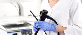
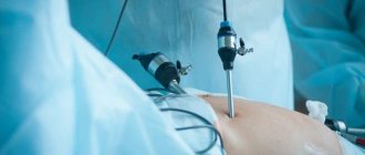
- colonoscopy - prescribed for cancer, Crohn's disease, ulcerative colitis; You can take a biopsy of the suspicious area;
- radionuclide diagnostics – used for inflammation of Meckel’s diverticulum;
- plain radiography of the abdominal cavity - reveals perforation of the organ;
- Ultrasound – used in Crohn’s disease to determine the thickness of the organ wall;
- CT, MRI - well visualize the growth of a cancerous tumor into neighboring organs, enlarged lymph nodes.
Author:
Selezneva Valentina Anatolyevna physician-therapist
How often can the procedure be performed?
Another exciting question for patients suffering from colon diseases and who are forced to constantly undergo diagnostics is: “How often can intestinal irrigoscopy be performed?”
The procedure is essentially an x-ray and the person is exposed to the usual radiation exposure in this case.
According to doctors, the maximum permissible radiation dose is 150 mSv per year. X-ray of the thoracic spine “pulls” 1.5 mSv.
The radiation exposure from irrigoscopy is approximately the same, so in total we get: 150: 1.5 = 100 procedures. Therefore, irrigoscopy can be done as often as prescribed by the doctor.
What to do after the procedure
At the end of irrigoscopy, the diet is completely canceled. No special events are needed.
There is no need to worry if the stool is too light in color; this is due to residual contrast material leaking out. In order for it to leave the body faster, the patient needs to drink more fluid.
In rare cases, constipation is noted. It is explained by the fact that the intestines were completely emptied before the study, and the diet was applied the day before. If it lasts for two days or more, then taking laxatives is recommended.
Types and capabilities of the method
There are 2 types of irrigoscopy: simple or double contrast. The first procedure involves the introduction of only one substance into the colon - either barium sulfate or air, and during the second procedure, a study with the first drug is replaced by a study with the second.
During irrigoscopy, a radiopaque substance gradually fills the colon, which allows the specialist to:
- study its structure, location relative to neighboring organs;
- assess the condition of the walls of the organ, identify the presence of ulcerations, scars, protrusions, fistulas, structural anomalies, and also detect neoplasms;
- assess the condition of the ileocecal valve, which is located between the small and large intestines and prevents the reflux of feces from the lower gastrointestinal tract to the upper;
- draw conclusions about how the large intestine functions.
Preparing the patient for barium enema plays an important role, regardless of the type of procedure.
What you can see during the study
When performing irrigoscopy, the doctor can observe the features of the structure and condition of the patient’s intestines, the location of the organ, its shape and the degree of preservation of the lumen.
The specialist, looking at the x-ray, analyzes the characteristics of its contractile activity and the functioning of the valve apparatus. Disturbances in the structure of the intestinal mucosa, various pathologies, malignant and benign neoplasms are also detected. The doctor determines the risk of developing a precancerous condition and finds possible diverticula and ulcers.
Decoding data: norm and pathology
Normally, the specialist observes a uniformly filled intestine along its entire length, with natural features of the anatomical structure. Its inner surface has a peculiar characteristic pattern.
Typically, barium gradually accumulates in the lowest section. When contrast is released into the external environment, the intestine takes on its normal shape.
An irrigogram allows you to diagnose adenocarcinoma or sarcoma. X-ray images very clearly reveal the degree of development of the tumor and its outline. They also determine the degree of intestinal patency.
In such cases, its inner surface acquires ragged outlines, becomes uneven and heterogeneous. Foci of severe inflammation are noted.
The doctor can also diagnose diverticulitis or ulcerative colitis. It determines its prevalence and the degree of neglect of the disease.
Typically, the pathological focus shifts towards the anus, and a pronounced inflammatory process and changes in the structure of the intestine are observed.
In case of oncology development, the study is supplemented with sigmoidoscopy.
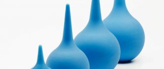
Enema cleansing
Giving an enema is a common method of deep cleansing the intestines. A few hours before irrigoscopy, an enema is performed. To do this, use Esmarch's mug. It is filled with water or chamomile decoction.
The tip is lubricated with Vaseline or fatty cream to minimize injury during insertion. After inserting the tube into the anus, they begin to fill the intestines with water. First, pour 1.5-2 liters.
After this, wait a while and after emptying, repeat the procedure again. You need to achieve almost clear water. Sometimes, during exacerbation of diseases, an enema is not recommended (hemorrhoids, colitis with severe pain). In such cases, they resort to medications that have a strong cleansing effect.
What does it feel like? Does it hurt?
Irrigoscopy is completely painless. The doctor doesn’t even use local anesthesia, let alone general anesthesia.
Some discomfort is felt only when a contrast agent is pumped into the intestines. Sometimes there is a desire to stool, a feeling of stretching, or occasionally mild, quickly passing spasms.
To monitor the normal course of irrigoscopy after barium administration, the doctor lightly palpates the patient’s anterior abdominal wall.
In general, a person does not experience any pronounced negative reactions from the body. If they appear, you should immediately report them to medical personnel.
Irrigoscopy, sigmoidoscopy or colonoscopy?
Irrigoscopy is often compared with endoscopic methods for studying the digestive system - sigmoidoscopy and colonoscopy. All of these methods are aimed at diagnosing pathologies of the large intestine. However, the information content of these studies differs.
Irrigoscopy is an x-ray examination. Despite the fact that it requires insertion of the apparatus tube into the rectum, the invasiveness of the examination is considered minimal. However, in terms of information content, irrigoscopy is inferior to sigmoidoscopy and colonoscopy. This is due to the fact that the assessment of the result is carried out “from the outside”. It is not always possible to obtain a good result on an x-ray image, even with the use of contrast.
Endoscopic research techniques are considered more informative.
They allow you to directly examine the colon mucosa using special video equipment that is inserted into the digestive system. This procedure is much less well tolerated by patients. However, in some cases it is impossible to do without conducting research of this type.
Colonoscopy and sigmoidoscopy differ in diagnostic capabilities:
- Sigmoidoscopy is the simplest endoscopic examination that allows you to evaluate the intestinal mucosa to a depth of 60 centimeters. In fact, during this procedure, only the anal canal and rectum are examined.
- Colonoscopy is a more comprehensive method that makes it possible to study the condition of the large intestine to a depth of 120-150 centimeters. Colonoscopy is the most informative way to diagnose diseases of the large intestine.
However, in most cases, the patient undergoes several studies at once, which make it possible to comprehensively assess the condition of the intestines and establish an accurate diagnosis. What studies will be included in the complex of diagnostic procedures will be determined by the attending physician during the examination of the patient.
In continuation of the topic, be sure to read:
- Diseases of the large intestine: symptoms and signs of the disease, treatment
- Details about the coprogram: preparation, conduct and interpretation of the analysis
- Details about intestinal colonoscopy: preparation and procedure
- How to check the intestines for oncology?
- Diagnosis of Helicobacter pylori: antibodies, breath test and biopsy
- Sigmoidoscopy and colonoscopy: what is the difference between the studies and which is better?
- Details about bowel cancer: stages, symptoms, treatment and prognosis
- Irrigoscopy and colonoscopy: which test is better?
- Checking the intestines for diseases: physical, laboratory and instrumental methods
- Preparation and performance of computed tomography (CT) of the intestine
Be sure to read:
What does an intestinal biopsy show and how is the procedure performed?