Quick transition Treatment of autoimmune hepatitis
Autoimmune hepatitis (AIH) is a non-infectious chronic inflammatory liver disease characterized by the presence of certain autoantibodies to liver cells.
Autoimmune hepatitis is more common in women (4:1) and can occur simultaneously with other autoimmune diseases, including type 1 diabetes, celiac disease, autoimmune thyroiditis, rheumatoid arthritis, and ulcerative colitis.
Approximately 25-50% of patients who already have a history of an autoimmune disease are likely to develop another autoimmune disease, including AIH.
There are two main forms or clinically significant types of the disease: the most common type 1 AIH is usually diagnosed in adulthood; Type 2 most often develops in childhood and young age, is characterized by a severe and aggressive course, and is less treatable. AIH in combination with another autoimmune liver disease (primary sclerosing cholangitis or primary biliary cholangitis) is classified as a variant type of the disease.
The prevalence of AIH worldwide is increasing annually and is currently <30 cases per 100,000 people.
Classification of autoimmune hepatitis
Autoimmune hepatitis is classified according to the type of antibodies to liver tissue (autoantibodies) circulating in the blood:
- Type I Antibodies against proteins of cell nuclei (antinuclear) and/or antibodies against proteins of smooth muscle cells predominate. With this type of autoimmune hepatitis, the body responds quickly and well to treatment; in approximately 20% of patients, remission continues after completion of therapy. Without treatment, cirrhosis of the liver develops in an average of three years.
- II type. Quite rare, detected in approximately 15% of patients. Antibodies are detected to fragments of membranes of intracellular structures (microsomes) of the liver and kidneys. It is difficult to treat and without therapy leads to cirrhosis approximately twice as fast as type I.
- III type. Antibodies to a protein called soluble liver antigen circulate in the blood; it is found inside hepatocytes. Some experts consider it a type I type of autoimmune hepatitis; the course of this disease and its features have not been sufficiently studied.
Therapy methods
Treatment of autoimmune hepatitis can be done in two ways: conservatively and surgically. Drug treatment involves the use of immunosuppressants, corticosteroids, and agents based on ursodeoxycholic acid (components of bile that prevent the death of liver cells). When used together (immunosuppressants and glucocorticoids), fewer side effects from the drugs are observed. Combined treatment is indicated for patients with obesity, skin diseases, osteoporosis, diabetes, and unstable blood pressure.
Drug treatment
Normalization of a person’s general condition, disappearance of symptoms, improvement of tests indicate the effectiveness of conservative treatment. If there is no response to medications, complications, or relapses, a liver transplant is needed. The operation is performed only if it is impossible to normalize the functioning of one’s own organ. Often, part of the liver is transplanted from a close relative. It is performed in the clinic by a specialized surgeon after a thorough examination.
During the period of stable remission, in consultation with your doctor, you can use traditional medicine. Recipes based on medicinal plants allow you to maintain organ health and improve its condition.
Autoimmune inflammation can be triggered by a virus
Doctors still have not found an exact answer to why exactly this disease occurs. Hereditary predisposition is quite clearly visible: five different types of mutations are known in the system of genes responsible for the immune response. In addition, it is known that autoimmune hepatitis is often combined with other chronic inflammatory diseases caused by an inadequate immune response: autoimmune thyroiditis, chronic ulcerative colitis, glomerulonephritis and others. However, the presence of a hereditary predisposition does not mean that the disease will certainly develop. Most likely, “triggering” triggers are needed for the onset of an autoimmune reaction. They could be:
- viral infections (herpes simplex, measles, hepatitis, cytomegalovirus, Epstein-Barr virus, etc.);
- exposure to certain medications (minocycline, diclofenac, methyldopa, infliximab, etanercept).
With an unsuccessful combination of genetic predisposition and external factors in the body, the number of cells that inhibit the immune response (T-suppressors) decreases. The immune system becomes overly active, begins to perceive liver tissue as foreign and, in an effort to get rid of the “aggressor,” produces antibodies. They bind to liver proteins and destroy liver cells.
Etiopathogenesis
The etiology of AIH is unknown, but the development of the disease is thought to be influenced by both genetic and environmental factors.
An important link in the pathogenesis may be certain alleles of the HLA II genes (human leukocyte antigen type II, Human leucocyte antigen II) and genes associated with the regulation of the immune system [7, 8].
It is worth mentioning separately about AIH, which is part of the clinical picture of autoimmune polyendocrine syndrome (autoimmune polyendocrine syndrome, autoimmune polyendocrinopathy-candidiasis-ectodermal dystrophy, APECED). This is a monogenic disease with autosomal recessive inheritance associated with a mutation in the AIRE1 gene. Thus, in this case, genetic determination is a proven fact [4, 9].
The autoimmune process in AIH is a T-cell immune response, accompanied by the formation of antibodies to autoantigens and inflammatory tissue damage.
Pathogenesis factors of AIH:
- pro-inflammatory factors (cytokines) produced by cells during the immune response. Indirect confirmation may be that autoimmune diseases are often associated with bacterial or viral infections;
- inhibition of the activity of regulatory T cells, which play a critical role in maintaining tolerance to autoantigens;
- dysregulation of apoptosis, normally a mechanism that controls the immune response and its “correctness”;
- molecular mimicry is a phenomenon where the immune response against external pathogens can also affect its own components that are structurally similar to them. Viral agents may play an important role in this. Thus, several studies have shown the presence of a pool of circulating autoantibodies (ANA, SMA, anti-LKM-1) in patients suffering from viral hepatitis B and C [2,4];
- factor of toxic drug effects on the liver. Some researchers associate the manifestation of AIH with the use of antifungal drugs and nonsteroidal anti-inflammatory drugs.
Symptoms of autoimmune hepatitis
In approximately a quarter of cases, autoimmune hepatitis is asymptomatic. An increase in AST and ALT (liver enzymes) in the blood is discovered accidentally during a biochemical study performed for another reason.
The disease can begin as an acute hepatitis: sudden weakness, high fever, jaundice, pain in the liver, dark urine.
Often the first manifestation of the disease is a detailed picture of liver cirrhosis: exhaustion, accumulation of fluid in the abdominal cavity, peripheral edema, dilation of the saphenous veins of the abdomen, etc.
Most often, with autoimmune hepatitis, a combination of hepatic and extrahepatic manifestations is detected.
“Liver” symptoms are quite typical for any damage to this organ:
- weakness;
- drowsiness;
- anorexia (loss of appetite);
- nausea;
- pain in the right hypochondrium;
- jaundice;
- skin itching.
In addition, extrahepatic manifestations are also possible:
- causeless fever;
- cutaneous vasculitis;
- joint and/or muscle pain;
- swollen lymph nodes;
- pleurisy;
- pericarditis;
- myocarditis.
These symptoms are often much more intense than manifestations from the liver, which misleads both the patient himself (who turns to other specialists rather than a hepatologist) and doctors. Hence the diagnostic errors.
Preventive measures
Since the exact causes of the disease are unknown, preventive measures remain questionable. Patients who have had the disease need to undergo a timely examination by a gastroenterologist, monitor the level of immunoglobulins, antibodies in the blood and the activity of liver enzymes.
Any liver pathology requires reducing the load on the organ. To do this, you should completely give up bad habits, minimize taking medications, and follow a strict diet. This is especially true for children who love “harmful” foods so much: chocolate, crackers, chips, carbonated sweet drinks. Adults should carefully monitor the child’s diet and avoid indulgences.
You should limit physical activity and alternate exercise with rest. If physiotherapeutic procedures are indicated for the patient, it is necessary to consult with the attending physician so as not to harm the liver. Preventive or routine vaccination is carried out only with the permission of the doctor. Autoimmune hepatitis is a poorly understood and severe liver disease. It is important to understand that this is not a death sentence. The earlier it is diagnosed and treatment started, the greater the chances of a positive outcome of the disease.
Diagnosis of autoimmune hepatitis
First of all, the doctor must exclude other causes of inflammation of the liver tissue: viral, toxic, alcoholic hepatitis.
Ultrasound and other imaging techniques of the liver can show changes in the organ's structure, but they do not provide insight into the cause of the damage. Therefore, a comprehensive diagnosis is necessary to exclude possible malignant pathologies.
Much more revealing are biochemical and immunological blood tests:
- high level of protein in blood plasma;
- increased levels of serum gamma globulins, IgG;
- increased levels of AST, ALT, alkaline phosphatase, LDH;
- antinuclear antibodies (ANA), antibodies to liver and kidney microsomes (anti-LKM-l) or antibodies to soluble liver antigen (anti-LKM-l) are detected in the blood;
- increased ESR.
The most diagnostic test is a histological examination of the liver - a biopsy. To do this, under ultrasound control, a certain amount of liver tissue is taken from the patient with a special needle and examined under a microscope.
Response to treatment
The results of treatment with prednisone and azathioprine for AIH may be as follows:
- A complete response is normalization of laboratory parameters, which persists for a year during maintenance therapy. At the same time, the histological picture is normalized (with the exception of small residual changes). The full effectiveness of treatment is also indicated in cases where the severity of clinical markers of autoimmune hepatitis significantly decreases, and during the first months of therapy laboratory parameters improve by at least 50% (and in the next 6 months they do not exceed the normal level by more than 2 times) .
- Partial response—there is an improvement in clinical symptoms and a 50% improvement in laboratory values within the first 2 months. Subsequently, the positive dynamics are maintained, but complete or almost complete normalization of laboratory parameters does not occur during the year.
- Lack of therapeutic effect (ineffectiveness of treatment) - an improvement in laboratory parameters by less than 50% in the first 4 weeks of treatment, and no further decrease (regardless of clinical or histological improvement) occurs.
- An unfavorable outcome of therapy is characterized by a further deterioration in the course of the disease (although in some cases there is an improvement in laboratory parameters).
Disease relapse is said to occur when, after achieving a complete response, clinical symptoms reappear and laboratory parameters deteriorate.
Typically, therapy has a good effect, but in 10–15% of patients it does not lead to improvement, although it is well tolerated. The reasons for the ineffectiveness of therapy can be [6]:
- lack of response to the drug;
- lack of compliance and adherence to therapy;
- drug intolerance;
- presence of cross syndromes;
- hepatocellular carcinoma.
Other immunosuppressants are also used as alternative drugs for the treatment of autoimmune hepatitis: budesonide, cyclosporine, cyclophosphamide, mycophenolate mofetil, tacrolimus, methotrexate [1, 2, 6, 11].
Clinical case
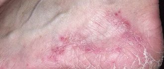
An eight-year-old girl was observed for skin rashes (erythematous and nodular elements without any discharge on the lower extremities) that had been bothering her for 5–6 months.
I used topical remedies for eczema for two months, there was no improvement. Later, discomfort in the epigastric region, weakness, and occasional nausea and vomiting arose. The rashes were localized on the legs. Histological examination of a skin biopsy revealed infiltration of subcutaneous fatty tissue with lymphocytes without signs of vasculitis. These phenomena were regarded as erythema nodosum. Upon examination, mild hepatosplenomegaly was revealed, otherwise the somatic status was without pronounced features, the condition was stable.
According to the results of laboratory tests:
- CBC: leukocytes - 4.5×10⁹/l; neutrophils 39%, lymphocytes 55%; signs of hypochromic microcytic anemia (hemoglobin 103 g/l); platelets - 174,000/μl, ESR - 24 mm/h;
- biochemical blood test: creatinine - 0.9 mg/dl (normal 0.3–0.7 mg/dl); total bilirubin - 1.6 mg/dl (normal 0.2-1.2 mg/dl), direct bilirubin - 0.4 mg/dl (normal 0.05-0.2 mg/dl); AST - 348 U/l (normal - up to 40 U/l), ALT - 555 U/l (normal - up to 40 U/l); alkaline phosphatase - 395 U/l (normal - up to 664 U/l), lactate dehydrogenase - 612 U/l (normal - up to 576 U/l).
Indicators of the hemostasis system (prothrombin time, international normalized ratio), the level of total protein, serum albumin, gamma globulin, the level of ferritin, ceruloplasmin, alpha-antitrypsin, gamma-glutamyl transpeptidase are within the reference values.
No HBs antigen, antibodies to HBs, HBc, or antibodies to hepatitis A and C virus were detected in the blood serum. Tests for cytomegalovirus, Epstein-Barr virus, toxoplasma, and brucella were also negative. The ASL-O titer is within the normal range. There is no evidence for diabetes mellitus, thyroiditis, Graves' disease, proliferative glomerulonephritis.
Ultrasound of the abdominal organs visualized an enlarged liver with an altered echostructure, without signs of portal hypertension and ascites. Ophthalmological examination did not reveal the presence of a Kayser-Fleischer ring.
Anti-SMA antibody titer - 1:160, ANA, AMA, anti-LKM-1 antibodies were not detected. The patient is positive for haplotypes HLA DR3, HLA DR4.
Histological examination of the liver biopsy revealed fibrosis, lymphocytic infiltration, formation of hepatocyte rosettes and other signs of chronic autoimmune hepatitis.
The patient was diagnosed with autoimmune hepatitis type 1 associated with erythema nodosum, and therapy with prednisolone and azathioprine was started. The IAIHG score was 19 points before treatment, which is characterized as “definite AIH.”
After 4 weeks of therapy, relief of general clinical symptoms and normalization of laboratory parameters were noted. After 3 months of therapy, the skin rashes completely regressed. 6 months after the end of therapy, the patient’s condition was satisfactory, laboratory parameters were within the reference values.
Adapted from Kavehmanesh Z. et al. Pediatric Autoimmune Hepatitis in a Patient Who Presented With Erythema Nodosum: A Case Report. Hepatitis monthly, 2012. V. 12. - N. 1. - P. 42–45.
Thus, AIH is a fairly rare and relatively treatable disease. With the introduction of modern immunosuppressive therapy protocols, the 10-year survival rate of patients reaches 90%. The prognosis is less favorable in patients with autoimmune hepatitis type 2, especially in children and adolescents, in whom AIH progresses much faster, and the effectiveness of therapy is generally lower. You should also be aware of the risk of developing hepatocellular carcinoma (4–7% within 5 years after diagnosis of liver cirrhosis).
List of sources
- Viral hepatitis and cholestatic diseases / Eugene R. Schiff, Michael F. Sorrell, Willis S. Maddray; lane from English V. Yu. Khalatova; edited by V. T. Ivashkina, E. A. Klimova, I. G. Nikitina, E. N. Shirokova. - M.: GEOTAR-Media, 2010.
- Kriese S., Heneghan MA Autoimmune hepatitis. Medicine, 2011. V. 39. - N. 10. - P. 580–584.
- Autoimmune hepatitis and its variant forms: classification, diagnosis, clinical manifestations and new treatment options: a manual for doctors. T. N. Lopatkina. - M. 4TE Art, 2011.
- Longhia MS et al. Aethiopathogenesis of autoimmune hepatitis. Journal of Autoimmunity, 2010. N. 34. - P. 7–14.
- Muratori L. et al. Current topics in autoimmune hepatitis. Digestive and Liver Disease, 2010. N. 42. - P. 757–764.
- Lohse AW, Mieli-Vergani G. Autoimmune hepatitis. Journal of Hepatology, 2011. V. 55. - N. 1. - P. 171–182.
- Oliveira LC et al. Autoimmune hepatitis, HLA and extended haplotypes. Autoimmunity Reviews, 2011. V. 10. - N. 4. - P. 189–193.
- Agarwal, K. et al. Cytotoxic T lymphocyte antigen-4 (CTLA-4) gene polymorphisms and susceptibility to type 1 autoimmunehepatitis. Hepatology, 2000. V. 31. - N. 1. - P. 49–53.
- Manns MP et al. Diagnosis and Management of Autoimmune Hepatitis. Hepatology, 2010. V. 51. - N. 6. - P. 2193–2213.
- Lohse AW, Wiegard C. Diagnostic criteria for autoimmune hepatitis. Best Practice & Research Clinical Gastroenterology, 2011. V. 25. - N. 6. - P. 665–671.
- Strassburg CP, Manns MP Therapy of autoimmune hepatitis. Best Practice & Research Clinical Gastroenterology, 2011. V. 25. - N. 6. - P. 673–687.
- Ohira H., Takahashi A. Current trends in the diagnosis and treatment of autoimmune hepatitis in Japan. Hepatology Research, 2012. V. 42. - N. 2. - P. 131–138.
Complications of the disease
- malignant neoplasms (hepatocellular cancer, as well as tumors of extrahepatic localization);
- cirrhosis of the liver (with the possible development of its complications);
- risk of increased incidence of infectious diseases during immunosuppressive therapy;
- complications associated with the occurrence of side effects during treatment (arterial hypertension, increased thrombus formation, osteoporosis, gastrointestinal ulcers, mental disorders and many others).
Story
Autoimmune hepatitis was first described in 1951 as chronic hepatitis in young women accompanied by hypergammaglobulinemia, which improves with adrenocorticotropic therapy. In 1956, a connection was discovered between AIH and the presence of antinuclear antibodies (ANA) in the blood, and therefore the disease was called “Lupus hepatitis.”
Between 1960 and 1980 A number of clinical studies have proven the effectiveness of AIH monotherapy with steroid drugs, as well as in combination with a cytostatic drug, azathioprine. AIH was the first liver disease for which drug therapy was proven to increase the life expectancy of patients.
Clinical manifestations
The disease manifests itself differently in each patient. Approximately 25% of patients diagnosed with autoimmune hepatitis have no symptoms at all until complications arise. Usually it begins either acutely and is very similar to the development of viral hepatitis, or manifests itself with signs atypical for liver damage.
In the first case, patients are mainly concerned about:
- weakness;
- dark color of biological fluids and yellowing of the skin;
- lack of appetite.
Since in the second case extrahepatic manifestations predominate, it is extremely difficult for doctors to immediately make a correct diagnosis. Therefore, the presence of a number of serious systemic diseases, in particular lupus erythematosus, rheumatoid arthritis, etc., is often mistakenly assumed.
In a number of situations, the disease begins acutely and proceeds extremely severely, which is accompanied by the development of fulminant hepatitis, in which most of the hepatocytes quickly die, and constantly produced toxins affect the brain, since it can no longer neutralize them. In such situations, the prognosis is extremely unfavorable.
Causes
The formation mechanism is poorly understood. The main provoking factor is considered to be loss of tolerance to antigens, which are normally present in the blood. One of the theories of the etiology of the disease is determined by hereditary origin. It is also taken into account that in many cases the autoimmune form occurs after acute hepatitis.
Other provoking factors include:
- Pernicious anemia.
- Ulcerative colitis.
- Inflammation of myocardial tissue.
- Pleurisy.
- Rheumatoid processes.
- Cushing's syndrome.
- Autoimmune form of thyroiditis.
- Long-term alcohol intake.
- Systemic administration of hormonal drugs.
Morphology
One of the main morphological signs of chronic autoimmune hepatitis is portal and periportal infiltration with the involvement of parenchymal cells in the process. At the early stage of the disease, a large number of plasma cells are detected, fibroblasts and fibrocytes are found in the portal fields, and the integrity of the border plate is disrupted.
Relatively often, focal necrosis of hepatocytes and dystrophic changes are detected, the severity of which can vary even within one lobule. In most cases, there is a violation of the lobular structure of the liver with excessive fibrogenesis and the formation of liver cirrhosis. The formation of macronodular and micronodular cirrhosis of the liver is possible.
According to most authors, cirrhosis usually has macronodular features and is often formed against the background of persistent activity of the inflammatory process. Changes in hepatocytes are represented by hydropic, less often fatty degeneration. Periportal hepatocytes can form glandular (glandular) structures, so-called rosettes.
Flow
Autoimmune chronic hepatitis is often characterized by a continuously relapsing course, the rapid formation of liver cirrhosis and the development of liver failure. Exacerbations occur frequently and are accompanied by jaundice, fever, hepatomegaly, hemorrhagic syndrome, etc. Clinical remission is not accompanied by normalization of biochemical parameters. Repeated exacerbations occur with less severe symptoms. Some patients may develop signs of other autoimmune liver lesions - primary biliary cirrhosis and (or) primary sclerosing cholangitis, which gives grounds to include such patients in the group of people suffering from overlap syndrome.
Mild forms of the disease, when biopsy reveals only stepwise necrosis and no bridging necrosis, rarely progress to liver cirrhosis; Spontaneous remissions alternating with exacerbations may be observed. In severe cases (about 20%), when aminotransferase activity is more than 10 times higher than normal, severe hyperglobulinemia is noted, and biopsy reveals bridging and multilobular necrosis and signs of cirrhosis, up to 40% of untreated patients die within 6 months.
The most unfavorable prognostic signs are the detection of multilobular necrosis in the early stages of the disease and hyperbilirubinemia that persists for 2 weeks or more from the start of treatment. Causes of death include liver failure, hepatic coma, other complications of cirrhosis (eg, bleeding from varicose veins) and infections. Against the background of cirrhosis, hepatocellular carcinoma can develop.