Ascites (accumulation of fluid in the abdominal cavity) is detected in 50% of patients in the early stages of cancer and in almost all patients in whom the cancer process is at the last stage.
The Oncology Clinic of the Yusupov Hospital is equipped with the latest diagnostic equipment from the world's leading manufacturers, with the help of which oncologists identify the early stages of oncological pathology. Chemotherapists, radiologists, and oncologists treat patients with ascites in accordance with international standards of medical care. At the same time, doctors take an individual approach to choosing a treatment method for each patient.
What it is?
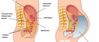
Ascites is a pathological accumulation of fluid in the abdominal cavity. It can develop rapidly (over a few days) or over a long period (weeks or months). Clinically, the presence of free fluid in the abdominal cavity is manifested when a fairly large volume is reached - from 1.5 liters.
The amount of fluid in the abdominal cavity sometimes reaches significant figures - 20 liters or more. By origin, ascitic fluid can be of an inflammatory nature (exudate) or non-inflammatory, resulting from a violation of hydrostatic or colloid-osmotic pressure in pathologies of the circulatory or lymphatic system (transudate).
Popular folk remedies
There are a huge number of folk recipes for combating ascites at home. Among them are lotions, baths, wraps, decoctions and infusions, medicinal mixtures. Diuretics constitute a separate category. They help quickly remove excess fluid from the body, eliminate swelling, tone and saturate the body with vitamins. Some formulations for internal use are very strong or may cause an allergic reaction, so it is recommended to consult a doctor before use.
Urine therapy
Treatment of ascites with urine is a method available to everyone, but its effectiveness has not been scientifically proven. It is based on the healing properties of urine, which contains more than 200 components. Among them are anti-inflammatory, bactericidal and nutritional. At the same time, toxins, metabolic products, hormones and microelements are removed from the body along with urine.
The statement that urine therapy has no contraindications is a myth. Those with gastrointestinal problems (ulcers, colitis), sexually transmitted diseases, chronic diseases of the pancreas, liver and kidneys should be wary of this method of self-medication.
Rules for taking urine:
- Only fresh mid-morning urine is taken orally.
- You should drink the liquid twice a day: 1 hour before lunch and in the evening before bed.
- Single dosage - from 0.5 to 1 glass. It is better to start with small portions, gradually working up to the norm.
- The course lasts 2-3 months.
In combination with urine therapy, it is advisable to first cleanse the body of toxins. It is also recommended to switch to a plant-based diet, stay hydrated, play sports and give up bad habits.
Therapeutic fasting
Fasting according to Breuss is a popular diet that helps eliminate ascites at home in case of oncology. The course is 42 days. This is exactly what the author of the technique needed to completely recover from cancer. Before this, he had undergone several operations, but the tumor returned again.
More than 40 thousand people have undergone successful treatment with the diet.
Principles of fasting:
- The diet consists of carrot, potato, beet, celery juices, infusions of sage and red geranium, and kidney tea.
- On an empty stomach after waking up, take 125-150 ml of kidney tea.
- After an hour, you should drink another 1-2 glasses of warm sage infusion.
- After an hour, you need to take a sip of vegetable juice. Then, with a break of 20-30 minutes, take another 10-15 sips.
- In between drinking vegetable juices, you are allowed to drink a chilled infusion of sage.
- At lunchtime you should drink half a glass of kidney tea again. And then repeat the intake of vegetable juice and infusion.
- Once a week you should take an infusion of geranium, drinking 1 glass of liquid per day. It contains radium.
- The maximum volume of vegetable juices per day is 0.5 liters. Sage infusion can be consumed in unlimited quantities.
- Sometimes vegetable juices can be replaced with fruit juices: lemon, orange, black currant.
- Mixing vegetable and fruit juices with each other is prohibited.
Recipes for making juices and infusions for the diet:
- Multivitamin vegetable juice. To prepare you will need 300 g of beets, 100 g of carrots, 100 g of celery, 30 g of black radish, 1 potato. Peel the vegetables and pass through a juicer. Strain the juice and take in small sips.
- Sage infusion. 2 tsp. dried flowers, pour 0.5 liters of boiling water and place on low heat until boiling. Cook for 3 minutes. Remove from heat. If desired, add 1 tsp to the broth. mint, lemon balm, St. John's wort. Leave for 10 minutes. Strain and take internally.
- Kidney tea. Mix 15 g of horsetail, 10 g of nettle, 8 g of knotweed, 6 g of St. John's wort. Pour a glass of boiling water over the dried flowers and leave for 10 minutes. Strain. Add another 0.5 liters of water to the clean liquid and take according to the instructions. The volume is designed for 3 weeks of fasting.
- Infusion of red geranium. 1 tsp. Pour a glass of boiling water over the dried plant and leave for 10 minutes. Take small sips throughout the day.
In the first weeks, the feeling of hunger can bring serious discomfort, since the diet contains very few calories. To dull it and gain vitality, you can cook onion soup. To prepare the dish, you need to take 1 onion, wash it and chop it together with the peel. Then fry in vegetable oil until golden brown, pour in 0.5 liters of cold water and bring to a boil. Add any light vegetable broth to the prepared broth and simmer for several minutes. The soup must be strained before use.
It is best to start Breuss fasting 2-6 months after surgery or a course of chemotherapy. When leaving the diet, familiar foods are gradually introduced into the diet. First courses should have a semi-liquid consistency, without salt.
Apricot compote
When used correctly, apricot is an excellent remedy for the treatment of ascites. The fruit has a gentle effect on the body, helps restore liver tissue and normalize metabolism. The product has no contraindications and cannot cause an overdose. Method for preparing compote:
- Pour 400 g of fresh or dried apricots into 1.5 liters of boiling water.
- Place the mixture on low heat and cook for 10-15 minutes.
- Taste the broth. If it seems sour, add 1 tsp. honey.
Drink about 400 ml of decoction every day. It is best to do this in the morning on an empty stomach and in the evening before bed.
Parsley for ascites
Parsley is a natural product that contains large amounts of vitamins, minerals and fatty acids. Herbal Recipes:
- Decoction. 2 tbsp. l. chopped greens, pour 1 liter of boiling water. Leave for 4-5 hours in a dark place. Take 2 tbsp. l. four times a day.
- Tea. Wash and chop 300 g of herbs. Pour 1 liter of clean water, put on low heat and cook for 20 minutes. Take small sips throughout the day.
- Milk drink. Bring 0.5 liters of milk to a boil. Add 200 g of chopped fresh herbs. Cook the mixture over low heat for 2 hours. Cool the product and take 2 tbsp. l. before every meal.
- Infusion. Mix 250 g of dry herbs and 1 chopped parsley root. Pour in 1 liter of boiled milk. Simmer the mixture in a water bath for 2 hours. Strain the medicine and take half a glass three times a day.
Folk remedies with parsley have a diuretic effect, which allows you to remove fluid from the abdominal cavity during dropsy.
The Internet is replete with reviews about traditional methods of treating ascites. Most of them are positive. The most popular diets and recipes based on parsley, beans and apricots. They are absolutely safe and have no contraindications. However, before starting the course, it is recommended to consult a specialist. During therapy and after recovery, you should undergo regular examinations and monitor your well-being.
Classification
Depending on the amount of fluid in the abdominal cavity, there are several degrees of the pathological process:
- Small ascites (no more than 3 l).
- Moderate (3–10 l).
- Significant (massive) (10–20 l, in rare cases – 30 l or more).
Based on the level of infection of ascitic contents, the following are distinguished:
- sterile (uninfected) ascites;
- infected ascites;
- spontaneous bacterial peritonitis.
According to the response to therapy, ascites occurs:
- transient. Disappears against the background of conservative treatment in parallel with the improvement of the patient’s condition forever or until the next exacerbation of the pathological process;
- stationary. The appearance of fluid in the abdominal cavity is not a random episode; it persists in a small amount even despite adequate therapy;
- resistant (torpid, or refractory). Large ascites, which cannot only be stopped, but even reduced with large doses of diuretics.
If the accumulation of fluid continues to increase steadily and reaches enormous sizes, despite the treatment, such ascites is called tense ascites.
Consequences and complications
bacterial peritonitis develops as a complication of ascites . This happens if the ascitic fluid becomes infected, which is usually a spontaneous process.
Ascites worsens the course of the underlying disease, as it provokes respiratory failure, hernia, hydrothorax , intestinal obstruction , etc.
It is important to realize that ascites is a deadly disease and must be treated promptly.
If dropsy has been cured, after that the person still needs to be very careful about his health and eat right, as there is a risk of relapse. If you experience discomfort or unpleasant symptoms, you should immediately consult a doctor and undergo testing.
If the disease is treated incorrectly or not practiced at all, very serious complications can develop: peritonitis , bleeding , respiratory failure , cerebral edema , cardiac dysfunction.
Reasons for the development of ascites
The causes of abdominal ascites are varied and are always associated with some serious disorder in the human body. The abdominal cavity is a closed space in which excess fluid should not form. This place is intended for internal organs - there is the stomach, liver, gall bladder, part of the intestines, spleen, pancreas.
The peritoneum is lined with two layers: the outer layer, which is attached to the wall of the abdomen, and the inner layer, which is adjacent to and surrounds the organs. Normally, between these sheets there is always a small amount of fluid, which is the result of the work of blood and lymphatic vessels located in the peritoneal cavity. But this fluid does not accumulate, since almost immediately after release it is absorbed by the lymphatic capillaries. The remaining insignificant part is necessary so that the intestinal loops and internal organs can move freely in the abdominal cavity and not stick together.
When the barrier, excretory and resorptive functions are disrupted, the exudate ceases to be absorbed normally and accumulates in the abdomen, resulting in the development of ascites.
TOP 10 causes of abdominal ascites:
- Heart diseases. Ascites can develop due to heart failure or due to constrictive pericarditis. Heart failure can result from almost all cardiac diseases. The mechanism of development of ascites in this case will be associated with the fact that the hypertrophied heart muscle is not able to pump the required volumes of blood, which begins to accumulate in the blood vessels, including in the inferior vena cava system. As a result of high pressure, fluid will leave the vascular bed, forming ascites. The mechanism of development of ascites during pericarditis is approximately the same, but in this case the outer lining of the heart becomes inflamed, which leads to the impossibility of its normal filling with blood. This subsequently affects the functioning of the venous system;
- Liver diseases. First of all, this is cirrhosis, as well as organ cancer and Budd-Chiari syndrome. Cirrhosis can develop against the background of hepatitis, steatosis, toxic medications, alcoholism and other factors, but is always accompanied by the death of hepatocytes. As a result, normal liver cells are replaced by scar tissue, the organ increases in size, compresses the portal vein and therefore ascites develops. A decrease in oncotic pressure also contributes to the release of excess fluid, because the liver itself is no longer able to synthesize plasma proteins and albumins. The pathological process is aggravated by a number of reflex reactions triggered by the body in response to liver failure;
- Kidney diseases. Ascites is caused by chronic renal failure, which occurs as a result of a wide variety of diseases (pyelonephritis, glomerulonephritis, urolithiasis, etc.). Kidney disease leads to increased blood pressure, sodium along with fluid is retained in the body, resulting in the formation of ascites. A decrease in plasma oncotic pressure, leading to ascites, can also occur against the background of nephrotic syndrome;
- Diseases of the digestive system can provoke excessive accumulation of fluid in the abdominal cavity. This could be pancreatitis, chronic diarrhea, Crohn's disease. This also includes any processes occurring in the peritoneum and preventing lymphatic outflow;
- Various lesions of the peritoneum can provoke ascites, including diffuse ascites, tuberculous and fungal peritonitis, peritoneal carcinosis, cancer of the colon, stomach, breast, ovaries, and endometrium. This also includes pseudomyxoma and peritoneal mesothelioma;
- Ascites can develop when lymphatic vessels are damaged. This happens due to injury, due to the presence of a tumor in the body that gives metastases, due to infection with filaria (worms that lay eggs in large lymphatic vessels);
- Polyserositis is a disease in which ascites appears in combination with other symptoms, including pleurisy and pericarditis;
- Systemic diseases can lead to the accumulation of fluid in the peritoneum. These are rheumatism, rheumatoid arthritis, lupus erythematosus, etc.;
- Protein deficiency is one of the factors predisposing to the formation of ascites;
- Myxedema can lead to ascites. This disease is accompanied by swelling of soft tissues and mucous membranes, manifests itself when the synthesis of thyroxine and triiodothyronine (thyroid hormones) is disrupted.
So, ascites can be based on a variety of inflammatory, hydrostatic, metabolic, hemodynamic and other disorders. They entail a number of pathological reactions of the body, as a result of which interstitial fluid sweats through the veins and accumulates in the peritoneum.
Ascites in oncology
As already mentioned, oncological (tumor) diseases are characterized by the uncontrolled proliferation of tumor cells. Roughly speaking, any tumor can cause the development of ascites if tumor cells metastasize to the liver, followed by compression of the hepatic sinusoids and increased pressure in the portal vein system. However, there are some tumor diseases that are complicated by ascites more often than others.
The cause of ascites may be:
- Peritoneal carcinomatosis. This term refers to damage to the peritoneum by tumor cells that metastasize into it from tumors of other organs and tissues. The mechanism of development of ascites is the same as with mesothelioma.
- Mesothelioma. This malignant neoplasm is extremely rare and originates directly from peritoneal cells. The development of a tumor leads to activation of the immune system in order to destroy tumor cells, which is manifested by the development of the inflammatory process, dilation of blood and lymphatic vessels and leakage of fluid into the abdominal cavity.
- Ovarian cancer. Although the ovaries do not belong to the abdominal organs, the layers of the peritoneum are involved in fixing these organs in the pelvis. This explains the fact that with ovarian cancer, the pathological process can easily spread to the peritoneum, which will be accompanied by an increase in the permeability of its vessels and the formation of effusion in the abdominal cavity. In the later stages of the disease, cancer may metastasize into the layers of the peritoneum, which will increase the release of fluid from the vascular bed and lead to the progression of ascites.
- Pancreas cancer. The pancreas is the site of production of digestive enzymes, which are secreted from it through the pancreatic duct. After leaving the gland, this duct merges with the common bile duct (which drains bile from the liver), after which they flow together into the small intestine. The growth and development of a tumor near the confluence of these ducts can lead to disruption of the outflow of bile from the liver, which can be manifested by hepatomegaly (increase in liver size), jaundice, skin itching and ascites (ascites develops in the later stages of the disease).
- Meigs syndrome. This term refers to a pathological condition characterized by the accumulation of fluid in the abdominal and other body cavities (for example, in the pleural cavity of the lungs). The cause of the disease is considered to be tumors of the pelvic organs (ovaries, uterus).
Pathogenesis
A certain amount of ascitic fluid is always present in the human peritoneum. During life, this fluid moves into the lymphatic vessels, and a new one appears in its place. However, in some pathological conditions, the absorption of this fluid stops, or it is produced in excess.
An important role in the development of ascites is played by functional liver failure, disruption of water-salt and protein metabolism, pathological changes in the vascular system of the peritoneum and its mesothelial cover.
Doctors identify the following pathogenetic mechanisms:
- Portal hypertension .
- Stagnation of blood in the systemic circulation in people with right ventricular heart failure.
- Local lymphostasis in the case of the development of filariasis of the lymphatic vessels that collect lymph from the peritoneal organs.
- Metastases to regional lymph nodes in cancer.
- Peritoneal carcinomatosis with the advancement of cancer cells of malignant formations of the peritoneal organs into its cavity.
- Exudation into the abdominal cavity during peritonitis.
- Hypoproteinemic edema in people with kidney disease or during fasting.
As a result, excessive fluid accumulation negatively affects the functioning of the circulatory system and internal organs. The digestive system suffers, the movements of the diaphragm are limited. Since the liquid contains salts and protein, metabolic processes are disrupted. Also, along with ascites, damage to the kidneys, heart, liver, etc. occurs.
Symptoms
The symptoms that manifest ascites (see photo), of course, greatly depend on the severity of the condition. If ascites is a mild disease, then no symptoms appear; it is difficult to identify even with the help of instrumental examinations; only ultrasound or CT scan of the abdominal cavity helps.
If ascites is severe, it is accompanied by the following symptoms:
- Bloating and heaviness of the abdomen.
- Bloating, swelling and increase in abdominal volume.
- Breathing problems due to pressure of abdominal contents on the diaphragm. Compression leads to dyspnea (shortness of breath, short and rapid breathing).
- Stomach ache.
- Flat navel.
- Lack of appetite and instant feeling of fullness.
- Swollen ankles (edema) due to excess fluid.
- Other typical symptoms of the disease, such as portal hypertension (resistance to blood flow) in the absence of cirrhosis.
In children
In newborns, the development of ascites may be associated with the development of hemolytic disease of the fetus or indicate hidden blood loss. In children in the first three years of life, ascites most often manifests itself as a consequence of liver disease. However, its connection with chronic nutritional disorders, congenital nephrotic syndrome, and exudative enteropathy cannot be ruled out. It is important to differentiate ascites in children from pseudoascites , that is, intestinal atony accompanied by bloating. This condition is possible with cystic fibrosis , celiac disease.
Diagnostics
The diagnosis of ascites can be detected already at the first examination:
- enlarged abdomen (similar to that during pregnancy), a protruding navel, in a supine position it spreads out on the sides due to drainage of fluid (“frog belly”), the saphenous veins on the front wall are dilated;
- when percussing (tapping) the abdomen, the sound becomes dull (like wood);
- During auscultation (listening with a phonendoscope) of the abdomen, bowel sounds will be absent due to significant accumulation of fluid.
A sign of fluctuation is indicative - one palm is placed on the patient’s side, the other hand makes oscillatory movements on the other side, as a result, the movement of fluid in the abdominal cavity will be felt.
For additional diagnostics, the following types of laboratory tests and instrumental studies are applicable:
- Ultrasound examination of the abdominal organs and kidneys (ultrasound). The examination method allows you to identify the presence of fluid in the abdominal cavity, space-occupying formations, will give an idea of the size of the kidneys and adrenal glands, the presence or absence of tumors in them, the echostructure of the pancreas, gall bladder, etc.;
- Ultrasound of the heart and thyroid gland - you can determine the ejection fraction (its decrease is one of the signs of heart failure), the size of the heart and its chambers, the presence of fibrin deposits (a sign of constrictive pericarditis), the size and structure of the thyroid gland;
- computer and magnetic resonance imaging - allows you to visualize even the slightest accumulation of fluid, evaluate the structure of the abdominal organs, identify anomalies in their development, the presence of neoplasms, etc.;
- plain X-ray of the chest organs - allows you to judge the presence of tuberculosis or lung tumors, the size of the heart;
- diagnostic laparoscopy - a minor puncture is made on the anterior abdominal wall, and an endoscope (a device with a built-in camera) is inserted into it. The method allows you to determine the fluid in the abdominal cavity, take part of it for further examination in order to find out the nature of the occurrence of ascites, and it may also be possible to detect the damaged organ that caused the accumulation of fluid;
- angiography is a method that allows you to determine the condition of blood vessels;
- general blood test - a possible decrease in the number of platelets due to impaired liver function, an increase in the erythrocyte sedimentation rate in autoimmune and inflammatory diseases, etc.;
- general urine test - allows you to judge the presence of kidney disease;
- biochemical blood test, thyroid hormones. The following are determined: protein levels, transaminases (ALAT, ASAT), cholesterol, fibrinogen to determine the functional state of the liver, rheumatic tests (C-reactive protein, rheumatoid factor, antistreptolysin) to diagnose rheumatoid arthritis, lupus erythematosus or other autoimmune diseases, urea and creatinine to determine kidney functions, sodium, potassium, etc.;
- determination of tumor markers, for example, alpha-fetoprotein for liver cancer;
- Microscopic examination of ascitic fluid allows us to determine the nature of ascites.
Execution technique
The surgeon initially determines the puncture point. It is located on the midline of the abdomen, three centimeters below the navel. The area is first thoroughly treated with a liquid antiseptic, after which anesthesia (1% novocaine solution or 2% lidocaine solution) is injected into the tissue layer by layer.
When the medications begin to take effect, the doctor makes a small incision with a scalpel, dissects the skin and tissue, then pierces the abdomen with a trocar. To create the necessary hole for the movement of the instrument, the umbilical ring is grabbed and the peritoneal wall is slightly raised. One end of a PVC tube is inserted into it, the other goes down into the pelvis. Liquid is released through it.
The first part is collected into sterile tubes and sent for cytological examination. The rest of the contents very slowly (one liter every five minutes) descends into a deep container. If we are talking about ascites, up to ten liters can be removed in one procedure. With rapid extraction, surges in blood pressure occur (even to the point of collapse), because the released vessels become overfilled with blood. The surgeon's assistant tightens the patient's abdomen with a towel as the fluid is removed. This technique helps to avoid hemodynamic disorders.
The surgeon is able to determine by the color of the extracted biological material what caused the accumulation of fluid in the peritoneum. If the resulting substance is the color of meat slop, the patient most likely has internal bleeding. In such a situation, the patient begins to urgently prepare for further extensive surgery. When transudate flows through the tube, the patient will be examined for cirrhosis of the liver, the presence of malignant tumors and tuberculous peritonitis. The appearance of turbid fluid with a predominance of neutrophils is characteristic of bacterial peritonitis.
The volume of pumped out fluid has great diagnostic value. The more it is, the more accurately the pathology can be determined by its qualitative composition. Even a minimal amount (300-500 ml) can help clarify the disease in 80%.
After completion of the manipulation, sutures and a tight sterile bandage are applied to the wound. The patient is turned onto the right side. The postoperative period proceeds favorably. The stitches are removed on the seventh day. Restrictions in the regime depend on the underlying pathology, the development of which led to the need to puncture the abdominal wall and pump out fluid. For example, with cirrhosis, the patient is prescribed a strict diet.
The restraining towel does not need to be removed immediately: it will help to form intrauterine pressure that is familiar to a healthy person and gradually adapt to new conditions of blood supply.
How to treat ascites?
Treatment of ascites should begin as early as possible and be carried out only by an experienced doctor, since otherwise the disease may progress and serious complications may develop. First of all, it is necessary to determine the stage of ascites and assess the general condition of the patient. If, against the background of intense ascites, the patient develops signs of respiratory failure or heart failure, the primary goal will be to reduce the amount of ascitic fluid and reduce the pressure in the abdominal cavity. If the ascites is transient or moderate, and the existing complications do not pose an immediate threat to the patient’s life, treatment of the underlying disease comes to the fore, but the level of fluid in the abdominal cavity is regularly monitored.
Free fluid can be easily removed from the abdominal cavity, but the causes of ascites will remain. Therefore, the complete treatment of ascites is the treatment of the diseases that provoked its occurrence.
Regardless of what triggered the ascites, general treatments are as follows:
- bed or semi-bed rest (with getting out of bed only if physiologically necessary) mode;
- restriction, and in advanced cases - complete exclusion of sodium from food. Achieved by limiting (or eliminating) the use of table salt.
If ascites occurs due to cirrhosis of the liver, then when the amount of sodium in the blood decreases, fluid intake in various forms (tea, juices, soups) is limited - up to 1 liter.
Drug therapy depends on the disease that provoked the ascites. The general purpose, regardless of the cause of ascites, is diuretics.
This may be either a combination with potassium preparations, or potassium-sparing diuretics. Also prescribed:
- for liver cirrhosis - hepatoprotectors (drugs that protect liver cells);
- if the amount of protein in the blood is low, protein preparations are administered intravenously. As an example, albumin, fresh frozen plasma (it is administered if, with ascites, disturbances in the blood coagulation system are observed);
- for cardiovascular failure - drugs that support heart function (they are selected depending on the cause of the failure)
Surgical methods for treating ascites are used for:
- significant accumulation of free fluid in the abdominal cavity;
- if conservative methods show low effectiveness or do not show it at all.
The main surgical methods used for ascites are:
- Laparocentesis. Exudate is removed through a puncture of the abdominal cavity under ultrasound guidance. After the operation, a drainage is installed. No more than 10 liters of water are removed in one procedure. In parallel, the patient is given drips of saline solutions and albumin. Complications are very rare. Sometimes infectious processes occur at the puncture site. The procedure is not performed for bleeding disorders, severe bloating, intestinal injuries, ventral hernia and pregnancy.
- Transjugular intrahepatic shunting. During the operation, the hepatic and portal veins are connected artificially. The patient may experience complications such as intra-abdominal bleeding, sepsis, arteriovenous shunting, or liver infarction. Surgery is not prescribed if the patient has intrahepatic tumors or cysts, vascular occlusion, bile duct obstruction, or cardiopulmonary pathologies.
- Liver transplantation. If ascites develops against the background of liver cirrhosis, an organ transplant may be prescribed. Few patients have the chance to undergo such an operation, since it is difficult to find a donor. Absolute contraindications to transplantation are chronic infectious pathologies, severe dysfunction of other organs, and cancer. Among the most severe complications is graft rejection.
Treatment of ascites in oncology
The reason for the formation of ascitic fluid in a tumor can be compression of the blood and lymphatic vessels of the abdominal cavity, as well as damage to the peritoneum by tumor cells. In any case, for effective treatment of the disease it is necessary to completely remove the malignant neoplasm from the body.
The following can be used in the treatment of cancer:
- Chemotherapy. Chemotherapy is the main treatment for peritoneal carcinomatosis, in which tumor cells affect both layers of the serosa of the abdominal cavity. Chemical drugs (methotrexate, azathioprine, cisplatin) are prescribed that disrupt the division of tumor cells, thereby leading to the destruction of the tumor. The main problem with this is the fact that these drugs also disrupt the division of normal cells throughout the body. As a result, during the treatment period, the patient may experience hair loss, stomach and intestinal ulcers may appear, and aplastic anemia may develop (lack of red blood cells due to a disruption in the process of their formation in the red bone marrow).
- Radiation therapy. The essence of this method is the high-precision impact of radiation on tumor tissue, which leads to the death of tumor cells and a decrease in the size of the tumor.
- Surgery. It involves removing the tumor through surgery. This method is especially effective for benign tumors or in cases where the cause of ascites is compression of blood or lymphatic vessels by a growing tumor (its removal can lead to a complete recovery of the patient).
Treatment of ascites in kidney disease
Treatment of chronic kidney diseases, which can cause ascites, is almost always a complex and lengthy process. Depending on the specific type of disease, the issue of the need to prescribe glucocorticosteroid hormones, surgery to correct defects, permanent hemodialysis or other therapeutic measures is decided. However, the general principles of treatment for these pathologies are the same. These include the following recommendations:
- Limiting salt. Since impaired renal function impairs the elimination of electrolytes, taking even small amounts of salt can lead to fluid retention and increased blood pressure. The maximum permissible dose for these diseases is no more than 1 g/day. This amount can be achieved by eating fresh food and unsalted drinks.
- Regular monitoring of toxic substances in the blood. This measure helps prevent the development of serious complications such as brain damage (encephalopathy).
- Maintaining adequate diuresis. With chronic damage to an organ, toxic substances begin to accumulate in a person’s blood. They lead to sleep disturbances, constant weakness, decreased performance and poor health. Therefore, it is important to regularly use diuretics to improve the elimination of “toxins”.
- Reducing the inflammatory process. In case of autoimmune diseases such as glomerulonephritis, lupus erythematosus, rheumatoid arthritis, it is necessary to reduce the body's immune functions. Due to this, the kidney tissue will be damaged significantly less. As a rule, glucocorticosteroid hormones (Prednisolone, Dexamethasone) or immunosuppressants (Sulfasalazine, Methotrexate) are used for this purpose.
- Taking nephroprotective drugs. ACE inhibitors and ARBs, in addition to their protective effect on the heart, have a similar effect on the kidneys. By improving the condition of their microvessels, they prevent further damage and remove hemodialysis from the patient.
Treatment of ascites in liver cirrhosis
One of the main stages in the treatment of ascites in liver cirrhosis is to stop the progression of the pathological process in it and stimulate the restoration of normal liver tissue. Without these conditions, symptomatic treatment of ascites (use of diuretics and repeated medical punctures) will give a temporary effect, but ultimately it will end in the death of the patient.
Treatment for liver cirrhosis includes:
- Hepatoprotectors (allochol, ursodeoxycholic acid) are drugs that improve metabolism in liver cells and protect them from damage by various toxins.
- Essential phospholipids (phosphogliv, essentiale) - restore damaged cells and increase their resistance to toxic factors.
- Flavonoids (gepabene, karsil) – neutralize free oxygen radicals and other toxic substances formed in the liver during the progression of cirrhosis.
- Amino acid preparations (heptral, hepasol A) - cover the need of the liver and the whole body for amino acids necessary for normal growth and renewal of all tissues and organs.
- Antiviral drugs (Pegasys, ribavirin) are prescribed for viral hepatitis B or C.
- Vitamins (A, B12, D, K) - these vitamins are formed or deposited (stored) in the liver, and with the development of cirrhosis, their concentration in the blood can significantly decrease, which will lead to the development of a number of complications.
- Diet therapy - it is recommended to exclude from the diet foods that increase the load on the liver (in particular fatty and fried foods, any types of alcoholic beverages, tea, coffee).
- Liver transplantation is the only method that can radically solve the problem of cirrhosis. However, it is worth remembering that even after a successful transplant, the cause of the disease must be identified and eliminated, since otherwise cirrhosis can affect the new (transplanted) liver.
Treatment
Drug therapy for ascites is not carried out due to low effectiveness. Aldosterone antagonists and diuretics normalize water-salt metabolism and prevent excess secretion of peritoneal fluid. Oncologists at the Yusupov Hospital offer palliative surgery to patients with ascites in the late stages of cancer:
- Omentohepatophrenopexy;
- Deperitonization;
- Installation of a peritoneovenous shunt.
Doctors at the Oncology Clinic carry out traditional or intracavitary chemotherapy for ascites - after removing the fluid, a chemotherapy drug is injected into the abdominal cavity. Laparocentesis is performed to remove fluid. The procedure is not performed if the following contraindications are present:
- Adhesive process inside the abdominal cavity;
- Severe flatulence;
- Perforation of the intestinal walls;
- Purulent infectious processes.
Laparocentesis is prescribed in cases where taking diuretics does not lead to a positive result. The procedure is also indicated for resistant ascites.
Laparocentesis is carried out in several stages using local anesthesia:
- the patient is in a sitting position, the doctor treats the subsequent puncture site with an antiseptic and administers painkillers;
- An incision is made in the abdominal wall along the linea alba at a distance of 2-3 centimeters below the navel;
- The puncture itself is performed using a trocar using rotational movements. A special flexible tube is attached to the trocar, through which excess fluid is removed from the body. The fluid is pumped out quite slowly, and the doctor constantly monitors the patient’s condition. As the exudate is removed, the nurse tightens the patient's abdomen with a sheet to slowly reduce the pressure in the abdominal cavity;
- After the fluid is pumped out, a sterile bandage is applied to the wound.
Using laparocentesis, up to 10 liters of fluid can be removed from the patient’s body. In this case, it may be necessary to administer albumin and other drugs to prevent the development of renal failure.
If necessary, temporary catheters can be installed in the abdominal cavity, through which excess fluid will gradually be removed. It should be noted that the use of catheters can lead to a decrease in blood pressure and the formation of adhesions.
There are also contraindications to laparocentesis. Among them:
- pronounced flatulence;
- adhesive disease of the abdominal organs;
- stage of recovery after surgery on a ventral hernia.
Diuretics are prescribed to patients with developing ascites in cancer over a long course. Such drugs as Furosemide, Diacarb and Veroshpiron are effective.
When taking diuretics, medications containing potassium must also be prescribed. Otherwise, there is a high probability of developing disturbances in water and electrolyte metabolism.
Dietary nutrition primarily involves reducing the amount of salt consumed, which retains fluid in the body. It is also important to limit the amount of fluid you drink. It is recommended to include more foods containing potassium in your diet.
After removal of fluid from the abdominal cavity, patients are provided with a balanced and high-calorie diet. This allows you to meet the body's needs for proteins, carbohydrates, vitamins and minerals. Fat consumption is reduced.
Forecast for life
The prognosis for ascites is largely determined by the underlying disease. It is considered serious if, despite treatment, the volume of fluid in the abdominal cavity continues to increase rapidly. The prognostic significance of ascites itself is that its increase aggravates the severity of the underlying disease.
Peritonitis Liver cirrhosis Liver diseases: symptoms and first signs Hepatitis C: first signs and treatment regimen Liver hemangioma - what is it? Treatment and symptoms Fatty liver hepatosis: symptoms and treatment
Answers to popular questions:
- How quickly does fluid accumulate with ascites?
The rate of fluid accumulation in the abdominal cavity directly depends on what disease is causing ascites. This process occurs slowest in cardiac pathologies, and fastest in malignant tumors and chylous ascites.
- How long do people live with abdominal ascites due to cancer?
Ascites itself does not directly affect the patient's life expectancy. However, its development due to cancer worsens the prognosis for survival. The patient's lifespan depends on the effectiveness of the treatment. It has been established that with frequent relapses of ascites resistant to therapy, more than 50% of patients die within a year.
- Is it possible to do an enema with ascites?
As a rule, an enema for ascites is performed only in a medical institution as a preparatory measure before surgery.
- Is it possible to eat watermelon with ascites?
Watermelon for ascites can be included in the menu, since its pulp has a diuretic effect and helps remove excess fluid from the body.
Author of the article:
Volkov Dmitry Sergeevich |
Ph.D. surgeon, phlebologist Education: Moscow State Medical and Dental University (1996). In 2003, he received a diploma from the educational and scientific medical center for the administration of the President of the Russian Federation. Our authors
What factors increase the risk of ascites?
The risk group includes individuals who have the highest likelihood of developing major diseases leading to ascites:
- alcohol abusers;
- smokers;
- drug addicts;
- survivors of acute hepatitis and those suffering from chronic forms;
- after a blood transfusion;
- those in need of hemodialysis support for renal filtration;
- those who are fond of tattooing;
- having excess body weight, obesity;
- patients with diabetes mellitus;
- with signs of impaired protein and fat metabolism according to blood tests;
- those who are keen on trendy diets for weight loss;
- having a hereditary burden of oncological pathology.
Read more about the features of ascites in liver cirrhosis in this article.
What is the danger of ascites?
Treatment of ascites requires constant attention and selection of optimal drugs. Lack of dynamics in the patient’s condition threatens him with complications:
- bacterial peritonitis;
- the emergence of resistance to diuretic therapy;
- development of the consequences of liver cirrhosis - encephalopathy with loss of a number of brain functions;
- hepatorenal syndrome, signs of renal failure;
- spontaneous leakage of ascitic fluid through the umbilical hernia.
What causes free fluid to accumulate?
The first and main cause of ascites is the appearance of any pathology in the pelvic organs. Initially, abdominal moisture collects in relatively small quantities (from 40 ml to 1 liter). Outwardly, it looks like swelling without inflammation.
The formation of the disease can also be influenced by a secretory disorder of proteins, the main task of which is to increase the degree of impermeability of the pathways and tissues that ensure the passage of circulating blood and lymph.
Such problems lead to the development of the following diseases:
- cirrhosis of the liver;
- chronic renal and heart failure;
- portal hypertension;
- lack of protein;
- lymphostasis;
- malignant or tuberculous lesions of the peritoneum;
- systemic lupus erythematosus;
- diabetes.
Often the problem appears in the presence of neoplasms in the area of the ovaries, mammary glands, on the serous walls of the abdominal cavity and pleura, as well as the digestive organs.
In addition, the accumulation of free fluid can be triggered by complications of amyloid dystrophy, hypothyroid coma, pseudomyxoma and the postoperative period.
Types of abdominal hydrops by ultrasound
The international classification of diseases does not identify ascites as a separate disease - this condition is a complication of the last stages of other pathological processes. Based on the severity of clinical symptoms, the following forms of ascites are distinguished:
- initial – the amount of water accumulated inside the abdomen reaches 1.5 liters;
- with a moderate amount of liquid - manifested by swelling of the legs, a noticeable increase in the size of the chest, shortness of breath, heartburn, constipation, a feeling of heaviness in the abdomen;
- massive (exudate volume more than five liters) - a dangerous condition characterized by tension in the walls of the abdominal cavity, the development of insufficiency of the cardiac and respiratory systems, and infection of the transudate.
When bacteriologically assessing the quality of free fluid, which is carried out under special laboratory conditions, a distinction is made between sterile (absence of pathogenic microorganisms) and infected (presence of pathogenic microbes) dropsy.
According to diagnostic forecasts, there is ascites, which is amenable to drug therapy, and a stable pathological condition (its reoccurrence or not amenable to treatment).