The disease belongs to lysosomal storage diseases (glucosylceramide lipidosis).
Characterized by deficiency of the enzyme glucocerebrosidase. This leads to metabolic disorders. Lipids are not broken down into re-consumption products, and glucocerebroside accumulates in macrophage cells. They enlarge, take on the characteristic appearance of soap bubbles and settle in the tissues of the body.
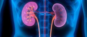
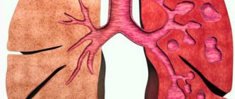
Gaucher syndrome develops: the liver, spleen, and kidneys enlarge, and the accumulation of glucocerebroside in the cells of bone tissue and lungs destroys their structure.
What it is?
In short, Gaucher disease is a genetic disease in which fatty substances (lipids) accumulate in cells and on some organs. Gaucher disease is the most common of the lysosomal storage diseases. It is one of the forms of sphingolipidosis (a subgroup of lysosomal storage diseases), as it manifests itself in the dysfunction of sphingolipid metabolism.
The disorder is characterized by fatigue, anemia, low levels of platelets in the blood, and an enlarged liver and spleen. This is caused by a hereditary deficiency of the enzyme glucocerebrosidase, which acts on the fatty acid glucosylceramide. When the enzyme is damaged, glucosylceramide accumulates, in particular in white blood cells, most often in macrophages (mononuclear leukocytes). Glucosylceramide can accumulate in the spleen, liver, kidneys, lungs, brain and bone marrow.
Pathological anatomy
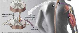
Small or extensive foci of accumulation of Gaucher cells are found in various organs. The spleen is most affected in both acute and chronic forms of the disease. It is increased in size and often has a bumpy surface. The tissue is gray-red, brick or chocolate in color, variegated when cut, with the presence of angiocavernous foci, infarctions, and scars. Microscopically, accumulations of Gaucher cells are found in the red pulp, trabeculae, and follicles; there is a sharp expansion of the sinuses (but, as a rule, there are no Gaucher cells here). Gaucher cells in the spleen contain a yellow-brown pigment. The liver is also enlarged. There are fewer Gaucher cells here than in the spleen; individual elements are transitional forms from stellate endothelial cells (Kupffer cells) to Gaucher cells. Gaucher cells are located diffusely in the liver lobules, disrupting the correctness of their structure, in the walls of capillaries, around the sinuses. In the bone marrow, focal accumulations of Gaucher cells are combined with the resorption of bone beams and the proliferation of connective tissue. In the adrenal glands, Gaucher cells are found primarily in the zona reticularis. In the lungs, they infiltrate the interstitial tissue of the alveolar septa and may be contained in the lumens of the alveoli. In the acute form of G. b. these cells are also found in the glial elements of the brain, the thymus, and the glomeruli of the kidneys. In the brain, dystrophic changes in ganglion cells, an increase in glial elements in the subcortical ganglia, the phenomenon of neuronophagia (see) and impaired myelination are detected. In the white matter of the brain, certain fractions of phospholipids, cholesterol and cerebrosides are sometimes reduced, which may be associated with demyelination.
Rice. 1. Microscopic specimen of the spleen in Gaucher disease: round Gaucher cells are visible (indicated by arrows).
Gaucher cells (Fig. 1) are round in shape, large (40-80 µm in diameter) with a small nucleus, often located eccentrically, and a wide zone of fibrillar, striated light gray cytoplasm. Gaucher cells are characterized by intense blue cytoplasm when stained using the Mallory method (see Mallory methods), a positive PAS reaction (see), high activity of acid phosphatase and nonspecific esterase. The cytoplasm often contains hemosiderin. Multinucleated cells are found. By electron microscopy in Gaucher cells and in cells that have not yet acquired morphol. features of Gaucher cells, but already accumulating glucocerebrosides, lipid cytosomes that are absent normally are found - formations in the form of a cluster of tubes containing glucocerebrosides, dia. 25-40 nm, delimited by a membrane. Using the negative contrast method, it was found that these tubes have a spiral structure. This distinguishes Gaucher cells from cells similar to them that are detected in other diseases, in particular in chronic myeloid leukemia (see Leukemia). Electron microscopy often reveals remnants of red blood cells in Gaucher cells.
Reasons for development
At the genetic level, mutations occur in the genes that are responsible for the production of the enzyme glucocerebrosidase. This gene with anomalies is localized on chromosome 1. These mutations cause low enzyme activity. Thus, glucocerebroside accumulates in macrophages.
Mesenchymal cells, called Gaucher cells, gradually grow and become hypertrophied. Since modifications occur in these cells, and they are located in the spleen, kidneys, liver, lungs, brain and bone marrow, they, in turn, deform these organs and disrupt their normal functioning.
Gaucher disease is an autosomal recessive disease. Therefore, any person can inherit a mutation of this enzyme with all the features in the same proportion, both from the father and from the mother. Thus, the degree of the disease and its severity will depend on the damage to the genes.
Theoretically, each person can inherit the glucocerebroside gene with lesions or completely healthy. As a result of inheriting a gene with abnormalities, a mutation of this enzyme occurs, but this does not yet indicate a disease. But when a child receives both affected genes, then a diagnosis of Gaucher disease is made. When inheriting one affected gene, a child is considered only a carrier of the disease, so there is a possibility of transmitting this trait, with hereditary pathology, to future generations. Thus, if both parents are carriers of the disease, a child can be born with Gaucher disease in 25% of cases, a carrier child in 50%, and a healthy child in 25%.
The frequency of occurrence of this hereditary pathology among ethnic races is 1:50,000, but it is much more often detected among Ashkenazi Jews.
Gaucher disease is also called a storage disease due to a deficiency of the enzyme, which should remove harmful metabolic products from the body, and not accumulate them. As a result of this, these substances accumulate in the macrophages of some organs and destroy them.
Classification and types of disease
The nature of the course of the disease varies in severity. Complications occur in childhood and adulthood. There are three types of disease:
- The first non-neuronopathic type. Sociology shows that it is common among Ashkenazi Jews. This pattern is called the Gaucher reaction. The clinical picture is characterized by a moderate, sometimes asymptomatic course of the disease. The psychology of behavior does not change, the brain and spinal cord are not damaged. Symptoms appear more often after thirty years of age. There are known cases of diagnosis in childhood. Timely treatment gives a favorable prognosis.
- The second type represents the neuronopathic infantile form and is rare. Symptoms appear in infancy as early as six months. Progressive damage to the child's brain occurs. Death can occur suddenly from suffocation. All children die before reaching two years of age.
- The third type (neuronopathic juvenile form). Symptoms have been observed since the age of 10 years. The intensification of symptoms is gradual. Hepatosplenomegaly - enlargement of the liver and spleen - is painless and does not impair liver function. Possible violation of behavioral psychology, the onset of neurological complications, portal hypertension, venous bleeding and death. Damage to bone tissue by Gaucher cells can lead to limited mobility and disability.
Symptoms of Gaucher disease
The clinical picture depends on the type of disease, but there are also general signs of this disease. You can suspect Gaucher syndrome (see photo) based on the following manifestations:
- pale skin;
- growth disturbance in children;
- general weakness;
- inflammation of the lymph nodes;
- enlarged liver and spleen;
- fractures in the absence of injuries;
- spontaneous nosebleeds;
- hemorrhagic stars on the skin.
Gaucher syndrome does not depend on the gender of the child. In addition, the symptoms of the disease often resemble the clinical picture of hematological pathologies. This makes diagnosing the disease difficult.
Characteristic signs of different forms of Gaucher syndrome:
| Type of disease | Clinical manifestations |
| First type |
|
| Second type |
|
| Third type |
|
Gaucher disease in children
Symptoms may begin to appear at different ages. The second type of disease often manifests itself at the age of six months. Patients in this case live up to 1-2 years. The third type is typical for children 2-4 years old, although it is sometimes observed in adolescence. The same applies to the first form. It can appear both in early childhood and adolescence. Symptoms of Gaucher syndrome in children:
- poor ability to suck and swallow;
- eye movement disorders;
- convulsions;
- breathing problems;
- whooping cough;
- yellow-brown skin pigmentation.
Notes
- CCHMC Molecular Genetics Laboratory Mutation Database - Gaucher Disease; glucosidase, beta, acid (GBA).
- Deegan, Patrick and Cox, Timothy
Imiglucerase in the treatment of Gaucher disease: a history and perspective. Drug Des Devel Ther. 2012;6:81-106. doi: 10.2147/DDDT.S14395. Epub 2012 Apr 18. PMID 22563238 (free full text) - Shire Announces FDA Approval Of VPRIV(TM) (velaglucerase Alfa For Injection) For The Treatment Of Type I Gaucher Disease. medicalnewstoday.com. Retrieved August 13, 2012.
- Yukhananov, Anna
. US FDA approves Pfizer/Protalix drug for Gaucher (1 May 2012). Retrieved May 2, 2012. (inaccessible link) - CenterWatch:Cerdelga (eliglustat).
- https://www.drugs.com/news/zavesca-approved-first-oral-option-type-1-gaucher-3351.html
Diagnostics
Collecting anamnesis and complaints of the disease (clarification of the time of appearance of the first symptoms of the disease, how they progressed over time).
The disease can be suspected if an enlargement of the liver and spleen is accidentally detected (for example, according to ultrasound), suppression of the hematopoietic system (changes in blood tests: decreased levels of hemoglobin, platelets, the appearance of atypical blood cells), and the appearance of symptoms of bone damage.
At the next stage, special studies are carried out to confirm the diagnosis:
- Enzyme analysis - determination of the level of enzyme (glucocerebrosidase) in leukocytes and skin cells, which makes it possible to establish a diagnosis with absolute accuracy;
- biochemical blood test (decreased β-glucocerebrosidase activity, increased chitotriosidase levels);
- bone marrow examination (presence of characteristic Gaucher cells);
- molecular studies at the gene level (detection of genetic disorders);
- radiography and computer diagnostics (magnetic resonance imaging) of bones, since there may be areas of lower density that have specific signs for this disease.
Third type of Gaucher disease
How to treat Gaucher disease?
Specialized care for patients with the first and third types of the disease is aimed at eliminating symptoms and compensating for the primary genetic defect - increasing the amount of the missing enzyme, increasing the catabolism of glycosphingolipids. With type 2 pathology, therapeutic measures are not effective enough; doctors’ efforts are reduced to alleviating clinical manifestations - pain, cramps, respiratory disorders.
The general scheme includes the following areas:
- Enzyme replacement therapy . The main treatment method is lifelong enzyme replacement therapy (ERT) using recombinant glucocerebrosidase. The effectiveness is quite high - the symptoms are completely relieved, the quality of life of patients increases. ERT is appropriate for the third and first types of the disease. The drugs are administered intravenously. Frequent infusions sometimes cause inflammatory diseases of the veins (phlebitis).
- Substrate-reducing therapy. This direction is new in the treatment of Gaucher disease and is relatively widespread in the USA and European countries. Aimed at reducing the rate of production of the substrate glycosphingolipids and accelerating the catabolism of accumulating macromolecules. The drugs used are specific inhibitors of glucosylceramide synthase. The method is indicated for type 1 disease with mild to moderate symptoms.
- Symptomatic therapy . In cases of osteoporosis, complex therapy is prescribed, including taking calcium-containing medications, vitamin D and following a diet enriched with calcium. These measures can slow down bone loss, increase bone strength, and prevent fractures. For skeletal complications, analgesics (NSAIDs) and antibacterial therapy are used. Symptoms of neurological disorders can be relieved with antiepileptic drugs, nootropics, and muscle relaxants.
Replacement therapy for this disease
There are also a number of medications that successfully help fight lysosomal disorders in the body. This is replacement therapy, the essence of which is to compensate for the lack of enzymes in the body, or to artificially supplement the missing parts of enzymes. Such drugs are created based on the latest advances in genetic engineering and help replace natural enzymes either partially or completely. A positive result of drug treatment is achieved in the early stages of the disease.
Forecast
With the first type of disease, early diagnosis, and timely initiation of replacement therapy for Gaucher disease, positive dynamics are possible. The second type of glucocerebrosidosis is the most unfavorable, as it is more severe. Sick children usually do not live more than two years. The third form of Gaucher disease, with timely diagnosis and adequate treatment, allows maintaining the patient’s vital functions. Otherwise, he dies quite quickly from complications that develop.
Fatty liver hepatosis: symptoms and treatment Liver diseases: symptoms and first signs Hepatitis C: first signs and treatment regimen Why is the liver enlarged - what to do and how to treat? Malabsorption syndrome: symptoms and treatment Liver cirrhosis
Additional information affecting the course and outcome of the disease
6.1 Forecast
In type I, it is favorable if ERT is prescribed in a timely manner.
For irreversible lesions of the osteoarticular system, surgical orthopedic treatment is indicated.
When vital internal organs are damaged, the prognosis is determined by the degree of dysfunction and complications.
6.2 Errors and unreasonable assignments
Splenectomy is not recommended , only for absolute indications (traumatic rupture of the spleen).
If splenomegaly and cytopenia are unclear and splenectomy is necessary, it is advisable to exclude Gaucher disease.
Repeated bone marrow punctures and other invasive diagnostics (liver biopsy, spleen) are not necessary if the diagnosis is proven.
Errors:
- surgical treatment of bone crises mistaken for osteomyelitis;
- prescription of glucocorticoids to relieve cytopenic syndrome;
- prescribing iron supplements to untreated patients with “anemia of inflammation”.
6.3 Gaucher disease and pregnancy
- Gaucher disease is not a contraindication for pregnancy.
- It is advisable to plan pregnancy after achieving the treatment goal.
- The issue of ERT during pregnancy and breastfeeding is decided individually.
- Pregnancy management by experienced obstetricians-gynecologists together with a hematologist.
- The method of delivery is determined by obstetric indications, taking into account cytopenia and the state of hemostasis.
external_script#827