Paget's disease
(J. Paget, English surgeon and pathologist, 1814-1899; synonym:
deforming ostosis, deforming osteitis, fibrous osteodystrophy, deforming osteodystrophy
) is a disease of the dysplastic skeleton with the development of pathological restructuring, which leads to its characteristic deformation. The disease was first described by J. Paget in 1877 and called it osteitis deformans, believing that it is based on an inflammatory process. Paget's contemporaries began to call the disease after him. Later, Paget's disease was combined into one group with Recklinghausen's disease (see Parathyroid osteodystrophy) due to certain similarities in histological data and was called fibrous osteitis. Stenholm (T. Stenholm, 1924) proved the dystrophic, rather than inflammatory nature of the process in these diseases and proposed a new name - fibrous osteodystrophy. K. Schmorl (1930) identified Paget's disease as an independent group and called it deforming osteodystrophy. A.V. Rusakov and other researchers believe P. b. postnatal dysplastic process.
The incidence of Paget's disease is 0.1 - 3%. The disease occurs in all countries of the world, more often in Australia, the USA, Western Europe, less often in China, Japan, the Middle East, India, and Africa. Men get sick more often. Sometimes P. b. occurs in several family members.
Causes of Paget's cancer
The causes of the disease are currently not fully known. There are two theories:
- Most experts are inclined to believe that Paget's cancer initially occurs inside the breast, and from there the cancer cells penetrate into the nipple and areola. This explains why 97% of patients have another tumor.
- According to the second theory, Paget's cancer develops independently as a result of malignancy of cells located in the nipple or areola. This is confirmed by the 3% of patients who do not have a second tumor in the chest.
The above-described changes in cells occur due to certain mutations. Each cancer cell carries a set of mutations that help it multiply and survive uncontrollably. Why such “cancerous” genetic changes occur in each specific case is impossible to say for sure. Doctors and scientists know some risk factors that increase the likelihood of developing Paget's cancer:
- Age. Over the years, mutations accumulate in the human body, and one day they can lead to the emergence of a malignant neoplasm in the mammary gland or other organ.
- Family history. If you have close family members who have been diagnosed with breast cancer, your risk is also increased.
- Hereditary mutations. For example, it is known that the likelihood of developing malignant breast tumors increases with mutations in the BRCA1 and BRCA2 genes. If parents have these genetic defects, they can pass them on to their children.
- Increased density of breast tissue. This can be detected during a mammogram, an x-ray examination.
- Burdened personal history. If a woman has previously been diagnosed with breast cancer, she has an increased risk of it recurring.
- Excess weight. This factor is especially dangerous if a woman begins to gain extra pounds after menopause.
- Alcohol. Excessive and frequent consumption of alcohol increases the likelihood of developing cancer.
- Hormone replacement therapy. Women who take estrogen medications are more likely to develop Paget's cancer.
If you find factors from the list, this does not mean anything. None of them are guaranteed to lead to cancer. Each only increases the probability to some extent. At the same time, the absence of all these factors does not guarantee protection against cancer.
Book a consultation 24 hours a day
+7+7+78
Advantages of Top Assuta
- The clinic’s specialists have unique experience in treating diseases of this type, have undergone many years of training and are leading experts.
- The examination in the clinic lasts only a few days, and the diagnosis is determined on the basis of accurate laboratory and hardware tests.
- A wide range of effective methods of conservative, surgical and physiotherapeutic treatment.
- Individual approach, personal curator-translator, attentive and responsive medical staff.
- 5
- 4
- 3
- 2
- 1
(2 votes, average: 5 out of 5)
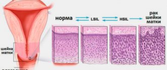
Stages of the disease
Paget's cancer, in which there are additional tumor nodes in the breast, is classified into stages according to the generally accepted TNM system. Depending on the indicators T (size and spread of the primary tumor), N (spread to regional lymph nodes) and M (presence of separated metastases), 5 stages are distinguished:
- Stage 0 - the tumor is contained within the tissue from which it developed and has not spread to adjacent tissue.
- Stage I - tumor no more than 2 cm in greatest dimension. Cancer cells do not spread to the lymph nodes, there are no distant metastases.
- Stage II - tumor is 2 to 5 cm, which can spread to the lymph nodes. In this case, mobile lymph nodes are palpated on the affected side.
- Stage III . At stage IIIA, the size of the tumor can exceed 5 cm. It spreads to the lymph nodes, which become fused with each other and surrounding tissues. At stage IIIB, the primary tumor grows into adjacent tissues: skin, chest wall.
- Stage IV - there are distant metastases. It does not matter what size the primary tumor is.
Complications
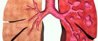
Neuritis of the trigeminal nerve (see), facial nerve (see), deafness (see), optic nerve atrophy (see), hypertensive syndrome (see), CSF disorders are caused by thickening of the bones of the base and vault of the skull, flattening of the cranial fossae and narrowing of the openings of the skull. If the spine is damaged, spinal compression syndrome may develop.
Involvement of the hip joints in the process leads to severe coxarthrosis (see Arthrosis) and sometimes to protrusion of the head into the pelvis. Patol, fractures are not uncommon (see). Against the background of P. b. Both benign tumors can develop - osteoblastoclastoma (see), and malignant ones - osteogenic sarcoma (see) and other types of sarcomas (see), multiple myeloma (see). Malignization is the most dangerous complication of P. b. Sarcomas occur in 3-15% of observations of P. b. By structure, these are osteogenic, spindle cell, polymorphic cell sarcomas, chondrosarcoma, fibrosarcomas, reticular sarcomas. The course of sarcomas complicating P. b. is particularly malignant.
Symptoms
In 40–50% of patients with Paget's cancer, a lump in the breast can be felt. In 60–90% of cases, such a symptom indicates invasive cancer, that is, growing into surrounding tissues.
Usually the first symptoms of the disease are associated with changes in the nipple and areola. They can easily be mistaken for manifestations of another pathology - eczema or psoriasis:
- redness;
- itching, tingling, burning, soreness;
- deformation, flattening of the nipple;
- discharge - yellow or bloody;
- ulcerations, weeping, crusts;
- the nipple is easily injured, begins to bleed, and then crusts form.
It is worth remembering about other manifestations of malignant breast tumors: induration, asymmetry, “lemon peel”-type skin changes, enlarged axillary lymph nodes.
These signs do not always indicate cancer. But, if they appear, it is important to immediately visit a doctor and get checked.
A very rare type of disease is Paget's pigmented cancer. In this case, it is necessary to carry out a differential diagnosis with melanoma.
Diagnostic methods
Diagnosis of the disease is carried out by an oncomammologist. During the initial appointment, he examines the mammary glands, nipple, areola, and palpates (feeling) to detect lumps.
Depending on the clinical manifestations, three forms of the disease are distinguished:
- There is damage to the nipple and areola, but there are no tumor nodes in the mammary gland.
- Nodal form.
- There is a tumor node, but there is no damage to the nipple and areola.
The main method for diagnosing Paget's cancer is biopsy. The essence of the study is that a sample of pathologically altered tissue is obtained and sent to the laboratory for examination under a microscope. This helps to reliably detect tumor cells and establish an accurate diagnosis.
Biopsy material can be obtained in different ways:
- Using scraping.
- Removing a thin layer of skin using a blade is a shave biopsy.
- Needle biopsy - using a special instrument in the form of a tube with sharp edges, it allows you to obtain a column of tissue.
- During a wedge biopsy, a wedge-shaped piece of tissue is removed.
- In some cases, the entire nipple is removed.
If the results of the biopsy reveal Paget's disease, the doctor prescribes additional tests: mammography, MRI, ultrasound. They help identify additional nodes in the chest.
Peculiarities
There are two more medical terms that define Paget's disease - osteodystrophy or osteitis deformans.
Osteitis deformans - what is it? This is a condition characterized by chaotic processes of bone resorption and restoration. In Paget's disease, old bone tissue is destroyed earlier than it should be normally, and new bone is formed abnormally quickly. Such unregulated processes lead to the formation of loose and soft bone, which is easily subject to fractures, curvatures and dystrophic changes.
In 30% of patients, lesions are found in one bone, but on average the disease affects 2 paired or 3 bones at once. In Paget's disease, degenerative processes are most often observed in the bones of the limbs, pelvis, spinal column and skull.
Modern methods and principles of treating Paget's cancer
For many years, surgeons used aggressive tactics for Paget's cancer: during surgery they removed the entire mammary gland (mastectomy) and axillary lymph nodes on the side of the tumor. This approach was argued by the fact that patients often have additional tumor nodes in the breast, and even if there is only one tumor, then after removal of the nipple and areola, part of it may still remain in the depths, which is fraught with relapse. Lymph nodes were removed just in case, since they may also contain cancer cells.
Currently, approaches have changed. Studies have shown that if there is only one tumor in the breast in the nipple area, and there are no other nodes according to mammography, only the nipple with the areola can be removed, subsequently prescribing a course of radiation therapy. After such organ-conserving surgery, there is no increased risk of relapse.
The attitude towards lymph nodes has also changed. Currently, there is a procedure that helps check whether there are any cancer cells left in them - sentinel biopsy . During the operation, the surgeon injects a special radiopharmaceutical into the tumor, which penetrates the lymphatic vessels, reaches nearby (“sentinel”) lymph nodes and causes them to “glow.” This “glow” is detected using a special apparatus - a gamma camera . This way, the doctor can find the lymph nodes that first receive lymph from the tumor, remove them and check for lesions. If the sentinel lymph nodes are “clean,” it means that the cancer cells did not have time to reach them, and there is no point in removing the remaining lymph nodes.
Sentinel biopsy helps to avoid unnecessary removal of lymph nodes and thereby prevent the development of complications associated with impaired lymph outflow - lymphedema .
After surgery, chemotherapy, hormonal, and targeted drugs may be prescribed. Typically, adjuvant chemotherapy is carried out in cases where a large tumor has been removed, foci have been found in the lymph nodes, and if the cancer cells were poorly differentiated and prone to aggressive growth. Hormonal therapy is indicated if there are hormone receptors on the surface of tumor cells.
Forecast
The prognosis for Paget's cancer depends on a number of conditions. The presence of additional nodes in the chest and the stage of the disease are of great importance. In the United States, statistics were studied and it turned out that the 5-year survival rate (the percentage of patients surviving 5 years from the time the tumor was diagnosed) among women who received treatment for Paget's cancer from 1988 to 2001 was 82.6%. However, the average five-year survival rate for other types of breast cancer is 87.1%.
If there are additional nodes in the breast, the five-year survival rate decreases as the stage increases:
- Stage I - 95.8%;
- Stage II - 77.7%;
- Stage III - 46.3%;
- Stage IV - 14.3%.
Thus, early diagnosis helps improve the prognosis and significantly increases the chances of successful treatment. As soon as any suspicious changes in the mammary glands appear, you should immediately visit a mammologist and undergo an examination. And in order to promptly detect these changes, every woman needs to regularly conduct breast self-examination once a month. Once a year you need to be examined by a mammologist, after 40 years - a mammogram.
Clinical picture
Paget's disease occurs more often over the age of 40 years, but is likely to be asymptomatic for a long time before detection. Some researchers identify the so-called. juvenile form of the disease (juvenilis Paget), others designate this lesion as hyperphosphatasia.
At the beginning of the disease, pain in bones and joints appears for no apparent reason. In advanced cases, bone deformation occurs.
Skeletal involvement is often asymmetrical. The long tubular bones thicken and become arched. The skull increases in size due to thickening of the bones of the arch and base. Hyperthermia of the soft tissues over the diseased bone is characteristic, caused by its hypervascularization and dilation of the subcutaneous vessels. Restriction of mobility in the joints and lameness develop in advanced cases.
The levels of calcium and phosphorus in the blood and urine may be normal, but high levels of alkaline phosphatase and high urinary excretion of hydroxyproline are often observed.