Urachal cyst – partial closure of the embryonic urinary duct (urachus) with the formation in its middle part of a cystic cavity filled with mucous secretion. If a urachus cyst becomes infected, a palpable tumor forms in the lower abdomen, pain, dysuria, fever, and symptoms of general intoxication appear. When abscess formation occurs, a urachal cyst can rupture into the bladder, abdominal cavity, or through the anterior abdominal wall to the outside, forming an umbilical fistula. Diagnosis includes cystography, ultrasound of the bladder, CT. In case of exacerbation, antibacterial and physiotherapeutic treatment is carried out; When the inflammation subsides, excision of the urachus cyst is indicated.
In urology, a urachal cyst is regarded as an embryonic malformation of the urinary system. The urachus serves as the embryonic urinary duct, which serves during intrauterine development to connect the bladder with the amniotic fluid. In the embryo, the urachus is a tubular formation, which normally overgrows by 5-6 months of development, forming the median umbilical ligament. In newborns and adults, at the site of the urachus, an obliterated acellular cord is determined, stretching from the apex of the bladder to the navel between the transverse fascia and the peritoneum.
If obliteration of the duct does not occur by birth, various pathological processes can develop in it: with complete non-closure - vesico-umbilical fistula; with obliteration of the proximal and distal ends, but nonunion of the middle section - urachus cyst; in case of non-closure of the peri-vesical section - bladder diverticulum.
The urachal cyst, as a residual embryonic formation, contains mucus, serous exudate, urine, and meconium. A urachus cyst may not increase in size for a long time and may not manifest itself clinically, being detected only in adulthood. Infection of a urachus cyst is dangerous due to the development of suppuration and septic complications.
According to statistics, urachal cysts occur 3 times more often in males than in females.
A urachal cyst can be as large as an adult man's fist. Most often the cavity is closed; less often it can communicate with the bladder or the external environment through a thin fistulous tract. In the latter case, urine is released out through the navel.
Urachal cyst - what is it?
The tumor is otherwise called a ureteral cyst. Formed in the duct of the bladder, it looks like a growth with a dense hollow capsule. The name is given according to the tubular organ formed during the development of the embryo. In fact, the additional growth is a ureter that is not fully formed.
Formed in the duct of the bladder, externally it looks like a growth with a dense hollow capsule
Urachal cysts in children are diagnosed in infancy. Adults live their entire lives with a congenital anomaly. But, if inflammation of the genitourinary system develops, then there is a risk of increased pathology.
The original dimensions are only 10-20 mm. Growth is relatively slow and does not cause any discomfort. It grows sharply up to 15 cm when exposed to infectious agents. It becomes malignant without timely treatment.
What is a tumor
A ureteral cyst is a convex formation, clear around the edges. The diameter is up to 20 mm. A problem develops in the ureter. In appearance, the disease appears as a hollow bulge. The cyst cavity is filled with fluid.
Structure of the neoplasm
The structure of the cystic bulge was divided into 3 regions: internal, external and middle. The inside of the tumor consists of a layer of epithelium. The outer part is expressed by the lining of the bladder along with muscle mass. And the middle zone of formation is represented by connective tissue.
Cystic formation of the ureter
Causes of pathology
The real cause of the development of the congenital anomaly is currently unknown. There is a theory associated with impaired intrauterine development. However, it has not been scientifically proven. What is known is that the cavity appears during the growth of the embryo.
Stones in the kidneys or urinary organ.
The sharp progression of the cyst is caused by several factors:
- use of a catheter after surgery;
- infections in the genitourinary system;
- stones in the kidneys or bladder;
- constant recurrence of infectious diseases.
A rare condition called cystic cystitis can provoke tumor growth. Constant inflammation of the bladder leads to the formation of many cavities. A urachus tumor is included among them if it was initially present in the body.
Complications
Other neoplasms in the genitourinary system resolve without serious consequences with proper treatment. But there are some consequences that are also inherent in urachus tumors:
- Complete blockage - the formation grows to such an extent that it blocks the ureter.
A large mass blocks the ureter. - Rupture - fluid from the ruptured cavity enters the bladder cavity, causing additional infection.
- Infection as a consequence of peritonitis, when a pathogenic environment develops in several parts of the urinary tract.
- Oncological disease.
In fact, an abscess or peritonitis develops inside, which requires surgical treatment, as with many other cystic diseases.
Diagnostic measures
If the cyst nevertheless grows to a sufficient size, then to diagnose it, the doctor only needs to palpate the umbilical area. But in order not to confuse the cyst with a hernia or other tumor, it is necessary to conduct an ultrasound examination using an ultrasound machine, which will confirm the diagnosis. In addition to ultrasound, patients also undergo tests such as MRI, CT, radiographic cystography and cystoscopy.
In moments of exacerbation, or excessive suppuration and subsequent perforation of the contents of the sac, the patient must be taken to the hospital. For acute and severe conditions, the laparoscopic method is used.
Symptoms of a urachus cyst
Neoplasms of the bladder and related organs are asymptomatic in most cases. Clinical signs appear when the cavity grows and interferes with normal functioning. Or a rupture occurs, the fluid spreads into the adjacent cavities and the bladder becomes infected.
Associated pathology symptoms:
Urine with blood.
- nagging pain in the pelvic organs (a common sign of urachus cysts in women);
- pain with the urge to urinate (more often appears as a sign of a urachus cyst in men);
- blood clots in urine;
- frequent urge;
- incontinence;
- large volume of urine at one time;
- acute pain in the lumbar region;
- cloudy urine color;
- sharp sour smell;
- swelling in the navel area;
- elevated temperature - with the development of complications.
The pathology is characterized by frequent urination and defecation of women, which is due to the anatomical feature - a short urethra. Painful sensations and a feeling of fullness are more often diagnosed in men. The release of purulent exudate indicates the risk of complications. These symptoms also indicate cystitis in any form.
Frequent urge to urinate and defecate in women.
The presence of an inflammatory process provokes a sharp growth rate of the cyst with accompanying symptoms:
- The tumor is felt when palpating the abdomen. The formation is dense to the touch, pressure causes pain.
- The abdominal organs (ureters, bladder, stomach, intestines) are compressed.
- Problems with the gastrointestinal tract develop.
Gastroesophageal reflux can develop as another complication of the disease. Food and hydrochloric acid from the stomach move back into the esophagus. This happens when the walls of the stomach are compressed, as a result its size decreases. The esophagus becomes inflamed, causing a constant feeling of nausea and heartburn.
Careful diagnosis is necessary because a urachal tumor may not be recognized until its acute presentation. Accordingly, suspicions will be directed towards other infectious diseases of the genitourinary system.
Consequences of formations
In addition to the states of discomfort arising from the presence of a cyst, the consequences of its existence may appear.
Usually this is its inflammation, fraught with the development of purulent complications:
- When pus breaks into the abdominal cavity, signs of peritonitis (inflammation of the peritoneum) appear.
- Purulent melting of the bladder wall forms a fistula of this organ.
- An umbilical fistula, the result of a perforation of the anterior abdominal wall, manifests itself by the outflow of purulent detritus through its mouth, the weeping of the skin around the navel for this reason, and other signs of omphalitis. The volume of leaking pus increases during moments of straining, coughing, or squeezing the involved area.
Suppuration of a urachal cyst is not particularly specific in its symptoms. It manifests itself:
- pain in the lower third of the abdomen, becoming more intense as the inflammation process increases;
- local changes in the form of redness and swelling of the skin of the pubis and peri-umbilical area, spreading over time to the entire anterior wall of the abdomen;
- rise in body temperature, chills;
- signs of intoxication in the form of weakness, sweating, palpitations, dizziness, nausea (both accompanied by vomiting and without it).
Diagnosis of urachus cyst
The disease does not manifest itself until it is affected by an infectious agent. A dangerous condition develops when the cyst puts pressure on the bladder. Symptoms become brighter, the functioning of surrounding organs is disrupted. Diagnosis is carried out using a full examination.
First, anamnesis is collected. Laboratory tests are prescribed:
- Blood tests, urine culture.
- Ultrasound.
Ultrasound of the genitourinary system. - Excretory urography (a contrast agent is injected, determines the rate of accumulation and output of urine).
- Uroflowmetry (deviations in urination).
- Cystoscopy (examination of the mucous membrane).
- Tomography.
Tumor on ultrasound.
A urachus cyst is detected by ultrasound by the occlusion of the ureter. The size of the organs of the genitourinary systems and kidneys is examined, the presence of inflammation and stones is determined.
Treatment
Timely diagnosed pathology allows you to quickly begin treatment. Most often, the tumor shows signs of life only when inflammation develops. Congenital pathology is not always diagnosed in childhood due to its small size. Discomfort occurs after infection of the cavity tissues.
Two methods are used - medication and surgery. There is no conservative treatment for urachus tumors. Therapy is carried out to relieve the main symptoms. The only way to get rid of a tumor is by complete excision.
Medication
Drinking plenty of fluids is recommended.
Antibacterial and physiotherapeutic treatment is carried out using medications. The main therapy is aimed at relieving inflammation. General antibiotics, antispasmodics and painkillers are prescribed to relieve pain. In addition, drinking plenty of fluids is recommended to ensure constant urination of the bladder. Due to frequent urination, bacteria do not have time to stick to the walls.
Surgical
The operation is permitted only after the inflammation has been eliminated. Laparoscopic cyst removal is more often used; it allows you to quickly and painlessly remove the tumor.
Laparoscopic removal allows you to quickly and painlessly remove the tumor.
The abdominal cavity is opened when an abscess is diagnosed:
- The lesion is opened and drainage is performed.
- The abdominal cavity is washed when the tumor breaks through.
- The cyst is removed - the tissue is excised, the place is stitched.
Medications are prescribed to speed up healing and strengthen the immune system.
A radical procedure, cystectomy, is performed when the cyst is malignant. Cancer is more likely to develop if there is an undetected cyst in the bladder in men. The affected organ and tissue are completely removed.
Traditional methods
It is impossible to cure a cyst, especially a congenital one, using only traditional methods. Recipes will help relieve inflammation and other symptoms. The tumor can only be removed surgically.
The following recipes are used:
- Herbal mixture: chamomile, calamus, peony, nettle, calendula, dill. Take 1 tbsp. spoon of dry collection, pour boiling water and heat over low heat for 15 minutes. Afterwards, the broth should be strained and left for 8 hours in a cool place. Drink half a glass before meals three times a day.
Drink half a glass of the decoction before meals three times a day. - Another collection of chamomile, St. John's wort and mint. Take 1 tbsp. spoon of the first ingredients and 1 teaspoon of mint (or 2-3 leaves). Boil water, throw the dry collection there. Let it brew over low heat for 20 minutes, then transfer to a water bath for 10 minutes. Allow to cool, strain before use and take 100 ml 5 times a day (about 1 shot glass).
A collection of chamomile, St. John's wort and mint. - Juice from fresh berries - viburnum, lingonberries. Squeeze out the juice and mix it with natural honey. Take 1 teaspoon three times a day before meals. Effective in relieving inflammation.
Squeeze out the juice and mix it with natural honey.
It is important to understand that traditional treatment only complements medication, but is not able to cure the cyst. The neoplasm will not stop growing and compressing the walls of surrounding organs only from medicinal decoctions. Consultation with a doctor is required to prescribe complete treatment.
Preventive measures
It is impossible to predict the development of a neoplasm, so all measures should be aimed at maintaining a healthy lifestyle.
Doctors recommend:
- do not neglect preventive examinations;
- In order to exclude a cyst or, conversely, to identify it, you can undergo diagnostics using modern equipment.
The simplest, safest and non-invasive method is ultrasound.
Prevention
It is important to follow prevention regardless of whether a cyst appears on the bladder of a woman or a man. It is important to have regular medical examinations. In addition, the following measures are required:
- Do not overcool the body, especially the lower part.
- Maintain personal hygiene: wash regularly, change underwear.
- Eat a balanced diet.
Cysts of the bladder, including urachus, are often benign and do not cause discomfort. However, if the infection is not treated in time, it can lead to the degeneration of the neoplasm into a malignant one and the development of cancer. It is important to seek medical help promptly and follow preventative measures.
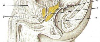
Anomalies of urachus development
Types of urachus anomalies: a - open urachus, b - sinus, c - bladder diverticulum, d - urachus cyst
The urachus is the embryological remnant of the allantois. Cysts account for a third of all urinary duct defects. The purpose of the urachus is to divert fetal urine into the amniotic fluid during intrauterine development. At 20-22 weeks of pregnancy, the canal begins to become obliterated and should normally be completely closed by the time the baby is born. Why this does not happen is unknown. Depending on the location of the cleft, urachal anomalies are distinguished:
• incomplete umbilical fistula or sinus, no obliteration of the part close to the umbilicus - 18%;
• completely open urachus or complete vesico-umbilical fistula - 48%;
• bladder diverticulum - non-closure of the part of the duct bordering the bladder - 3%;
• urachus cyst - absence of obliteration in the middle part of the canal - 31%.