Gallstone disease (GSD) is a disease characterized by the formation of stones in the gall bladder and/or bile ducts as a result of impaired metabolism of cholesterol and/or bilirubin in the body.
GSD is one of the most common human diseases and ranks third in the structure of morbidity after cardiovascular diseases and diabetes mellitus. In developed countries, the incidence of cholelithiasis averages 10-15% of the adult population. In Russia, the prevalence of cholelithiasis ranges from 3-12%. In women, cholelithiasis occurs 3-4 times more often than in men. Due to the high prevalence of cholelithiasis and the steady trend towards its growth, the number of operations for cholelithiasis is also increasing. Currently, in terms of the number of surgical interventions, cholecystectomy has taken second place in the world after appendectomy.
According to their chemical composition, they are distinguished: cholesterol (pure and mixed) and bilirubin (pigment), brown and black. Depending on the degree of saturation with calcium salts, stones are divided into two groups: calcified (calcified) and non-calcified (non-calcified).
The composition of stones is important to consider to assess the possible effectiveness of drug dissolution with ursodeoxycholic acid preparations.
Classification of cholelithiasis (3rd extraordinary congress of the Scientific Society of Gastroenterologists of Russia, 2002).
Stage I – initial or pre-stone.
A. Thick heterogeneous bile.
B. Formation of biliary sludge: with the presence of microlites, with the presence of putty-like bile, a combination of putty-like bile with microliths.
Stage II – formation of gallstones.
A. By localization: in the gallbladder, in the common bile duct, hepatic ducts.
B. By the number of stones: single, multiple.
B. By composition: cholesterol, pigment, mixed.
D. According to the clinical course:
- latent flow,
- with the presence of clinical symptoms:
- painful form with typical biliary colic,
- dyspeptic form,
- under the guise of other diseases.
Stage III – chronic recurrent calculous cholecystitis.
Stage IV – complications.
The mechanism of stone formation in gallstone disease
Stones are formed as a result of a violation of the chemical composition of bile. The gallbladder functions as a storage reservoir for bile, where bile “ripens”, acquiring the functions necessary for the digestive process, and from where it enters the duodenum. If the chemical balance of the components is disturbed, the bile secretes a sediment - small solid particles that settle to the bottom of the gallbladder. If the gallbladder does not work properly and the bile stagnates, these particles become nuclei for subsequent crystallization; that is, a “speck of dust” (microlith), remaining in the gallbladder, begins to grow and gradually turns into stone.
Gallstones vary in number, size and chemical composition. One large stone may form, but more often many (tens or even hundreds) of smaller stones form. The size of the stones varies from millet grain (and smaller) to a chicken egg. In 80% of cases, cholesterol (so-called cholesterol stones) is the main component of the stones; pigment (bilirubin) stones, calcareous stones and stones of mixed nature are also found.
Forecast
With early detection of cholelithiasis, the probability of complete recovery (adjusted for the need for constant diet) is maximum, and is more than 80%.
If the disease has gone too far, the likelihood of recovery is close to zero, while the risk of complications increases exponentially.
In addition, even a successful operation to remove the gallbladder implies a total change in lifestyle, mainly food preferences.
Causes of gallstone disease
The main reason for the formation of gallstones is a combination of two factors - 1) an increased content of certain substances in the bile (primarily cholesterol), when their solubility limit is reached and a precipitate begins to form, and 2) stagnation of bile. The occurrence and development of gallstone disease can be facilitated by:
- pregnancy
(hormonal changes cause liver cells to release increased amounts of cholesterol); - taking oral contraceptives
(hormone replacement therapy); - obesity
. Even slight weight gain due to fat accumulation is accompanied by an increase in cholesterol content in the bile; - rapid weight loss
, for example, due to fasting or following a diet inconsistent with doctors, can lead to changes in the composition of bile and stimulate the formation of stones; - a sedentary lifestyle
contributes to the development of dyskinesia of the gallbladder and biliary tract and can lead to stagnation of bile; - genetic predisposition
; - various diseases (diabetes mellitus, Crohn's disease, liver cirrhosis, blood diseases, etc.)
With age, the likelihood of developing gallstone disease increases. People over 60 years of age are at increased risk of developing this disease.
Causes of stones
When a lot of cholesterol begins to accumulate in the bile, the disease gradually begins to develop. A large accumulation of cholesterol can result from:
- obesity;
- frequent consumption of fatty foods;
- stagnation of bile.
Bile stagnation can be functional or mechanical. In the latter case, the pathology can be caused by:
- tumor;
- an increase in the size of neighboring organs;
- the appearance of scars;
- inflammatory reactions leading to swelling of the walls of the organ.
Stages of development and forms of gallstone disease
Gallstone disease is a chronic disease that develops gradually over a long period of time (years). The following stages can be distinguished in its development:
- change in the composition of bile (physico-chemical stage);
- asymptomatic stone carriage
(latent, hidden form). While the stones are small, the patient may not notice their presence in the gallbladder. At this stage, the disease is most often detected during an ultrasound scan of the gallbladder (for example, during a preventive examination); - clinical stage
.
At this stage, two forms of the disease are distinguished - biliary colic
(acute form) and
calculous cholecystitis
(chronic form).
Gallstones injure the mucous membrane and often cause inflammation of the gallbladder (cholecystitis). Cholecystitis in most cases occurs precisely against the background of cholelithiasis.
Development mechanism
The initial process of gallstone formation is the formation of putty-like bile (biliary sludge). In 80-85% of cases, biliary sludge disappears, but most often returns. The causes of biliary sludge are: pregnancy, taking hormonal medications, sudden weight loss, etc.
But in some situations it is necessary to take medications, which is decided individually in each case. Gallstones are formed from the basic elements of bile. Normal bile, secreted by hepatocytes, in an amount of 500-1000 ml per day, is a complex colloidal solution with a specific gravity of 1.01 g/cm³, containing up to 97% water. The dry residue of bile consists primarily of bile salts, which ensure the stability of the colloidal state of bile, play a regulatory role in the secretion of its other elements, in particular cholesterol, and are almost completely absorbed in the intestine during enterohepatic circulation.
There are cholesterol, pigment, calcareous and mixed stones. Stones consisting of a single component are relatively rare. The overwhelming majority of stones have a mixed composition with a predominance of cholesterol. They contain over 90% cholesterol, 2-3% calcium salts and 3-5% pigments, and bilirubin is usually found in the form of a small core in the center of the stone. Stones with a predominance of pigments often contain a significant admixture of calcareous salts, and they are called pigment-calcareous.
The structure of stones can be crystalline, fibrous, layered or amorphous. Often, one patient’s bile ducts contain stones of different chemical composition and structure. The sizes of the stones vary greatly. Sometimes they are fine sand with particles less than a millimeter, in other cases one stone can occupy the entire cavity of the enlarged gall bladder and weigh up to 60-80 g. The shape of gallstones is also varied. They are spherical, ovoid, multifaceted (faceted), barrel-shaped, subulate, etc.
To a certain extent, two types of stone formation in the bile ducts are conventionally distinguished:
- primary
- secondary
The formation of stones in unchanged bile ducts is the beginning of a pathological process, which for a long time or throughout life may not cause significant functional disorders and clinical manifestations. Sometimes it causes disturbances in the patency of various parts of the biliary system and the addition of a chronic infectious process prone to exacerbations, and, consequently, the clinic of cholelithiasis and its complications.
Secondary stone formation occurs as a result of the fact that already during cholelithiasis, disturbances in the outflow of bile (cholestasis, biliary hypertension) occur due to obstruction of the “narrow” places of the biliary system by primary stones (neck of the gallbladder, terminal part of the common bile duct), as well as secondary cicatricial stenoses, as a rule, localized in the same places, which contributes to the development of ascending infection from the lumen of the gastrointestinal tract. If disturbances in the composition and colloidal structure of bile play a major role in the formation of primary stones, then secondary stones are the result of cholestasis and associated infection of the biliary system.
Primary stones form almost exclusively in the gallbladder, where bile under normal conditions stagnates for a long time and is brought to a high concentration. Secondary stones, in addition to the bladder, can also form in the bile ducts, including intrahepatic ones.
Symptoms of gallstone disease
Acute calculous cholecystitis (biliary colic)
caused by a violation of the outflow of bile from the gallbladder. The stone blocks the entrance to the bile duct or enters the bile duct and irritates the mucous membrane of its walls. Biliary colic (also called hepatic colic - based on the localization of pain in the liver) is manifested by symptoms such as:
- severe pain in the right hypochondrium. The pain begins suddenly, often at night. The duration of an attack can vary from several minutes to several hours and even days. The nature of the pain is acute at first, then the pain becomes constant and dull;
- strong bitterness in the mouth;
- nausea, vomiting. An attack of vomiting does not bring relief; a high content of bile is found in the vomit;
- temperature increase. With the development of a purulent form of the disease, the temperature can rise to 38-39°C;
- Yellowing of the skin and whites of the eyes, bloating, and constipation may occur.
If such symptoms occur, you should call an ambulance.
Symptoms of chronic calculous cholecystitis
appear less acutely. They may occur or worsen after eating large and fatty foods. However, it should be borne in mind that similar symptoms are characteristic of a number of other diseases.
The main symptoms of chronic calculous cholecystitis:
Flatulence
There is discomfort and bloating in the abdomen. In some cases, stool disorder is observed.
More about the symptom
Bitterness in the mouth
There may also be attacks of nausea after eating.
More about the symptom
Abdominal pain
Pain in the gallbladder area is a typical manifestation of gallstone disease. The pain is localized in the upper middle or upper right part of the abdomen. Sometimes the temperature may rise against the background of pain.
Relieving an attack of biliary colic
At the outpatient stage or before transfer from the therapeutic department to surgical relief of an attack of biliary colic, it is carried out according to the following scheme:
- bed rest;
- use of painkillers (2–5 ml of 50% analgin solution intramuscularly or 2–5 ml of baralgin solution intramuscularly or intravenously, or Tramal solution 1–2 ml (50–100 mg) intramuscularly or intravenously; for severe pain, 1–2 ml is administered 2 % promedol solution subcutaneously);
- antispasmodics (2–3 ml of 2% solution of no-shpa or 2 ml of 2% solution of papaverine intramuscularly);
- anticholinergic drugs (1 ml of 0.1% atropine solution subcutaneously);
- cold on the right hypochondrium in the form of a bubble with cold water or ice;
- hunger.
Further treatment is carried out in the surgical department of the hospital, where most patients undergo surgical treatment.
Methods for diagnosing cholelithiasis
When the first signs of discomfort appear in the right hypochondrium, you should consult a gastroenterologist. It will be necessary to undergo an examination, including laboratory and instrumental studies.
General blood analysis
In case of cholelithiasis, attention is paid, first of all, to such indicators of a general blood test as the number of neutrophils in the blood and ESR. An increase in the number of neutrophils (neutrophilic leukocytosis) and ESR indicates the development of an inflammatory process (acute cholecystitis).
More information about the diagnostic method
Blood chemistry
A biochemical blood test is used to assess the condition of the liver (indicators such as ALT, AST, bilirubin, protein, alkaline phosphatase, CRP). If you have gallstones, this is important because the stone can block the bile duct and cause cholestasis (stagnation of bile), which can lead to liver damage.
More information about the diagnostic method
General urine analysis
The most important indicator of a general urine test for cholelithiasis is bilirubin.
More information about the diagnostic method
Ultrasound of the abdominal organs
Ultrasound of the abdominal organs and, in particular, the gallbladder can detect stones in the gallbladder and cystic duct (their size and location). Stones in the general bile flow cannot always be seen using ultrasound. The study can also reveal inflammation or destruction of the walls of the gallbladder, expansion of the intra- and extrahepatic bile ducts. Ultrasound is the basic method for diagnosing cholelithiasis (cholelithiasis).
More information about the diagnostic method
Endoscopic retrograde cholangiopancreatography
In some cases, ultrasound data is not enough (for example, if there is reason to suspect the presence of stones in the common bile duct). And then additional instrumental research is required. To clarify the diagnosis, the method of endoscopic retrograde cholangiopancreatography (ERCP) is often used, which involves the introduction of an X-ray contrast agent into the extrahepatic bile ducts using endoscopic equipment. Next, an X-ray examination is performed to identify the stones.
Magnetic resonance imaging (MRI)
Currently, MRI cholangiography is increasingly used, the information content of which is comparable to ERCP.
MRI cholangiography is a non-invasive method. No contrast agent is required. The method allows you to obtain a computer reconstruction of a three-dimensional image of the biliary tract.
More information about the diagnostic method
Sign up for diagnostics To accurately diagnose the disease, make an appointment with specialists from the Family Doctor network.
A little history
Biliary colic, provoked by the movement of stones in the bladder, was mentioned in the treatises of medieval doctors. It was believed that the stones formed in the liver and from there migrated to the gallbladder. Until the end of the 19th century, this very inconvenient disease in all respects was treated in one way - Carlsbad waters and herbal decoctions, consumed internally and externally.
The effectiveness of these methods from the standpoint of modern medicine was zero, and in the case of spasm of the bile ducts and the development of jaundice or acute inflammation, patients often died.
Only in the 80s of the 19th century, the German surgeon Langenbuch invented a very effective method for eliminating problems with a gallbladder filled with stones - its complete removal through a 30-centimeter incision in the anterior abdominal wall. It was then that the era of successful treatment of cholelithiasis began, which, by the way, did not lead to total relief from it, since no one suspected the need to follow a special diet after cholecystectomy.
Prevalence of the disease and its significance
Currently, at least 15% of the entire world population suffers from cholelithiasis. Most of them are adults, especially women.
Cholelithiasis is also diagnosed in children, but these cases are very rare and are associated with more complex problems, such as significant disruptions in metabolic processes.
Gallstone disease has an uneven distribution, since its development requires certain conditions of human existence. The disease is most often diagnosed in residents of Europe and North America. Aborigines of the African continent and Southeast Asia suffer least often from calculous cholecystitis.
Gallstone disease often provokes the occurrence of other pathologies, but in itself it is a consequence of changes that are difficult to treat. Timely treatment, and even better, prevention of the disease, allows you to avoid significant and irreversible changes in physiological processes.
Risk factors
Literally every person has a risk of developing calculous cholecystitis, since the list of phenomena that can trigger the onset of the formation of stones from the bile fraction consists of a dozen points:
disruptions in the functioning of the endocrine system;- metabolic changes;
- hormonal changes;
- improper (unbalanced) nutrition;
- pathology of the pancreas;
- chronic intestinal diseases;
- decreased contractile functions of the bile ducts;
- excess body weight;
- hormone replacement therapy;
- physical inactivity.
Also, the occurrence of gallstone disease can be affected by pregnancy. During the growth of the gradual enlargement of the uterus, the gallbladder is displaced, and the hormonal background changes.
In childhood, stones in the gallbladder are formed mainly due to genetic defects and congenital anomalies in the structure of the gallbladder and gastrointestinal tract.
Treatment methods for gallstone disease
Treatment of gallstone disease with conservative methods can be effective only at an early stage - before stones appear. Unfortunately, at this stage, rarely does anyone see a doctor. If the stones have already formed, treatment is usually possible only with surgical methods. The search for conservative methods of treating gallstone disease is often just an attempt to avoid the need to make a decision about surgery. Meanwhile, surgical treatment should not be delayed, since cholelithiasis is a serious disease.
If multiple stones or stones of significant size are identified, surgical treatment of cholelithiasis is recommended, namely:
Conservative treatment at the initial stage
If the process of stone formation has not yet begun (the disease is at the physicochemical stage), it can be avoided. In this case, treatment is aimed at normalizing the composition of bile and preventing its stagnation. Already formed stones, as a rule, do not dissolve (in a small number of cases, small, recently formed cholesterol stones dissolve). Therefore, when gallstones are detected, you need to be especially careful about your health.
Cholecystectomy
If multiple stones or stones of significant size are identified, it is recommended to treat cholelithiasis surgically, namely cholecystectomy (removal of the gallbladder). The indication for cholecystectomy is an inflammatory process in the gallbladder (calculous cholecystitis).
At Family Doctor, cholecystectomy surgery is performed laparoscopically.
More information about the treatment method
Make an appointment Do not self-medicate. Contact our specialists who will correctly diagnose and prescribe treatment.
Rate how useful the material was
thank you for rating
Diagnosis and treatment
If you notice symptoms of the disease, you should immediately consult a doctor. Initially, a physical examination and medical history are prescribed (they are preliminary procedures). Based on the examination, the specialist can refer the patient for certain examinations, namely: blood test (clinical or biochemical), abdominal ultrasound, cholecystography. For maximum clarity, MRI or CT is prescribed.
The disease can be managed using medical and conservative methods. Initially, they resort to therapeutic methods.
- Taking medications. Many doctors prescribe Urolesan.
- Using a shock wave, which will destroy the formed stones. It is created due to the action of special medications. The procedure is practically painless. Stones are passed out of the body during bowel movements.
- Special diet. During the entire treatment course, you should adhere to fractional meals. You need to eat small portions 5-6 times a day. It is worth giving up fried and spicy foods, sweets, and spices. You need to add more dairy and plant products to your diet. Wheat bran is especially useful.
In advanced cases, surgery cannot be avoided.
When choosing a particular treatment method, it is worth taking into account the patient’s age and the presence of other diseases.
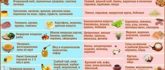
What can and cannot be eaten?
During exacerbation of cholelithiasis, there are a number of products that are recommended and prohibited for use.
| Can | It is forbidden |
|
|
Remote destruction of stones
Shock wave lithotripsy has proven itself well. If the size of gallstones is less than 20 mm, and the activity of the smooth muscles of the gallbladder is preserved, then the influence of the wave can eliminate the cause of the pathology. Impact can be carried out using various methods:
- electrohydraulic shock;
- electric shock;
- exposure to a magnetically restrictive generator.
Special equipment generates several shock waves simultaneously and delivers them to the area where the stone is located from different sides; as a result, the total force is concentrated in the area of the stone and it is destroyed, while the “associated” tissues remain intact. Despite its high efficiency, the method is characterized by a large number of complications:
- biliary colic – 50%;
- acute pancreatitis – 3%;
- acute cholecystitis – 2%;
- obstructive jaundice – up to 5%;
- hematoma of the liver, gall bladder and right kidney – 1%.
How the disease develops
The main, initial link in the pathogenesis of cholelithiasis is the increased secretion of cholesterol into bile, which occurs as a result of an unbalanced diet, oversaturated with cholesterol and calories, taking certain medications (in particular, clofibrate) or oral hormonal contraceptives, especially against the background of obesity. If a person has diseases, such as Crohn's disease, a condition after resection (removal of part) of the small intestine, or others that impair the functioning of the liver, its secretion of phospholipids and bile acids worsens, which also increases the tendency of bile to form stones.
The second stage of stone formation is the crystallization of bile, which is oversaturated with cholesterol. This process is accelerated by a glycoprotein contained in the mucus produced by the mucous membrane of the gallbladder. Also at this stage, hypomotor dyskinesia of the bladder plays an important role, that is, a violation of the organ’s motility, which contributes to the stagnation of bile in it.
It is worth mentioning congenital anomalies of the gallbladder. Every 5-6 people have a septum in the bladder that separates the bottom and body of the organ. This feature also contributes to the stagnation of bile in the bladder, and hence its subsequent crystallization.
Nucleation centers form in cholesterol, after which it crystallizes and transforms into stones of various sizes.
The process of formation of pigment stones occurs as follows: under the influence of a number of factors, the content of unconjugated bilirubin in the bile increases, which then precipitates and forms stones.
Basic drugs
| Name | Pharmacological group | Mechanism of action | Mode of application | average cost |
| Drotaverine | Antispasmodic | Blocks phosphodiesterase enzyme, leading to muscle relaxation. | Intramuscularly 1 ml of 0.25% solution 2 times a day. | 100 rubles |
| Platifilin | Antispasmodic | Blocks M-cholinergic receptors, which leads to disruption of the efferent innervation of smooth muscles of the gastrointestinal tract. | 0.04 orally 4 times a day. | 200-250 rubles |
| Ursofalk | Ursodeoxycholic acid drug | Reduces cholesterol synthesis and increases its dissolution in bile, thereby reducing the risk of crystallization. | 2 capsules 2 times a day, orally. | 1,500-2,000 rubles |
| Domperidone | Prokinetic | Blocks central and peripheral dopamine receptors, eliminating the inhibitory effect of dopamine on the smooth muscles of the gastrointestinal tract. | Orally 0.01 4 times a day 20-30 minutes before meals. | 150 rubles |
Types of stones
Gallstones are formed as a result of different components of bile sticking together. They have a crystalline structure.
Depending on the composition of the stones, they are divided into 3 types:
- cholesterol (more than 70% composed of cholesterol);
- pigment (their main component is calcium bilirubinate, and cholesterol is only 10%);
- mixed (contain impurities of calcium salts, pigments, bile and fatty acids, phospholipids).
What to do if an attack occurs?
For cholelithiasis, the treatment used depends on the stage at which the exacerbation occurred.
At an advanced stage, the use of independent methods and traditional medicine is prohibited. Otherwise, the person risks his life.
If an attack of illness does not stop for several minutes, it is necessary to adhere to a certain algorithm of actions to alleviate the condition.
In order to relieve pain you need to:
- take a horizontal position, it is strictly forbidden to bend over;
- Tablets that have an antispasmodic and vasodilator effect help relieve pain; when the spasm is relieved, the movement of the stone accelerates and it moves much easier;
- use a heating pad, it should be applied to the sore spot, the water temperature should be slightly warm, applying a hot heating pad is strictly prohibited;
- if there is severe pain, it is recommended to take a warm bath; the procedure should not exceed 15 minutes;
At the same time, it is recommended to drink in small portions, but quite often, especially in cases where there are attacks of nausea.
The best option for drinking would be to use mineral water; drinking it makes it possible to relieve nausea.
What mineral water can you drink for the liver with gallstone disease? The best option is to use Borjomi mineral water
Mineral water should be consumed warm, this will eliminate the urge to vomit.
The list of drugs used to relieve pain includes the following:
- But-shpu.
- Drotaverine.
- Papaverine.
- Antispasmodics of any order. Such a drug may be Spazmalgon and similar medications.
After completing all the steps and ensuring a calm environment around the patient, you need to call an ambulance. This is required because if you do not seek help in a timely manner, the gallbladder may rupture. Penetration of bile into the abdominal cavity leads to death.