Eczema is a skin disease accompanied by signs of acute inflammation of the epidermis and dermis. Pathology can affect both the face and body. It also has another name - atopic dermatitis. Eczema on the body and face usually manifests itself as rashes in the form of blisters, redness of the skin, itching and burning. The disease can appear at any age, but most often affects children under 5 years of age, especially if they live in large cities. Atopic dermatitis affects up to 20% of children from developed countries, and only 1-3% of adults.
Eczema can cause a lot of discomfort to the patient and reduce the quality of life. In addition, it is often accompanied by other diseases (asthma, allergies, etc.). Therefore, it is important to diagnose the disease as early as possible and begin treatment. The easiest and fastest way is to make an appointment at the Otradnoe Polyclinic. You will see a dermatologist at a time convenient for you and without having to wait in lines. You will be prescribed the necessary diagnostic procedures and will develop an optimal treatment plan.
Eczema and stages of its development
The term eczema comes from the Greek word ekzeo - to flare up, to boil. It was first mentioned in 541 in the scientific works of Aetius, a Byzantine physician. However, in those days, this term was used as a general collective name for dermatological diseases with acute development - erythema, urticaria, erysipelas, etc. It was only in the 18th century that eczema was identified by scientists as a separate, independent disease.
This is a pathology of allergic origin. It can be caused by various reasons, depending on its type. There are more than 20 types of eczema. All of them manifest themselves in a similar way, the differences are only in the cause of the disease and the duration of the rash.
Eczema has 4 stages of development:
- Erythematous - reddish-pink spots appear on the skin, which rapidly increase in number and size.
- Papular - small papules (nodules) appear, which are accompanied by swelling.
- Vesicular – vesicles (bubbles) are formed.
- Weeping - the vesicles open, and bright red erosions (wounds) appear, from which a clear serous fluid (exudate) is released.
Reference! With eczema, the general protective functions of the body are reduced.
This leads to both aggravation of atopic dermatitis and the development of other pathologies.
Scaly layer at the border of the scalp
The so-called seborrheic eczema often appears in the scalp area. At the initial stage, it looks like single yellowish nodules. The number of these nodules is rapidly increasing, they merge into spots, on the surface of which whitish scales form.
The merging of individual foci of seborrheic eczema often leads to the formation of a “seborrheic crown” at the border of the hair - a continuous scaly ring, along the edge of which areas of hyperemic skin are visible. In the absence of treatment, in advanced cases, this form of eczema spreads from the sides of the head to the folds behind the ears, and from the back to the neck.
Seborrheic eczema is very difficult to distinguish from true seborrhea, and some experts consider seborrheic eczema only as a type of seborrhea with some nuances in the course of the pathological process.
Classification of eczema
According to the nature of the course, eczema can be:
- Spicy.
- Sub-acute.
- Chronic.
The acute form by the presence of rashes, exudate, redness and swelling of the skin. These symptoms are accompanied by severe itching and flaking of the skin. Then the exudate dries out, and crusts form on the pathological lesions. After this, epithelization (growth of new skin cells, formation of connective tissue) of the wounds occurs. In the subacute form, there are rashes, redness, peeling and itching, but there is no weeping (exudate). Chronic eczema is characterized by alternating periods of exacerbations and remissions. New weeping rashes are adjacent to dry crusts. Less exudate is released than in the acute form. In the chronic stage, there are more dry crusts on the body than weeping rashes, which is why this form is also called dry eczema. The crusts often crack, causing itching, which often causes patients to experience insomnia. Also in the chronic stage, severe peeling of the skin and the development of pigment spots may be observed. As a rule, exacerbations develop in the cold season, and remissions occur in the warm season.
Eczema is also classified according to the cause of its occurrence. As mentioned above, there are more than 20 types of this disease. The most common types of eczema are:
- Idiopathic (true).
- Microbial.
- Seborrheic.
- Professional.
- Children's room.
Idiopathic eczema develops due to a combination of several causes and can affect patients of any age. Usually the disease occurs abruptly and develops in spurts. Pathological foci, as a rule, have unclear boundaries, different sizes and are located symmetrically. The rashes alternate with areas of healthy skin. Most often, idiopathic eczema develops on the skin of the palms (on the back), feet and forearms. The acute form in most cases gradually turns into chronic.
Microbial eczema occurs when immunity decreases around chronic foci of infection: abrasions, wounds, fistulas, scratches, trophic ulcers. Pathological foci are located asymmetrically, clearly defined and covered with purulent crusts. This type of disease is characterized by the absence of areas of healthy skin between the rashes. Manifestations of eczema are usually observed on the skin of the arms and legs, but can also affect any part of the body, including the scalp.
Seborrheic eczema is a form of seborrhea (a disease that develops against the background of increased activity of the sebaceous glands). It appears as clearly defined red spots, which can have different shapes. The disease usually develops in people with increased sebum production and occurs in those areas where there are the most sebaceous glands: on the scalp and behind the ears, on the face, between the shoulder blades, in the nasolabial folds and on the chest. The causes of the disease are the activity of Malassezia fungi, hormonal disorders, and lack of vitamins. Emotional shocks are considered a provoking factor.
Occupational eczema is similar in clinical manifestations to idiopathic eczema. It develops due to regular contact with substances that cause allergic reactions (usually this occurs in industrial conditions). Occupational eczema goes away quite quickly if contact with the provoking substance is avoided. However, if the patient is not protected from its exposure, subsequent outbreaks of the disease will be more severe.
Infantile eczema is a type of idiopathic atopic dermatitis. In young patients, the disease proceeds a little differently than in adults: it is characterized by a variety of rashes and the release of a large amount of exudate.
Important! Eczema is not a contagious disease!
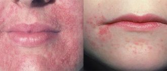
Weeping and itchy skin on the face and hands
The appearance of multiple rashes on the skin in the form of blisters and bumps against the background of reddened skin may be a manifestation of true eczema. Characteristic is the subsequent opening of the microvesicles with the release of serous fluid. The blisters are gradually replaced by shallow point erosions (ulcers). The released liquid dries, forming soft crusts.
Content may be difficult to view
Constant accumulation in the area of the pathological focus leads to the fact that vesicles, ulcers, and crusts are simultaneously visible on the skin - this is the so-called polymorphism of elements. All skin rashes are accompanied by severe skin itching. Sometimes the itching is such a strong sensation of irritation that a person suffers from insomnia.
True eczema is characterized by symmetry, the absence of clear boundaries at the lesions. There is a tendency to spread to other parts of the body: chest, torso, abdomen and back.
Eczema: causes
Idiopathic eczema is a disease that develops due to a combination of reasons, the main ones of which are:
- Genetic predisposition – it has been proven that gene mutations that affect the development of the disease are inherited.
- Impaired epidermal barrier function.
- Immune system failures.
- Provoking factors.
Provoking factors include:
- General decrease in immunity.
- The presence of allergies in the patient or his close relatives.
- Hyperhidrosis (increased sweating).
- Use of products that irritate the skin (inappropriate cosmetics and perfumes).
- Endocrine disorders.
- Gastrointestinal diseases.
- Varicose veins.
- Stress.
- The presence of chronic foci of infection (for example, caries or chronic sore throat).
Reference! The presence of allergic rhinitis, bronchial asthma, and neurodermatitis in the mother increases the risk of eczema in the child by 40%. If similar diseases are observed in both parents, the risk increases to 60%.
Callus-like rashes on the palms
The appearance of callus-like rashes on the palmar surfaces of the hands in places where the appearance of calluses is uncharacteristic is a sign of tylotic eczema. In this case, hyperemia due to the thickness of the stratum corneum is not very noticeable, but vesicles continue to appear, and they may not open due to the thickness of the skin.
When such lesions appear, you need to pay attention to other areas of the skin, since tylotic eczema most often accompanies the appearance of true eczema in other places.
Diagnosis of eczema
The disease has a characteristic clinical picture, so the diagnosis can be made based on examination of the patient and history taking. However, to identify the cause of eczema and develop an effective treatment plan, the following may be prescribed:
- General blood and urine tests.
- Determination of the level of immunoglobulin IgE in blood serum.
- Allergy and immunological tests.
- Bacteriological culture of the discharge (carried out when a secondary infection is attached and is necessary to determine the sensitivity of its pathogens to antibiotics).
- Biopsy.
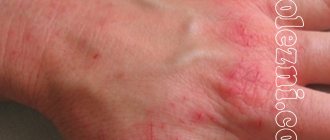
Round rashes on the skin of the hands
Itchy and itchy rashes on the upper extremities can be symptoms of a wide range of diseases. One of them is eczema on the hands, in 80% of cases this localization is characteristic of true eczema, another 15% is due to occupational eczema and the remaining 5% to other varieties.
Round shapes are characteristic of eczema at the initial stage; as the disease progresses, they lose their regular shape. The roundness of the outline is also characteristic of mycotic eczema, in which the primary lesion has large scalloped edges.
It is quite rare that microbial eczema appears on the hands, but it is worth paying attention to the nature of the weeping - if purulent crusts form on the surface, then with a high probability it is microbial eczema.
Eczema: treatment
You cannot self-medicate for eczema: there is a risk of provoking the development of a secondary infection. In addition, the occurrence of pathology may be due to the presence of other diseases, so it is necessary to treat them. The patient may need the help of an immunologist, allergist, endocrinologist, nutritionist, phlebologist and other specialized specialists.
General therapy for eczema includes the following medications:
- Antipruritic.
- Anti-inflammatory.
- Antibacterial.
- Antifungal.
- External corticosteroids.
- Immunomodulators.
- Desinsibilizing and antihistamine.
- Antiseptic and astringent drugs.
- Vitamins (groups B, C).
In severe cases, physical therapy (exposure of affected areas to ultraviolet radiation) may be prescribed.
Treatment will be more effective if the patient follows a diet. First of all, any foods that can provoke an exacerbation of the disease are excluded from the diet: allergens, fried, spicy, fatty, canned, salty foods. It is necessary to eat light food rich in vitamins, minerals, essential fatty acids and protein: vegetables and fruits, herbs, cereals, boiled lean meat, dairy products, vegetable oils. It is also recommended to stop drinking alcohol and smoking during treatment.
Symmetrical scaly rashes on the body
The symmetry of the lesion is more characteristic of true eczema. Despite the fact that the disease most often manifests itself with the appearance of a rash on the extremities, in some cases the lesions initially appear on the body.
In the initial stages, these lesions have the typical appearance of “boiling skin.” With a long-term recurrent (repeating) course, weeping lesions are replaced by areas of thickened skin with lichenification (increased skin pattern). With long-standing eczema, the lesions during the healing stage are covered with flaky skin, their weeping is significantly reduced - in this case they speak of dry eczema.
Traditional methods
Coin-shaped eczema and treatment with folk remedies worries all patients. You can combine unconventional methods with the main therapy prescribed by a specialist.
A very effective ointment can be prepared from eggs and vinegar. To do this, one egg should be mixed with vinegar and water in equal proportions. The ingredients must be beaten in a mixer until a homogeneous mass emerges. Apply the resulting composition to sore spots at night. In order not to stain the linen, you need to bandage the treated skin with gauze bandages. Ten sessions are enough to get rid of itchy spots.
Nummular eczema is treated with a decoction of coltsfoot. To do this, take the coltsfoot herb, brew it with hot water and boil over low heat in a water bath. Then cool and strain. You can also mix fresh coltsfoot leaves in a blender with sour cream in a 1x1 ratio. Apply the resulting slurry to a napkin and apply to the plaques. The course of treatment is 10 procedures.
Plaque eczema will recede if you prepare an ointment from badger or goose fat to treat it. In the absence of such components, you can replace them with regular baby cream. Add fir oil to the fat or cream and mix well. Apply to plaques three times a day for three weeks. This is an excellent remedy for the weeping form of the disease.
Redness of the areola and rash around the nipple
Mostly in women, extremely rarely in men, eczema can affect the skin around the nipple. In women, this process is quite often associated with injury to the nipples due to improper breastfeeding. In men, nipple eczema can develop against the background of scabies.
Typical manifestations are the appearance of crimson lesions near the nipple, often covered with crusts or large scales. Wetting is noted, and quite deep cracks often appear. When cracks appear, in addition to itching, severe pain also occurs.
Content may be difficult to view
The addition of a bacterial infection often leads to the fact that nipple eczema is microbial in nature. Difficulties in treating eczema of this localization in women are associated with the process of lactation, against the background of which inflammation is excessive.
Areas of depigmentation after peeling on the skin
Peeling skin with eczema is a sign of either recovery or chronicity of the process. Recovery is usually accompanied by the complete disappearance of skin manifestations of eczema. At the site of the lesions, skin remains that is lighter than the intact skin. This is a normal phenomenon called depigmentation, that is, a decrease in the amount of pigment. Typically, such depigmented spots disappear on their own within 3-5 weeks.
Foci of depigmentation after peeling form in most types of eczema, but are more common in true and microbial eczema. With seborrheic, tylotic and dyshidrotic, they are not so noticeable due to the localization of these forms of pathology.
Relapse
The main complication when the neck is affected by dermatosis is its re-development. After stopping the inflammatory process and healing the damaged dermis, experts advise not to relax - the disease can return at any time, so you should always remember about prevention. Proper skin care, a hypoallergenic diet, avoiding contact with the allergen and physiotherapeutic techniques in the form of ultraviolet irradiation, radon and sulfide baths, magnetic therapy have a positive effect and may well prevent relapse.
If an exacerbation does occur, there is no need to hesitate to consult a doctor. Timely treatment can prevent complicated eczema and speed up the healing process.
Skin thickening on the arms and body
In the initial stages of any eczema, the skin is swollen and hyperemic, but in consistency it differs little from healthy skin. The appearance of foci of compaction may indicate transformation of the disease into a chronic form.
This process is characterized by a decrease in the process of weeping and the appearance of large areas of thickened and flaky skin. During the healing process, all manifestations regress - the foci of eczema disappear completely.
It should also be remembered that some autoimmune diseases and other skin pathologies are manifested by changes in the nature of the skin, so an examination by a doctor is required to carry out a differential diagnosis.
Bibliography
- Current problems of dermatovenerology [Text]: materials of the conference. / State Educational Institution of Higher Professional Education SamSMU. - Samara: SamSMU, 2007. - 132 p.
- Baumann, L. Cosmetic dermatology: principles and practice [Text] = Cosmetic dermatology: principles and practice / L. Baumann; lane from English; edited by N. N. Potekaeva. - Moscow: Medpress-inform, 2012. - 688 p.: ill.
- Skin diseases [Text]: atlas / ed. N. V. Kungurova; Ministry of Health of the Russian Federation, Federal State Budgetary Institution Ural. Research Institute of Dermatovenereology and Immunopathology of the Ministry of Health of the Russian Federation. - Ekaterinburg: Publishing house "VIP-Ural", 2014. - 176 p.: ill.
- Macharadze, D. Sh. Atopic dermatitis in children [Text] / D. Sh. Macharadze. - Moscow: GEOTAR-Media, 2007. - 392 p.: ill. - (Current medical issues).
- Morgan, M. B. Atlas of deadly skin diseases [Text] = Deadly dermatologic diseases / M. B. Morgan, B. R. Smoller, S. K. Somach; lane from English; edited by A.V. Molochkova. - Moscow: GEOTAR-Media, 2010. - 304 p.: ill.
- White, G. Atlas of dermatology [Text] = Color atlas of dermatology / G. White; lane from English N. G. Kochergina; edited by O. L. Ivanova, N. G. Kochergina. - Moscow: GEOTARMEDIA, 2009. - 384 p.: ill.
- Dermatology [Text] = Dermatology. Vol. 1./ed. by Jean L. Bolonia; associate and artwork ed. JV Schaffer; section ed. JP Callen. — 2nd ed. - London: Mosby Elsevier, 2008. - ill.
- Fitzpatrick`s dermatology in general medicine [Text] = Fitzpatrick's dermatology in general medicine. Vol. 2. / ed.: K. Wolff. – 7th ed. – N.York [etc.]: McGraw-Hill Medical, 2008. – ill.
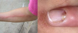
The skin around wounds on the legs and arms festers and itches
Redness of the skin and the appearance of purulent crusts around the edges indicate the development of microbial eczema. It appears in places where skin trauma occurs more often.
Usually, against the background of existing hyperemia with typical eczematous elements, the redness of the skin intensifies significantly, the transparent serous secretion on the surface of the lesions is replaced by a yellowish, purulent one. When the pus dries, it forms fairly rough crusts that are easily removed, exposing the skin that continues to become wet.