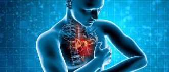
Endocarditis is an inflammation that occurs in the inner lining of the heart - the endocardium. The disease does not always occur with obvious signs: it is characterized by mild malaise, an increase in temperature to low levels, and, less often, discomfort in the heart. At the same time, it is characterized by an unpredictable course: at any moment, inflammation of the endocardium can cause thromboembolism of the arteries of vital organs, acute heart failure, dangerous arrhythmias, and damage to internal organs. In addition, the disease can recur.
Typically, endocarditis occurs as a complication of inflammation of the tonsils, kidneys, lungs, myocardium and other diseases, and therefore is rarely diagnosed. But there is also an independent pathology - infective endocarditis. It develops when microorganisms enter the endocardium.
Most often these are bacteria, which is why the disease was previously called “bacterial endocarditis”. Now that fungi have become more frequently detected in blood cultures, this name for the disease is considered obsolete. Infectious endocarditis is also called septic because here, as in sepsis, microorganisms are found in the blood, which should normally be sterile.
What is the endocardium and why is its inflammation dangerous?
The endocardium, which becomes inflamed during endocarditis, consists of several layers of cells:
- the innermost layer consists of endothelial cells. They are similar to those that form the mucous membranes of all internal organs, and identical to the cells that line the inside of blood vessels. Endothelial cells lie on a basement membrane, which gives them signals to grow and divide;
- subendothelial layer. It is built of connective tissue rich in poorly differentiated cells;
- muscle-elastic layer. Consists of muscle fibers that are “packed” into connective tissue. The layer is analogous to the middle tunic of blood vessels;
- outer connective tissue layer. Consists of connective tissue and is identical to the outer lining of blood vessels.
The endocardium lines the inside of the heart wall, forms folds - valve flaps, as well as chordae tendineae attached to them and papillary muscles that pull on the chordae. It is this lining of the heart that is the separator between the blood and the internal structure of the heart. Therefore, in the absence of inflammation, it is designed so that there is no significant friction of the blood against the heart walls, and there is no deposition of blood clots here. This is achieved by the fact that the surface of the endothelium is covered with a layer of glycocalyx, which has special atrombogenic properties.
The endocardium of the heart valves on the atrium side is denser. This is ensured by a large number of collagen fibers in the muscular-elastic layer of the membrane. On the side of the ventricles, the muscular-elastic layer is 4-6 times thinner and contains almost no muscle fibers. The valves between the cavities of the heart and the vessels (pulmonary trunk, aorta) are thinner than the atrioventricular valves. The endocardium covering them is thicker at the base of the valve, but any layering is no longer visible on the valves themselves. There are very few muscle fibers on the valves that close the entrance to the vessels.
The nutrition of the deepest endocardium, bordering the myocardium, comes from the vessels that make up its structure. The remaining sections receive oxygen and necessary substances directly from the blood, which is located in the cavities of the heart.
Directly below the endocardium is the heart muscle - the myocardium. It is responsible not only for the contractions of the parts of the heart, but also for the correct rhythm of these contractions: “paths” of cells are laid in the myocardium, some of which produce, and others transmit, electrical impulses that cause the necessary parts of the heart to contract.
When enough microbes (bacteria or fungi) enter the blood, they naturally end up inside the cavities of the heart. If a person’s immunity is sufficiently weakened, then microorganisms settle on the endocardium (especially on the valves between the left atria and the ventricle, as well as at the entrance from the left ventricle to the aorta) and cause inflammation there. The inflamed endocardium grows and thrombotic masses are deposited on it. This form of the disease is called “warty endocarditis” and is more characteristic of the rheumatic process
Thrombotic masses can break off at any time and enter the arteries that feed the internal organs through the bloodstream. This is how a stroke, infarction of the spleen, intestines, lungs and other organs can develop.
Due to the increase in the mass of the valve due to blood clots and scar tissue, it ceases to normally perform its function - to prevent the reverse flow of blood. Because of this, a condition called chronic heart failure develops.
Microorganisms deposited on the valves, chordae, or the surface of the papillary muscles can cause the formation of endothelial ulcers (ulcerative endocarditis). If this leads to the development of a “hole” in the valve or separation of the chord, the heart “loses control” of its own processes. This is how acute heart failure develops, occurring according to one of the scenarios: either pulmonary edema, shortness of breath and a feeling of lack of air, or a sharp decrease in pressure, increased heart rate, panic state with possible loss of consciousness.
The presence of bacteria or fungi in the blood causes activation of the immune system, as a result of which antibodies are formed to these microorganisms and the complement system (several immune proteins) is activated. Microbial antigens combine with antibodies and complement proteins, but are not destroyed (as should be normal), but are deposited around the vessels of many organs: kidneys, myocardium, joints, individual vessels. This causes inflammatory and allergic reactions, resulting in the development of glomerulonephritis, arthritis, myocarditis or vasculitis.
Causes of endocarditis and its classification
Depending on the initial state of the inner lining of the heart, infective endocarditis of the heart can be primary or secondary. Both of them are caused by the following microorganisms:
- bacteria: viridans (the main cause of subacute endocarditis) and pneumonic streptococci, Staphylococcus aureus and Enterococcus (cause an acute inflammatory process), Escherichia coli, Mycobacterium tuberculosis, Treponema pallidum (with syphilis), Brucella, some gram-negative and anaerobic bacteria;
- mushrooms, usually Candida. Such microflora usually appears when a person has been treated with antibiotics for a long time, or has had a venous catheter in place for a long time (in the treatment of any disease);
- some viruses;
- some protozoa.
Only primary endocarditis is one that occurs on normal, healthy valves, and secondary endocarditis occurs on valves affected by rheumatism or prolapse, on artificial valves and those near which there is a pacemaker. Recently, the incidence of primary endocarditis has begun to increase. It reached 41-55%.
Microorganisms enter the human blood in the following ways:
- through a wound of the skin or mucous membranes when it has become contaminated with microbes in a person with reduced immunity or with an installed artificial valve or pacemaker;
- when performing various invasive methods of examination and treatment: catheterization of peripheral veins to introduce contrast into them (for angiographic studies), endoscopic and open interventions, abortions, cystoscopy and even extraction (pulling out) of teeth when a foreign surface comes into contact with blood;
- from any source of bacterial or fungal inflammation (for example, from the lungs with pneumonia, abscess of the tonsils, gangrene of the extremities) - in conditions of reduced immunity, especially if it is combined with pathology of the valve apparatus;
- for any infection (microorganisms always enter the blood and pass through the heart): respiratory tract, maxillary sinuses, kidneys, joints, intestines, and so on, if a person has an artificial valve or pacemaker;
- when using injecting drugs (the endocardium of the right side of the heart is most often affected), when sterility is not maintained;
- during the installation of prostheses or implants, especially when it comes to the installation of artificial heart valves or a pacemaker;
- during any heart surgery.
There is a greater chance that the microbe will “stick” to the endocardium and cause an inflammatory process in it in older people, drug addicts, and people with immunodeficiency conditions, including those who have immunodeficiency as a result of cancer treatment. People who constantly drink alcohol are also more susceptible to developing endocarditis.
There are also local factors that contribute to the development of this disease. These are heart defects - congenital and acquired (especially ventricular septal defects and coarctation of the aorta), artificial valves. There is evidence that in the presence of valve pathology, any entry of a certain amount of bacteria into the blood (even with a tooth root cyst or sore throat) can cause infective endocarditis in 90% of cases.
If everything is fine with the heart valves, then if bacteria enter the blood, it is more likely that endocarditis will develop in older people with arterial hypertension, coronary artery disease, cardiomyopathies, and Marfan syndrome. There is a higher risk of developing endocarditis in a person who has already had this disease once, even if it did not leave visible, ultrasound-detectable marks on the inner lining of the heart.
If the disease occurs when a pathogen is detected in the blood and there is already damage to internal organs, this is septic endocarditis, which is also called infectious and bacterial. When it occurs as a complication of streptococcal lacunar or follicular inflammation of the tonsils, or glomerulonephritis caused by streptococcus, it is called rheumatic endocarditis. There are also tuberculous, syphilitic, traumatic and post-infarction inflammation of the myocardium.
Depending on the course, any endocarditis can be:
- acute: lasts about 2 months;
- subacute, which lasts 2-4 months, is usually a consequence of an untreated acute process;
- chronic (protracted), “lasting” for more than 4 months. This is a rare type of infective endocarditis, but a fairly common type of disease of rheumatic origin.
According to the damage to the valves, there are:
- mitral valve endocarditis;
- inflammation of the aortic valve;
- endocarditis of the tricuspid (tricuspid) valve;
- inflammation of the pulmonary valve.
The last 2 valves, located in the right side of the heart, become inflamed most often in injection drug addicts.
The diagnosis may also include the activity of the process. Endocarditis will be considered active if a person experiences an increase in temperature in combination with the isolation of microorganisms during blood culture or bacteriological examination of the valves (if heart surgery was performed). If the first episode of endocarditis is over, and no symptoms have been observed for a year or more, then the re-development of inflammation of the endocardium, with the release of another pathogen from the blood or valves, will be called “recurrent endocarditis”. If, despite treatment, symptoms of the disease persist for 2 months or more, and the same microbe is sown from the blood, this is called persistent endocarditis.
If endocarditis develops after heart surgery, it is divided into:
- early: occurs in the first year after the intervention. Means that the infection occurred nosocomially;
- late: developed when a year passed after the operation. Caused by community-acquired microflora.
The selection of antibacterial therapy and prognosis depend on the latter classification. So, if infection occurs with nosocomial microflora, in the first 72 hours of hospital stay, mortality can reach 40-56%.
Endocarditis in children has an additional classification. It is divided into:
- congenital, which is formed in the prenatal period when the fetus is infected;
- acquired, arising after childbirth: either due to the same reasons as in adults, or due to infection during childbirth or immediately after it.
In children over 2 years of age, most cases of endocarditis develop against the background of congenital or acquired heart disease.
Preventive measures
To reduce the risk of endocarditis, you need to follow simple preventive measures. It includes the following rules:
- Caries, pathology of the nasopharynx are conditions that need to be treated on time. They are foci of infection, which becomes one of the factors in the septic form of endocarditis.
- After surgery, people at risk are required to take a short course of antibiotics.
- Observation by a doctor after endocarditis (dispensary registration).
- Vaccination against influenza and other infections.
Throughout their lives, patients at risk or those who have been ill must adhere to these rules. Daily stress and overexertion create additional stress on the body, which reduces its defenses.
Patients at high risk of developing endocarditis should take care of their future and receive timely treatment if necessary. Taking care of yourself will help keep your heart and other organs healthy.
Symptoms
Signs and symptoms of endocarditis depend on its type (infectious, rheumatic, syphilitic, tuberculous) and are dictated by the course of the disease. So, if acute endocarditis has developed, the symptoms will be as follows:
- high body temperature (up to 39.5°C);
- during the ascent, a person’s temperature rises with severe chills;
- profuse sweating;
- pain in all joints and muscles;
- lethargy;
- headache;
- the skin becomes grayish with a slight yellowness, sometimes red spots appear on it;
- reddish painful nodules appear on the fingers;
- There are hemorrhages in the conjunctiva.
Subacute infective endocarditis occurs with the following symptoms:
- elevated body temperature – up to 38.5°C;
- chills;
- worsening sleep;
- weight loss;
- skin color becomes “coffee with milk”;
- red rash on the body;
- small painful nodules appear under the skin,
but the main difference from the acute process is that these symptoms are observed for 2 months or more.
The chronic process is characterized by the same symptoms (only the temperature is usually up to 38°C) for six months or more. During this time, the person loses a lot of weight, his fingers take on the appearance of drumsticks (widened in the area of the nail phalanges), and the nails themselves become dull and become convex (reminiscent of watch glasses). Hemorrhages may appear under the nails, and painful reddish nodules the size of a pea are always found on the fingers, toes, palms and soles.
When a heart defect develops, shortness of breath appears: first during physical activity, then at rest, pain in the chest, the heart beats faster (up to 110 beats per minute or more often) regardless of temperature.
If glomerulonephritis or kidney infarction develops, swelling appears on the face, urination is impaired (usually there is less urine), urine changes color to reddish, and lower back pain appears.
If, against the background of the main symptoms, severe pain develops in the left hypochondrium, this indicates that one of the branches of the arteries feeding the spleen is clogged, and part or all of this organ dies.
With the development of pulmonary embolism, there is a sudden feeling of lack of air and pain in the chest. Against this background, impaired consciousness quickly increases, and the skin (especially on the face) acquires a purple tint.
Symptoms of infective endocarditis develop in three stages:
- Infectious-toxic: bacteria enter the blood, “land” on the valves, and begin to multiply there, forming growths - vegetations.
- Infectious-allergic: due to activation of the immune system, internal organs are affected: myocardium, liver, spleen, kidneys.
- Dystrophic. At this stage, complications develop both from the internal organs and from the myocardium (sections of the heart muscle die in 92% of cases of prolonged inflammation of the endocardium).
Infectious endocarditis in children develops as an acute process and is very similar to ARVI. The difference is that with ARVI, the complexion should not change to yellowish, and there should be no pain in the heart.
If endocarditis is rheumatic, then it usually develops after a sore throat or glomerulonephritis, in which beta-hemolytic streptococcus was isolated (in the first case, from the surface of the tonsils, in the second, from urine). After the disease has subsided, after a while the person notices weakness, fatigue, and malaise. Again (after a sore throat or kidney inflammation), the temperature usually rises to 38°C, but can be higher. There are also unpleasant sensations in the heart area. Against this background, other signs of rheumatism may be observed: temporary enlargement and pain in large joints, which goes away on its own.
Complications
One of the most dangerous complications of endocarditis is embolism - separation of a section of an overgrown valve, a blood clot, or a blood clot with a section of the valve with the further “travel” of this particle through the arteries. The embolus (or thromboembolus) will stop where it exactly corresponds to the diameter of the artery.
If a particle breaks away in the left side of the heart, then embolization of the systemic vessels develops - one of the internal organs may be damaged: the intestines, spleen, kidneys. They develop a heart attack (that is, the death of the area).
If a thrombus or unstable (poorly fixed) vegetation is located in the right sections, the embolus blocks the vessels of the small circle, that is, the pulmonary artery, resulting in a pulmonary infarction.
The following complications may also occur due to endocarditis:
- Acute heart failure.
- Formation of heart disease.
- Myocarditis.
- Pericarditis.
- Chronic heart failure.
- Kidney damage: glomerulonephritis, nephrotic syndrome, renal failure.
- Lesions of the spleen: abscess, enlargement, rupture.
- Complications from the nervous system: stroke, meningitis, meningoencephalitis, brain abscess.
- Vascular lesions: inflammation, aneurysms, thrombophlebitis.
Diagnostics
Diagnosis of endocarditis is based on the following data:
- listening to the heart: first the systolic murmur is determined, then the diastolic murmur;
- determining the boundaries of the heart: they expand to the left (if the valves are damaged in the left parts of the heart) or to the right (if vegetations are found in the right parts);
- ECG: if irritation of the myocardial pathways by the inflamed endocardium occurs, the cardiogram determines the rhythm disturbance;
- Ultrasound of the heart (echocardioscopy): this is how vegetations (growths) on the valves and thickening of the endocardium and myocardium are determined. Ultrasound with Doppler can be used to judge the function of the heart and, indirectly, the pressure in the pulmonary circle;
- bacteriological examination of blood (inoculating it on various nutrient media);
- blood tests using the PCR method: this is how some viruses and bacteria are determined;
- rheumatic tests: in order to distinguish infective endocarditis from rheumatic;
- If necessary, a magnetic resonance or computed tomography scan of the chest with a targeted examination of the heart can be performed.
An accurate diagnosis of infective endocarditis is made when there is a specific ultrasound picture of the heart, and the pathogen is determined in the blood. If all the symptoms indicate this disease, a microbe is detected in the blood, but there are no significant changes on echocardioscopy, the diagnosis is called into question.
When the pathogen is not detected in the blood, but the ultrasound picture is beyond doubt, the diagnosis states that infective endocarditis is either “culture-negative” (that is, bacteriological culture did not reveal anything), or “PCR-negative” (if it was not isolated by PCR). pathogen).
Prevalence of the disease
Septic myocarditis is divided into two types:
- primary – develops on intact valves (Chernogubov’s disease);
- secondary – develops on valves that previously had pathologies.
The secondary type of the disease is found in 70-80% of patients, most often having previously had rheumatic heart disease. Much less commonly, it occurs in patients with atherosclerotic, syphilitic or congenital defects.
The primary type of disease is found in 20-30% of all cases.
Treatment
Since the disease in question is characterized by unpredictability and unexpected development of complications, treatment of endocarditis should be carried out only in a hospital. It includes the mandatory intravenous administration of antibiotics according to the latest orders of the Ministry of Health. Usually these are broad-spectrum antibiotics, with a special focus against viridans streptococcus and Staphylococcus aureus (Vancomycin, Zyvox); A combination of 2-3 drugs is often used.
Before starting antibiotic treatment, three blood samples are taken from a peripheral vein to ensure sterility. Based on its results (they are obtained approximately on the 5th day), the antibacterial drug can be changed.
The course of antibiotics is from 4 to 12 weeks. Their cancellation is carried out only after the temperature and laboratory parameters have normalized and after three negative bacteriological cultures have been obtained against the background of a trial withdrawal of antibacterial drugs.
In addition to antibiotics, the following are prescribed:
- blood thinners (heparin);
- glucocorticoids;
- antifungal agents;
- proteolytic enzyme inhibitors;
- antistaphylococcal plasma or immunoglobulin;
- drugs necessary to treat one or another complication of endocarditis;
If drug treatment is ineffective within 3-4 weeks, then surgery is performed to remove foci of infection inside the heart and avoid the progression of heart failure and the development of thromboembolism. The intervention involves removing the affected valves and then installing their prostheses.
Surgical intervention can also be performed urgently (within 24 hours after diagnosis). This can be life-saving if you develop:
- acute heart failure,
- the valve walls came off,
- valve perforation occurred
- fistulas, abscesses or pseudoaneurysms of the valves have developed,
- during the first week of therapy, movable growths on the valves more than 10 mm in diameter appeared,
but the risk from such an operation is also extremely high.
After surgery, the person receives antibiotics for 7-15 days. He is in the hospital, on semi-bed rest.
After endocarditis, the motor regime expands, but physical activity remains prohibited. Diet - table No. 10 with restriction of salt, liquid, complete exclusion of alcohol, cocoa, chocolate, coffee, as well as spicy, fatty and smoked foods.
SEE OTHER CLINICAL GUIDELINES
Approval year 2021
Professional associations:
- Association of Cardiovascular Surgeons of Russia;
- American Association of Thoracic Surgeons;
- European Society of Cardiology.
Table of contents
1. Brief information 2. Diagnosis 3. Treatment 4. Rehabilitation 5. Prevention 6. Additional information
Forecast
Infective endocarditis is a disease whose prognosis is conditionally unfavorable. In people without immune deficiency, defects and diseases of the heart and its valves, it is more favorable, especially if the disease is diagnosed early and powerful antibacterial therapy is started immediately. If a person develops endocarditis, has chronic heart disease or a suppressed immune system, life-threatening complications may develop.
The prognosis also worsens if:
- symptoms of the disease began to appear after admission to the hospital (where either invasive diagnostics or operations, including heart surgery) were performed - within the first 72 hours;
- if gram-negative flora, Staphylococcus aureus, antibiotic-insensitive Cochiella or Brucella, fungal flora are sown from the blood (from the valves).
With infective endocarditis affecting the right side of the heart, a better outcome can be expected.
Rheumatic endocarditis is more favorable for life: acute heart failure and thromboembolism are less typical for it. But heart disease with this pathology develops in the vast majority of cases.
general information
In the septic form of the disease, there is a sharp increase in the body's reactivity, which is expressed by acceleration and intensification of local and general reactions to the allergen, therefore such endocarditis can be considered bacterial septicemia (blood poisoning).
The general picture of septic endocarditis depends on the pathogen. Fungal infections and gram-negative microflora rarely cause this disease, and such endocarditis most often develops in drug addicts or people who have artificial valves . Streptococci can cause subacute or acute forms of septic endocarditis, and the treatment of these forms differs in similar tactics.
Acute septic endocarditis has a very rapid development (from 3 to 14 days) and is extremely difficult. The subacute form of septic endocarditis and its symptoms develop over a longer period of time (3 months), while the chronic (protracted) form can last for several years.
Subacute infectious-septic endocarditis is characterized by symptoms such as general weakness, fatigue, weight loss, and the appearance of low-grade fever. Immune complex organ damage (nephritis, arthralgia) and the development of embolic complications (strokes, renal infarctions) also
Prevention
Prevention of endocarditis is as follows:
- you need to adhere to sufficient physical activity and follow the rules of a healthy diet in order to be examined and treated with invasive methods as little as possible;
- it is important to promptly sanitize foci of infection: treat diseased teeth, wash the lacunae of the tonsils in case of chronic tonsillitis, ensure the outflow of contents from the sinuses - in case of chronic sinusitis;
- if you still have to undergo treatment, you need to do it not at home or dubious offices, but in specialized clinics;
- If work or everyday life involves frequent trauma, you need to take care of maintaining sufficient immunity. To do this, it is important to eat right, move enough, maintain the hygiene of your skin and external mucous membranes;
- in case of injury, proper antiseptic treatment of the wound and, if necessary, a visit to the doctor is required;
- if, as a result of heart disease, heart surgery, installation of an artificial valve or pacemaker was necessary, after which blood thinning drugs were prescribed, you cannot stop taking them without permission;
- If the doctor prescribes antibiotics for any reason, you need to take them for as many days as prescribed. From the 5th day of taking antibacterial therapy, you need to ask your doctor about the need to prescribe antifungal drugs;
- It is important to administer antibiotic prophylaxis before starting any invasive treatment. So, if the operation is planned, it is better to start administering drugs 12-24 hours before it (especially if the intervention will be carried out on the oral cavity or intestines). If you had to resort to emergency surgery, the antibiotic should be administered as soon as possible after admission to the hospital.
Author:
Krivega Maria Salavatovna resuscitator