Patau syndrome is a genetically determined disease that develops in individuals who have an extra 13th chromosome. This chromosomal anomaly manifests itself as signs of damage to the nervous system, visual analyzer, bones, muscles, heart, and organs of the urinary system. The appearance of the patients is characteristic. They have a reduced size of the skull and eyes, the brain is not divided into hemispheres, there are additional fingers, cleft lip and palate, a hernia of the umbilical cord, defects in the structure of the heart and large vessels. Such anomalies cause antenatal fetal death and short life expectancy for sick children.
For the first time, clinical manifestations of pathology were described by doctors back in the 17th century. Several centuries later, the scientist Patau determined that the disease is associated with the appearance of an additional 13th chromosome in the human karyotype. Thanks to this discovery, the syndrome got its name. Modern scientists have established that one of the causes of pathology is ionizing radiation.
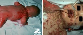
surviving child with Patau syndrome
The diagnosis of Patau syndrome is made based on the results of prenatal screening or karyotyping of the newborn. Among all genetic mutations, this disease is relatively common and is second in prevalence only to Down syndrome. According to statistics, racial, geographic and gender factors do not affect the incidence of pathology. Postnatal diagnosis is quite simple: serious deformations of body parts are visible to the naked eye.
Since Patau syndrome is a genetic disease, special treatment methods designed to eliminate it have not been developed. Patients are prescribed conservative treatment to improve their general condition, as well as surgical intervention to eliminate congenital defects.
Etiology
The main cause of occurrence is a disorder of chromosome divergence during cell division of one of the parents. Predisposing factors to the progression of such a karyotype disorder are:
- maternal age – there is a high probability of having this genetic disorder in children born to mothers over forty-five years of age;
- genetic predisposition. One of the parents has a pathology such as Robertson's karyotype. Outwardly, such people look healthy with a normal set of chromosomes, only they, for unknown reasons, give birth to children with Patau or Edwards syndrome;
- sexual contacts between relatives;
- high degree of environmental pollution.
Errors in the formation of sperm and eggs, which are directly involved in conception, cannot be ruled out. But this reason cannot be corrected or predicted, since it is a completely random factor.
Karyotype of Patau syndrome
Causes and mechanism of development
Patau syndrome is on a par with other chromosomal diseases - Down syndrome (trisomy 21 pairs) and Edwards syndrome (trisomy 18 pairs). Compared to them, the disease is somewhat less common, but is more severe.
According to statistics, 1 out of 7,500 newborns suffer from Patau syndrome.
Boys and girls get sick with the same frequency. All children have severely underdeveloped organs and tissues, but this cannot be explained by prematurity.
Symptoms
Patau syndrome entails multiple disorders of both internal organs and systems, so the symptoms of the disease are divided depending on the location of the pathology. Signs of this disorder in terms of structural changes in the skull and skeleton are:
- the baby’s body weight is critically low – less than two kilograms;
- deformation of the skull – the child’s head is much smaller than the body;
- the location of the ears is much lower than in a healthy child;
- pronounced bilateral clefts on the palate and upper lip;
- palpebral fissures are significantly narrowed;
- in some cases there is an increase in the number of fingers on the upper and lower extremities;
- short neck;
- skin imperfections in the back of the head;
- irregular foot shape.
Symptoms of this disorder related to the nervous system:
- reduced brain volumes;
- underdevelopment or complete absence of some components of the brain, for example, the cerebellum;
- delay in mental, physical and mental formation of personality.
Signs of Patau syndrome, characterizing anomalies in the structure of vital organs:
- structural pathologies of the heart;
- changes in the normal volumes of large and medium-sized vessels;
- underdevelopment or duality of the ureter;
- neoplasms on the kidneys in the form of cysts;
- umbilical cord deformities, such as hernia;
- changes in the natural position of all organs.
Manifestation of such a karyotype disorder in the formation of the genital organs:
- underdevelopment – reduction in penis size;
- detection of a testicle in the abdominal cavity;
- duplication of the genital organs in girls, in particular the uterus or vagina.
Symptoms of Patau disease from damage to the organs of vision and hearing:
- possible lack of one eyeball;
- defects of the membranes of the eye and lens;
- complete deafness caused by irregularly shaped ears;
- congenital cataract.
Due to the presence of multiple congenital defects in children, the prognosis for them is quite sad - on average, 90% of infants will not live their first year of life completely. Only a sixth of children live to be five years old, and only three percent of the total live to be ten years old. In most cases, babies with Patau syndrome die inside the womb.
Patau syndrome
Causes of Patau syndrome
As with other similar diseases, the main cause of Patau syndrome is an extra chromosome in the 13th pair. Due to an excess of genetic material, the normal development of the body becomes impossible, and numerous anomalies arise. Until now, scientists cannot say exactly why it occurs. This issue is the subject of scientific debate. Not in all cases the influence of risk factors or other unfavorable circumstances is visible.
According to geneticists, such a chromosomal defect can provoke:
- pregnancy of a woman over 40 years of age;
- taking certain medications in the early stages of pregnancy;
- poor environmental conditions, especially radioactive contamination;
- exposure to industrial poisons, radiation and other adverse factors;
- consanguineous marriages;
- family history - the likelihood that Patau syndrome may develop increases significantly if any of the couple’s relatives suffered from this pathology.
Trisomy 13 of chromosome leads to disruption of the formation of internal organs and all body systems even during intrauterine development of the fetus.
Diagnostics
An important role in the diagnosis of this karyotype disorder is played by the timely distinction of this disorder from other ailments with similar symptoms using prenatal examinations of the mother. For Patau syndrome it is absolutely the same:
- examination using ultrasound - malformations become noticeable starting from the twelfth week of pregnancy;
- establishment of a biochemical marker;
- calculating the risk of having a baby with this disease.
Those women who fall into the risk group described above are recommended to undergo examinations such as:
- biopsy of the embryonic membrane - this is carried out during the period from the eighth to the twelfth week of pregnancy;
- puncture of amniotic fluid - often the sample is taken from the fourteenth to the eighteenth week of gestation;
- study of blood from the umbilical cord - must be performed after the twentieth week.
The resulting fluids will be searched for violations of the thirteenth chromosome. Such studies are not abandoned in cases of fetal death - this will help prevent or prevent the occurrence of the syndrome in cases of repeated pregnancy. Additional diagnostic methods are:
- Ultrasound of all organs;
- CT scan of the brain;
- consultations with such doctors as an ophthalmologist, otolaryngologist, neurologist, pediatric surgeon, genetic specialist, endocrinologist.
Based on the results of an external examination, analysis of the course of pregnancy and the research results obtained, the newborn baby is given a final diagnosis - Patau or Edwards syndrome.
Diagnostic measures
Diagnosis of this pathology, like any other genetic disease, is carried out prenatally and postnatally. Prenatal detection of such anomalies allows us to compare the possible risks of giving birth to a sick child. Prenatal diagnostic methods are divided into invasive and non-invasive.
Non-invasive methods are considered safe. Fetal material is not required to detect a genetic abnormality. Ultrasound examination of the fetus and Dopplerography of the uteroplacental blood flow are recommended for all pregnant women. Diagnosis using ultrasound is 100% reliable.
Standard prenatal screening includes a blood test for biochemical markers. The results obtained are correlated with the age of the pregnant woman and gestational age. If the data obtained is outside the normal range, doctors recommend terminating the pregnancy. The analysis requires venous blood from a pregnant woman, which contains fragments of the genetic structure of the fetus.
Invasive methods include:
- Chorionic villus biopsy, which allows diagnosing the disease from 8 to 12 weeks of pregnancy. One of the membranes is taken for analysis using a puncture needle. A small amount of material is sufficient for research.
- Amniocentesis is the collection of amniotic fluid with a special needle through the peritoneum for cellular analysis. The method is relevant from 14 to 18 weeks of pregnancy. The study is carried out under ultrasound control. Cells containing fetal DNA are tested for genetic diseases.
- Cordocentesis is a study of fetal cord blood that allows one to determine genetic abnormalities with high accuracy. It is carried out after the 20th week of gestation.
Despite the high accuracy and reliability of invasive methods, they are used only in extreme cases. Introduction into the body of a pregnant woman and penetration into the membranes of the fetus carries a certain risk. Any incorrect movement by the specialist performing the procedure can lead to intrauterine death of the fetus.
The diagnosis is made after determining the baby's karyotype by qf-PCR. Only DNA analysis can confirm or refute the alleged diagnosis. Using chromosomal studies, it is possible to determine whether a child has trisomy 13, whether the anomaly has a hereditary etiology or is caused by a spontaneous intrauterine mutation. To assess the risks and be able to prevent such mutations, parents must undergo a detailed genetic study, which gives a 100% result. Genetic testing is carried out even if intrauterine death of the fetus has occurred, in order to prevent the syndrome during a second pregnancy.
Postnatal diagnosis consists of examining the newborn by a neonatologist and identifying clear and specific symptoms. Sick children are advised to undergo a thorough comprehensive examination, including the following instrumental techniques:
- Cardiography – electrocardiography, phonocardiography, coronary angiography, magnetocardiography;
- Ultrasound of internal organs located in the abdominal cavity and pelvis: pancreas, kidneys, ureters, organs of the reproductive system;
- Neurosonography - ultrasound of the brain;
- Tomographic examination of brain structures - CT or MRI.
Sick children usually require consultation with doctors - specialists in the field of ophthalmology, otolaryngology, neurology, pediatric surgery, genetics, endocrinology, and psychiatry.
Treatment
Due to the fact that karyotype disorder is characterized by chromosomal abnormalities, there is no specific treatment. Parents who decide to keep their baby will have to provide him with proper care and treatment for the rest of his life, which includes:
- carrying out plastic surgery to eliminate gaps in the palate and lip, and eliminate extra fingers;
- performing multiple surgical interventions to remove pathologies of internal organs;
- careful care, proper nutrition and constant monitoring of the baby;
- general strengthening measures to maintain the functioning of affected organs;
- limiting the child from contracting infectious or inflammatory diseases.
Babies with this karyotype pathology who remain alive after birth suffer from idiocy until their death and are completely disabled. If the cause of such an illness is a random factor, subsequent pregnancies can be successful. In cases where one of the future parents has Robertson's pathologies, the possibility of having a healthy baby is excluded.
Prevention of Patau syndrome
Despite the fact that Patau syndrome is a serious disease with very high mortality rates, no specific preventive measures have been developed for it. The main prevention of Patau syndrome is careful pregnancy planning, diagnosing the disease in the early stages of intrauterine development and deciding on the advisability of continuing the pregnancy.
A far from secondary role is played by nonspecific prevention of Patau syndrome, which consists of proper nutrition, elimination of bad habits, and timely treatment of infectious diseases. These simple measures will help prevent not only chromosomal pathologies, but also many other diseases.
During IVF, this chromosomal abnormality can be detected even before pregnancy occurs. At the stage of embryo cultivation, most clinics offer parents PGD - preimplantation genetic diagnosis.
This is an analysis aimed at identifying mutations - both gene and chromosomal. If the disease is detected in one of the embryos, it is not implanted into the uterus. For this purpose, another, healthy embryo is selected. Thus, parents who use IVF to conceive a child are in a better position, as they are able to prevent many congenital diseases in their offspring.
Genetics
Basically, there are two genetic forms of trisomy on the thirteenth chromosome:
- Simple trisomy - three chromosomes of the same type exist freely and realize their genetic potential.
- Robertsonian translocation - two chromosomes remain free, and the third merges with long arms with another acrocentric chromosome (for example, with 14 or 21). Since acrocentric chromosomes in short arms have only rRNA genes that are duplicated many times in the karyotype, their functionality is preserved in most cases.
In any case, the karyotype of a patient with Patau syndrome is determined by the formula 47 XX (XY) 13+. Sometimes cases of non-Robertsonian translocations, isochromosomal and mosaic forms of trisomy 13 are recorded.
The pathological picture and symptoms do not differ in the genetic varieties of Patau syndrome. In 3/4 of patients, an extra 13th chromosome is found, the remaining quarter of patients suffer from the involvement of the 13th pair of chromosomes in a translocation, which in 75% of cases occurs due to a denovo mutation (new, independently occurring) - the so-called “genetic error”. Studies of the heritability of Patau syndrome indicate that the remaining 25% of trisomy 13 cases are due to the transmission of a translocalized thirteenth chromosome from one parent. At the same time, the risk of recurrence of the case in the next child (recurrent risk) is 14%.
Risk factors
The main risk factors are age (especially significant for Down syndrome), as well as exposure to radiation and certain heavy metals. It should be borne in mind that even without risk factors, the fetus can have pathology.
As can be seen from the graph, the dependence of the risk on age is most significant for Down syndrome, and less significant for the other two trisomies.
Forecast
If a woman nevertheless chooses to continue the pregnancy, or if abortion is already contraindicated due to timing, then, with the exception of rare cases, she will have to refuse termination. In addition, serious attention should be focused on exactly how to give birth.
A fetus with Patau syndrome in most cases dies antenatally (during pregnancy) or is born dead. This is explained by the significant severity of congenital anomalies of internal organs. Babies with this pathology, who remain alive after birth, are completely disabled throughout their lives and suffer from idiocy.
If the cause of the disease was a random factor, then subsequent pregnancies in this woman can be successful.
Historical facts
Trisomy 13 was first described in 1657 by Erasmus Bartholin. And in 1960, Dr. Klaus Patau revealed the chromosomal nature of this disease. It was named in his honor. The syndrome has been described in Pacific Island tribes. It is also believed that the anomaly was caused by radiation contamination resulting from nuclear weapons testing.
Approximately one in 7–10 thousand babies is born with this syndrome. As a rule, it occurs equally in both boys and girls.
Frequency of occurrence of the genetic disease Synrom Patau
According to statistics available from WHO, every year in the world there is 1 newborn with Patau syndrome per 7000-10000 children. It is currently not determined whether Patau syndrome is a hereditary disease. It has not been fully revealed whether its occurrence is influenced by the mother’s age and her lifestyle.
Possible complications during childbirth
Women carrying children with a genetic abnormality usually do not carry the child to term. It all ends with a miscarriage or the birth of a dead baby.
If a child is born alive, this happens early, at approximately 29 weeks. Doctors, in addition to the main diagnosis, have to deal with prematurity. The diagnosis of the child does not affect the course of the mother's labor, with the exception of premature delivery.