Intestinal obstruction is a complex of symptoms that develop against the background of a complete or partial disruption of the movement of food through the intestines. Intestinal obstruction is a dangerous condition, which, if left untreated, is inevitably complicated by peritonitis, intestinal necrosis and leads to the death of the patient. Therefore, you need to start treating this disease as early as possible.
- Causes and types of acute intestinal obstruction
- Consequences of intestinal obstruction
- Symptoms of intestinal obstruction
- Diagnosis of intestinal obstruction
- Treatment of intestinal obstruction and first aid
What is intestinal obstruction?
Intestinal obstruction
– a pathology in which the process of evacuation of substances from the intestines is disrupted. It is especially characteristic of vegetarians and can be dynamic or mechanical.
At the first suspicion of intestinal obstruction, you should immediately contact a surgeon for help. Only he can make a final diagnosis and recommend the necessary treatment. Deciding solely on traditional methods on your own is very dangerous.
Acute obstruction can cause death, which is why it is so important to know its main symptoms and causes.
Statistics:
- After emergency surgery for intestinal obstruction, about 20% of patients die. If the pathology was severe, then these figures increase to 40%.
- Among all acute conditions requiring surgical treatment, ACI (abbreviation for acute intestinal obstruction) occurs in 8-25% of cases.
- If the cause of intestinal obstruction is a tumor, then the number of deaths is 40-45%.
- When the cause of acute intestinal obstruction is adhesions, death occurs in 70% of cases.
- In men, pathology occurs more often than in women - in 66.4% of cases.
- At risk are older people, whose likelihood of developing ACI increases 4 times.
Content:
Prevention
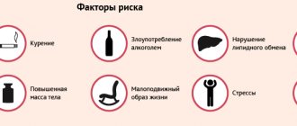
To avoid a dangerous condition, great attention should be paid to preventive measures. These include the following:
identification and elimination of formations in the intestines;- treatment of infectious processes in the peritoneum;
- prevention of adhesions;
- maintaining a healthy lifestyle;
- increased movement (gymnastics);
- getting rid of worms (if any);
- complete and balanced nutrition.
Causes of acute intestinal obstruction
Acute intestinal obstruction can be caused by various reasons. They are divided into predisposing and producing. Predisposing causes contribute to increased mobility of intestinal loops or immobilization. This leads to the fact that the organ occupies the wrong position, and feces cannot move through it normally.
Predisposing factors can be anatomical and functional.
Anatomical reasons include:
- The presence of adhesions in the peritoneal cavity.
- Meckel's diverticulum.
- The mesentery is too narrow or too long.
- The presence of a hole in the mesentery.
- Presence of a hernia. The danger is represented by a hernia of the white line of the abdomen, inguinal and femoral hernia, as well as internal protrusions.
- Malformations of the organ, for example, mobile cecum, dolichosigma, etc.
- Peritoneal pockets.
- Tumors of the intestine or organs that are in close proximity to it.
Functional causes that can cause acute intestinal obstruction include:
- Excessive eating after a long period of abstinence. If a person has been on a diet for a long time and then eats a large amount of roughage, the intestines will begin to contract strongly. This can cause intestinal obstruction or “hunger man’s disease” (according to Spasokukotsky).
- Colitis of various origins.
- Previous spinal cord injuries, TBI.
- Trauma of a psychological nature.
- Strokes.
- Dysentery and other conditions that contribute to increased intestinal contractility.
Producing causes lead to spasms and intestinal paresis. A similar situation can be provoked by an excess of food, a sharp increase in intra-abdominal pressure, physical inactivity (forced bed rest and paralysis).
How is an ultrasound of the stomach performed?
Preparation for an ultrasound of the stomach is similar to an ultrasound of the intestines: the patient adheres to a strict diet for 3 days, and the night before does not eat any food from 18.00. If there is a tendency to form gas, the patient drinks 2 capsules of Espumisan before bed. In the morning, half an hour before the procedure, you should drink a liter of water so that the walls of the stomach straighten.
There is also a method of ultrasound examination with contrast. Water is an excellent conductor of ultrasound, and without it, scanning an organ is somewhat difficult.
The procedure is carried out on an empty stomach. The doctor assesses the condition and thickness of the walls on an empty stomach, looking for the presence of free fluid. Then he asks the patient to drink 0.5-1 liter of liquid, and uses an ultrasound machine to evaluate changes in the expanded stomach. A third ultrasound scan is performed 20 minutes later when the stomach begins to empty. The doctor evaluates the motility of the organ and the rate of fluid loss. Normally, a glass of water (250 ml) comes out of the stomach in 3 minutes.
The patient lies on the couch on his side, the specialist applies gel to the peritoneal area and moves the sensor over the surface. Periodically, he tells the patient to change position or change his posture slightly. The doctor pays attention to the following indicators:
- position of the stomach and its size
- Has the mucous surface of the stomach expanded?
- is there thickening or thinning of the walls
- what is the state of the circulatory system of the stomach?
- contractility of the stomach
- are there inflammations and neoplasms?
The entire examination takes a maximum of 30 minutes and does not cause discomfort or pain. Ultrasound, unlike FGDS, is much easier to tolerate for children and the elderly.
Symptoms of acute intestinal obstruction
Intestinal obstruction, as a rule, begins with a sharp, increasing, cramping pain in the abdomen and is accompanied by nausea and vomiting. Over time, the contents of the intestines begin to enter the stomach, and the vomit has an unpleasant odor characteristic of feces. Constipation and increased gas formation occur. Intestinal peristalsis is preserved at the initial stage and can be observed through the abdominal wall. The abdomen takes on an irregular shape and bloating rapidly increases.
Symptoms of acute intestinal obstruction:
- Abdominal pain.
It occurs in all patients without exception. At an early stage of development of the pathology, pain occurs like contractions. It will be concentrated in the place of the abdominal cavity where the collapse has formed. The pain is constant, becomes dull, and spreads throughout the abdomen. When the pathology reaches its peak, the pain decreases, but this symptom cannot be called favorable.
- Nausea and vomiting.
These symptoms occur in 60-70% of people. They will be more intense, the more severe the intestinal obstruction. First, bile will be present in the vomit, and then intestinal contents. The masses coming out of the mouth begin to smell like feces. Vomiting does not develop immediately, but once it starts, it is not expected to stop.
- No gas or feces.
Even at an early stage of development of intestinal obstruction, stool will be absent. If the intestinal lumen is not completely blocked, then gases and stool can escape, but only partially. At the same time, the person does not experience relief; he does not have the feeling that the intestines have been completely cleansed.
- Bloating
, changing its natural contours. Most often, this symptom characterizes intestinal obstruction. If the blockage occurs in the area of the small intestine, the peritoneum will be distended evenly. When the large intestine is affected, the abdomen acquires asymmetrical features and swells in one of the areas.
- Forced body position
- a person lies down and pulls his knees to his stomach. The patient cannot lie quietly; he constantly turns over, as he is haunted by severe pain.
- The general health of a person at an early stage of the development of pathology can be called satisfactory. However, if he experiences intestinal strangulation, then his health worsens within the first few hours of the development of the disorder.
- The blood pressure level drops and the pulse rises, indicating the development of a state of shock.
- The tongue becomes dry, a yellow coating forms on it, and an unpleasant odor emanates from the mouth. If the blockage occurs in the small intestine area, the smell will be fecal. The terminal stage of development of the pathology leads to the appearance of cracks in the tongue, after which ulcers form on it. This indicates severe poisoning of the body, dehydration and developing peritonitis.
When the patient goes to the doctor, the doctor notes the following clinical picture:
- Palpation of the peritoneum gives a person painful sensations.
With deep palpation, a tumor or intussusception can be determined. The doctor may also palpate the hernia. The abdominal wall will be stretched, but the abdominal muscles will not be tense. The doctor also notes intense contraction of the intestines.
- Thevenard's sign.
When pressing on the root of the mesentery of the small intestine (it is located 2 cm below the umbilical fossa), a person experiences painful sensations.
- Val's symptom.
When palpating the anterior wall of the abdomen, the adductor loop can be clearly felt. Its outline can be visualized.
- Anstutz syndrome.
The abdomen will be distended in the right iliac region.
- Palpation of the intussusception.
It is determined in the area of the ileocecal angle and has a shape resembling a sausage.
- Schlange's sign.
Even without special devices, you can notice how the patient’s stomach is swollen with gases.
- Sklyarov's symptom.
If you shake the anterior wall of the peritoneum with your hand, you can feel its seething.
- Listening to the abdomen.
The doctor will hear a loud noise. If the pathology has already provoked the death of the intestinal walls, then the seething stops, giving way to silence.
- Loteyson's sign.
When listening to the anterior wall of the peritoneum, you can hear heart and respiratory sounds.
- Obukhov hospital syndrome (Grekov's sign).
The anus is dilated, as is the rectum, but no feces are observed there. This symptom is characteristic of intestinal volvulus.
- Kivul's symptom.
When you tap the front wall of the abdomen, you can hear a ringing sound. If it has a metallic tint, then the balloon symptom is indicated (Kivul's symptom). If you tap the side of the abdomen, the sound will be somewhat muffled.
- Tsege-Manteuffel sign.
It is characterized by the fact that when performing an enema, more than 1.5 liters of liquid does not enter the intestines. This symptom is diagnosed with volvulus of the sigmoid colon.
- Mondor's syndrome.
When palpating the rectum, you can feel a tumor in it and visualize the stool, which will have a crimson color.
Characteristic manifestations of intussusception are:
- Severe abdominal pain like an attack (Tiliax symptom).
- False urge to defecate and palpation of a formation in the peritoneal cavity (Roush's symptom).
- The appearance of blood from the anus (Crovele's symptom).
- Giving an enema results in the resulting contents resembling the appearance of meat slop.
Folk remedies
Intestinal adhesions, the symptoms and treatment of which are given in this article, can be treated with folk remedies. If signs of intestinal obstruction occur, you should immediately consult a doctor, as this is a life-threatening condition.
The basis for treating adhesions with folk recipes are herbs with antibacterial, anti-inflammatory and regenerating effects. Comprehensive use of medications, adherence to the diet and other recommendations listed above helps improve the quality of life of patients and avoid relapses of adhesive disease.
Fruit and berry and herbal preparations
The most effective fruit and berry and herbal infusions, their regimen and duration of treatment are given in the table below.
| Components | Quantity in mixture, tbsp. l. | Cooking method | Quantity per single appointment, art. | Number of appointments per day | Total course duration, days. |
| Raspberries | 4 | 2 tbsp. l. mixture pour 1 tbsp. boiling water, keep in a water bath for 10 minutes. leave in a dark place for 2 hours. | ½ | 2 | 30 |
| Black currant berries | 4 | ||||
| Dog-rose fruit | 4 | ||||
| Lingonberries | 1 | 2 tbsp. l. mixture pour 1 tbsp. boiling water, leave in a thermos for 3 hours. | ¼ | 4 | 30 |
| Dog-rose fruit | 2 | ||||
| nettle leaves | 2 | ||||
| Sweet clover leaves | 2 | Similar to the previous recipe | ¼ | 4 | 30 |
| Yarrow leaves and flowers | 2 | ||||
| Coltsfoot flowers | 2 |
Healing decoctions and infusions
The recipe for decoctions and infusions used in folk medicine for adhesions is listed in the table below.
| Main component | Cooking method | Quantity per single appointment | Number of appointments per day | Course duration, days. |
| Bergenia root | 1 tbsp. l. pour 1 tbsp, simmer in a water bath for half an hour, leave for 4 hours, strain. | 1/3 tbsp. | 3 | 14 |
| St. John's wort herb | 1 tbsp. l. pour 1 tbsp. boiling water, keep in a water bath for 10 minutes, leave for 2 hours, strain. | 2 tbsp. l. | 2 | 10 |
| Evading Peony Root | ½ tbsp. of ground raw materials pour ½ tbsp. vodka, leave in a dark place for 1 week. | 30 drops | 3 | 30 |
Castor oil compresses
In addition to paraffin and ozokerite applications, compresses with castor oil can be applied to the abdominal area. This substance contains fatty acids, which help increase the elasticity of adhesive cords.
To treat with this remedy proceed as follows:
- Apply oil to the projection of the adhesions and massage in a circular motion.
- Cover with gauze folded in several layers.
- Place a warm cloth over the gauze.
- Leave the compress on for 2-3 hours (if possible, leave it overnight).
Comfrey and calendula oil massage
Comfrey and calendula oil have a regenerating, antiseptic and softening effect.
It is prepared according to the following recipe:
- Mixture 1 tbsp. calendula flowers and 1 tbsp. comfrey leaves are poured with castor and olive oil (1 tbsp of each).
- The jar is tightly sealed and the product is infused in a dark place for 5 days.
- Strain to obtain a homogeneous composition.
The resulting oil is rubbed into the wall of the abdominal cavity in a circular motion. This procedure must be performed daily for a long time (2-3 months).
Formed intestinal adhesions are difficult to treat.
The best way to prevent them in the postoperative period is to follow the doctor’s recommendations, a diet that helps improve intestinal motility, and physical activity. If symptoms of intestinal obstruction (retention of gases and stool, vomiting, etc.) appear, you must seek urgent medical help.
Stages of acute intestinal obstruction
The pathology, despite its acute course, has a certain staged nature.
Experts distinguish 3 phases:
1st phase - reactive
The duration of the reactive phase is 10-16 hours. During this period, a person experiences intense pain similar to contractions. At first they are paroxysmal, with periods of calm, but later they become constant. Often the pain is so severe that the person goes into shock. Doctors call the reactive phase the “Jelius cry.”
When the reactive phase has just manifested itself, intervals without pain will be frequent, at which time the patient’s well-being returns to normal. However, when the intestines are strangulated, no clear spaces are observed. The pain goes from moderate to acute. High intestinal obstruction is accompanied by nausea and vomiting. With low intestinal obstruction, gas formation increases and there is no stool.
The pain is visceral, radiates to other organs, develops against the background of a spasm, in which the intramural nerve plexuses are irritated. Subsequently, intestinal motor function becomes depleted. The intestines become swollen and greatly stretched. As swelling increases, the pain becomes constant and intense. There are no periods of enlightenment.
Phase 2 - intoxication
After 12-36 hours, a toxic phase develops, in which paresis of the organ is observed. The pain becomes constant, the intestines stop contracting, the stomach swells and takes on an irregular shape.
The person develops vomiting, it is profuse, and it is impossible to stop it. The intestines will be full during this period, as will the stomach.
A man refuses water because he feels sick all the time. This leads to dehydration of the body, minerals, electrolytes, and enzymes are removed from it. The face becomes like a mask (the face of Hippocrates), the oral mucosa dries out, and the patient himself is very thirsty. He gets severe heartburn. Neither feces nor gases come out.
During this period, Valya's symptom, Sklyarov's symptom, Kivul's symptom, and symptom of peritoneal irritation appear. The lists function suffers. Since a lot of fluid accumulates in the intestines, it begins to leak through its walls. This becomes the first step towards the development of inflammation of the peritoneum.
Phase 3 - terminal
After 36 hours, the final stage of pathology develops. In this case, all organs are affected. The person begins to breathe quickly, body temperature rises to feverish levels, and there is no urination. The abdomen is no longer protruded forward, blood pressure drops, and the pulse becomes very rapid, but weak.
From time to time the patient vomits, which gives off the smell of feces. Then the patient develops blood poisoning, internal organ failure and death.
Preparation and performance of intestinal ultrasound
Preparation for the procedure begins 3 days in advance, the patient refuses food that causes constipation or flatulence (legumes, sweets, flour products, smoked and spicy foods).
The day before, from 18.00, the patient completely refuses any food, having first taken a laxative (Guttalax, Regulax, Duphalac, Bisacodyl). If there are problems with peristalsis, the patient is given an enema, and in special cases, a special cleansing enema is performed using a Bobrov apparatus (a glass vessel for introducing a large amount of liquid inside).
In the morning, the patient goes for an ultrasound examination until 11.00 am. This is due to the fact that the procedure is carried out only on a well-cleansed intestine and a completely empty stomach, while long breaks in food intake are contraindicated.
In the ultrasound diagnostic room, the patient lies on the couch on his side with his back to the machine, having first removed his clothes below the waist and lowered his underwear. The legs are tucked with the knees to the chest. Ultrasound begins in the direction from the lower parts to the higher ones. In parallel with this, the doctor moves the probe in such a way as to examine the intestine in the transverse, longitudinal and oblique planes. When the echogenic picture is not entirely clear, the doctor asks the patient to change position (lean on his knees and elbows, stand up).
Ultrasound of the large intestine is performed using a transabdominal probe. A contrast liquid (barium sulfate solution) is first injected into the empty intestine. Thanks to this, a clear picture is obtained on the monitor screen.
To examine the rectum, 3.5-5 MHz sensors are used. Ultrasound of a given length passes through the soft tissue of the intestine, being reflected back. The built-in receiving sensor picks up the signal and transmits it in processed form to the monitor screen. Various compactions, neoplasms and erosions are expressed in the form of white, black or mixed areas of varying echogenicity. An experienced doctor does not make a diagnosis immediately, but correlates the data obtained with the results of tests and other studies.
Diagnosis of acute intestinal obstruction
If a person experiences symptoms of acute intestinal failure, one should not hesitate to consult a doctor. The violation is identified by a surgeon who examines the patient and listens to his complaints.
Laboratory methods
In addition to external examination and palpation of the abdominal cavity, the doctor directs the patient to undergo laboratory tests, including:
- General blood analysis. A shift in the leukocyte formula to the left, an increase in ESR and hematocrit are detected. This occurs due to increasing dehydration of the body, against the background of which the blood becomes thick.
- Donating blood for biochemical analysis. The level of nitrogen, urea, and glucose will increase in the analysis. At the same time, the values of potassium and sodium, calcium, chloride and proteins fall.
- The urine becomes cloudy and dark in color. The laboratory technician discovers red blood cells and albumin in it.
- A coagulogram shows blood thickening, the prothrombin index increases, and the blood clotting time decreases.
X-ray
X-ray of the intestine is the most accessible and very informative method for detecting intestinal obstruction. It has low cost and is also easy to implement. The procedure is performed using barium as a contrast agent. Separately, an X-ray of the intestine and an X-ray of the abdominal cavity are performed. If it is not possible to clarify the diagnosis, then resort to irrigoscopy or intestinoscopy. These studies allow us to assess the condition of different parts of the intestine. Alternatively, endoscopy of the lower intestine is performed.
During fluoroscopy, the patient must lie (on his side or back) or stand.
A typical picture that the doctor visualizes:
- Kloiber bowls.
This symptom consists of accumulations of gases that look like upside-down bowls. It is this clinical sign that is one of the first to be detected. When the intestines are cut off, Kloiber's cups are visible on x-ray after 5 hours, and when the organ is strangulated - after an hour. The bowls can be multiple, they can be layered one on top of the other, so they resemble the appearance of a staircase.
- Intestinal arcades.
They are formed in the small intestine. Due to pathology, it swells and becomes overfilled with gases. In the lower parts of the arcades, horizontal levels of liquid are noticeable.
- Symptom of featheriness.
It develops with high obstruction, since in this case the small intestine is greatly stretched. It is its walls that form the folds. In the photo it looks like a spring that has been stretched.
- X-ray with contrast
involves the patient absorbing 50 ml of barium suspension. Then the doctor takes an image of the gastrointestinal tract. There are several of them done at certain time intervals. If barium remains in the intestines for a long time (longer than 4 hours), it may be a sign of obstruction.
Depending on where the intestinal obstruction developed, the x-ray picture will be as follows:
- If there is obstruction in the area of the small intestine, the Kloiber cups will be small in size. The width of the liquid level exceeds the height of the gas. Regardless of the section of the intestines, the fluid levels in them will be the same. The rosary shows spirals and arcades represented by the mucous membrane of the organ.
- If the jejunum is obstructed, fluid levels will be located in the epigastric region and in the area of the right hypochondrium.
- If there is a distal ileal obstruction, fluid levels will be located in the center of the abdomen.
- With a large intestinal obstruction, fluid levels are located along the sides of the abdomen, but they are much less than in the case of a small intestinal obstruction.
- With dynamic intestinal obstruction, fluid levels are visualized in the small intestine and colon.
If the doctor assumes that the patient is developing large intestinal obstruction, then he prescribes sigmoidoscopy and colonoscopy. These diagnostic techniques allow you to determine the cause of the disorder and identify a tumor, foreign bodies or fecal debris.
And your gastrointestinal tract is healthy: stomach health is a matter of time
The content of the article
Let's look at medical statistics on diseases of the stomach and intestines. Alas, it is frightening, even without taking into account hidden patients who have not been examined and residents of the poorest countries where there is no access to medical services.
According to statistics:
- Almost 90% of the population of developed countries suffers from gastritis of varying degrees of neglect.
- 60% of the world's inhabitants are infected with Helicobacter pylori, a bacterium that causes inflammation of the mucous membrane of the stomach and intestines, and is the cause of gastritis and stomach ulcers.
- In Western countries, up to 81% of citizens, according to statistics, periodically experience heartburn, which is a symptom of gastroesophageal reflux disease - a disease of the esophagus that leads to disruption of the gastrointestinal tract.
- About 14% of people have stomach ulcers.
At the age of over 60 years, the quality and length of life depends on the condition of the stomach and intestines, but it is possible to get rid of existing pathology only in the initial stages of the disease. That is why it is so important to be attentive to your health and not bring the problem to a chronic stage.
What diseases can be confused with intestinal obstruction?
Symptoms of intestinal obstruction may resemble those of other diseases. Therefore, there is a possibility of confusing obstruction with disorders such as:
- Acute appendicitis. This disease also causes severe abdominal pain, vomiting and constipation may develop. However, with appendicitis, the pain originates in the epigastric region, moving to the right iliac region. With obstruction, the pain occurs like contractions, it is intense, followed by pain-free periods. Such intense contraction of the intestines does not occur with appendicitis. If a general blood test for both pathologies indicates the presence of inflammation, then an X-ray of the intestine will show no signs of obstruction.
- Perforated stomach ulcer. The disease develops as suddenly as intestinal obstruction, the patient has no stool or gas, and the stomach hurts greatly. If a perforation occurs, the person will feel very bad. The anterior abdominal wall is very tense and does not take part in breathing. If you try to palpate the intestines, the person will feel severe pain. In case of obstruction, the organ, on the contrary, contracts strongly, and an enlarged loop can be palpated. With an ulcer, a person does not vomit and the intestines do not contract. During the X-ray, Kloiber's cups are not detected, but free gas is visible in the peritoneal cavity.
- Acute inflammation of the gallbladder. A person experiences intense pain, feels nauseous, and his stomach swells. However, the pain will be concentrated in the right side, radiating to the shoulder and scapula. If there is obstruction, it will not be possible to clearly limit the location of the pain. If you palpate the area of the right hypochondrium, then in a patient with cholecystitis you can detect tense muscles, while the contractile activity of the intestine does not increase, and pathological sounds are not heard. When the gallbladder becomes inflamed, body temperature rises and jaundice develops.
- Acute inflammation of the pancreas. The pain manifests itself suddenly, vomiting occurs several times in a row, gases do not pass, the stomach is swollen, the intestines are in a state of paresis. The stomach hurts in the upper part, the pain surrounds the body. With intestinal obstruction, pain occurs like contractions. If you palpate the abdomen of a patient with pancreatitis, you can feel a distended colon. In addition, bile will be present in the vomit. After some time, the gases will begin to pass and stool will appear. In a blood test, the level of diastase increases.
- Myocardial infarction accompanied by abdominal syndrome. A patient with a heart attack has a swollen abdomen, severe pain in the upper part, and increased weakness. The person feels nauseous, may vomit, but there is no stool or gas. However, additional signs that indicate a heart attack are: hypotension, dullness of heart sounds, percussion expansion of the borders of the heart, the abdomen does not become asymmetrical, the intestines do not contract strongly, and murmurs do not appear. To clarify the diagnosis, you need to perform an electrocardiogram.
- Kidney failure. Similar signs are: intense pain similar to contractions, bloating, lack of stool and gas, increased anxiety of the patient. Distinctive characteristics of renal colic: pain radiates to the genitals, to the lower back, urine is retained, a person may have difficulty urinating, and there is blood in the urine. X-rays can detect stones in the kidneys and ureters.
- Pneumonia concentrated in the lower lobes of the lungs. Similar symptoms: abdominal muscle tension, abdominal pain. Distinctive characteristics of pneumonia: pink cheeks, shallow breathing, shortness of breath, chest pain. If you listen to the lungs, you can hear wheezing, crepitations, and noises. X-rays help make the correct diagnosis.
Advantages and disadvantages of ultrasound of the stomach when examining the gastrointestinal tract
The doctor prescribes an ultrasound examination of the stomach to the patient as a primary auxiliary diagnostic method.
The advantages of ultrasound are as follows:
- the exit section most susceptible to diseases is examined;
- ultrasound “sees” any foreign bodies in the cavity;
- Ultrasound accurately assesses the thickness of the walls of the organ;
- thanks to the method, venous blood flow is clearly visible;
- using diagnostics, benign and malignant tumors of minimal size are identified;
- stomach ulcers are well assessed;
- the degree of inflammation of the gastric mucosa varies;
- the method allows you to see reflux disease - the reflux of the contents of the lower sections back into the stomach;
- the organ is examined from different points and in different sections, which is impossible with x-rays;
- Ultrasound sees what is happening in the thickness of the stomach wall;
- thanks to the echo structure, ultrasound can easily distinguish a polyp from an oncological neoplasm;
- in addition to diagnosing the stomach, ultrasound diagnostics reveals concomitant pathologies of other organs (usually with gastritis, diseases of the biliary tract and pancreas develop);
- Ultrasound is performed on newborns and small children for whom it is impossible to undergo an FGDS or x-ray.
The main advantage of ultrasound over FGDS is the ability to detect forms of cancer developing in the thickness of the organ wall (infiltration forms), which cannot be detected using fibrogastroscopy.
Despite all the advantages, ultrasound has some disadvantages that do not allow the method to become widespread as an independent examination of the stomach.
The disadvantages include the following:
- Unlike endoscopic examination, ultrasound does not allow tissue samples to be taken for further study (for example, gastric juice;
- scraping of the mucous membrane, tissue biopsy);
- Ultrasound cannot assess the degree of changes in the mucous membrane;
- limitation of the areas studied (it is possible to examine only the outlet zone of the stomach).
Treatment of acute intestinal obstruction
If a person develops symptoms that indicate intestinal obstruction, they should be taken to a medical facility as soon as possible. Until the patient is examined by a doctor, he should not have an enema, take laxatives, painkillers, or perform gastric lavage. Treatment may involve either medication or surgery. It all depends on the characteristics of the course of the disease. With dynamic obstruction, it is possible to correct it with medication, but with mechanical blockage of the intestine, it will not be possible to do without the help of a surgeon. Often, to save the patient's life, surgery is performed on an emergency basis.
When the obstruction has just begun to develop, it is difficult to identify its form. Therefore, the doctor postpones the intervention for several hours. If taking medications does not lead to an improvement in well-being, the patient is sent to the surgical table. Provided that the patient has already been diagnosed with peritonitis or intestinal strangulation, the operation is performed immediately.
Drug correction often allows one to overcome coprostasis, as well as cope with obstruction due to a neoplasm in the intestine.
Medication correction
- Reducing pain intensity, influencing intestinal contractions.
Perinephric novocaine blockade is performed to reduce pain. Antispasmodics (Atropine, Spazgan, Drotaverine) are administered intravenously. If the patient is diagnosed with intestinal paresis, then to eliminate it, Neostigmine, a hypertonic sodium chloride solution is prescribed, and an enema is performed.
- Decompression of the gastrointestinal tract.
The stomach contents should be removed using a tube and a siphon enema, through which 10 liters of water are administered. Such measures can only be carried out if the patient has not developed peritonitis. If chyme is found in the stomach, this indicates a severe intestinal obstruction. Also, the volumes of injected fluid allow us to make an assumption about the level of obstruction. Carrying out decompression makes it possible to normalize the contractility of the intestine and improve microcirculation in its walls.
- Prevention or elimination of dehydration.
Patients with intestinal obstruction are prescribed infusion therapy. Patients are administered Ringer's solution, glucose, insulin, and potassium solution. The volumes of injected solutions are large and cannot be less than 3 liters. Soda is prescribed to patients with metabolic acidosis. During infusion therapy, blood pressure and urine output must be monitored. The patient is given a catheter in the bladder and subclavian vein.
- Bringing blood supply to the digestive organs back to normal.
For this purpose, albumin, plasma, protein, rheopolyglucin, pentoxifylline, and amino acids are used. If there are indications, the patient is prescribed cardiotropics. If the patient begins to pass gas, stool appears and the pain goes away, then this is a good sign. If after 2-3 hours the person’s well-being does not improve, he is prepared for surgery.
Surgery
If a patient develops a mechanical blockage of the intestine, surgery is required in 95% of cases. The remaining 4% of patients do not undergo it because they are in serious condition. Another 1% of patients simply do not seek medical help and die.
Contraindications to surgery for mechanical blockage of the intestine are only agony and pre-agony of the patient.
Indications for surgical intervention:
- Developing peritonitis.
- Intoxication and dehydration of the body, which corresponds to phase 2 of obstruction.
- Signs indicating intestinal strangulation.
Measures to prepare the patient for intervention:
- Placement of the probe into the stomach.
- Introduction of drugs that will ensure the functioning of the circulatory and respiratory systems.
- Carrying out massive infusion therapy.
The patient is given a catheter in the bladder, stomach and central vein. Endotracheal anesthesia is performed, and the operation is laparotomy with a midline incision. If the patient's obstruction is caused by a strangulated hernia, then spinal anesthesia can be performed.
The goals pursued by the surgeon:
- Determining the type of obstruction and performing an examination of the abdominal organs.
- Elimination of the cause that caused the blockage. The adhesions or hernia gates are dissected, and when the intestines volvulus or when a node is formed, they are eliminated. Disinvagination is also carried out, or resection of part of the affected area is planned.
- Assessment of the condition of the intestine and its possibility of further functioning. If the organ is not dead, it will have a burgundy or blue color, its mesentery is smooth, and hemorrhages are visible in some of its zones. The vessels continue to pulsate, there are no blood clots. The intestine reacts to exposure to warm saline with hyperemia, increased pulsation and contractions. Removal of the organ is required if blood clots form in the vessels; if it becomes black or dark blue, the mesentery will be dull and covered with hemorrhages. The intestine does not respond to treatment with a warm solution.
- Removal of the affected area. The part of the organ that has undergone necrosis, as well as the intestine at a distance of 40 cm from the zone of death, must be removed. Then anastomosis is performed.
- Unloading. When the organ loops are subject to excessive stretching, intestinal decompression is performed using nasogastric intubation of the small intestine with a probe. Drainage is performed through an enterostomy or cecostomy.
- Drainage and sanitation of the peritoneum. The abdominal cavity is washed with special compounds and dried. The drainage is removed through the anterior abdominal wall.
Period after surgery
If the operation is successful, the patient is placed in the intensive care unit. He must spend at least 3 days there.
Main areas of treatment and care:
- Avoiding dehydration, intoxication and infection of the body.
- Elimination of disorders of the respiratory system and cardiovascular system.
- The use of electrolytes to normalize the acid-base environment.
- Improvement of rheological blood parameters.
- Preventing the formation of blood clots. Fraxiparine is used for this purpose.
- To strengthen the body, vitamins and immunomodulators are administered.
- To prevent intestinal paralysis from occurring, its work is supported with the help of enemas, Proserin, Cerucal, electrical stimulation, etc.
In the first 3 days, the person must be in the intensive care unit. He undergoes percussion massage of the sternum and performs breathing exercises. This is necessary to normalize the functioning of the respiratory system.
The patient should be lifted out of bed as early as possible. This is done so that the intestines begin to contract and stagnation does not develop. It is recommended for the patient to get up for 2-3 days, if there are no contraindications.
For the first 3 days, a person receives parenteral nutrition. It is important to monitor his pulse, breathing rate, and the quality of the drainage discharge. During the same period, he receives antibiotics and anti-inflammatory drugs.
On days 4-7, the patient is transferred to the general ward. There he must observe semi-bed rest. The tube is removed from the stomach. From this time on, a person should receive food as usual, but it is served in semi-liquid and ground form (table 1A).
The bandage is changed on the Adam's apple for 2 days, the drainage is removed on the 4th day if there is no discharge from it.
To prevent the seams from coming apart, the patient should use a bandage. From day 5 you need to start doing gymnastics under the supervision of a doctor. The patient continues to receive vitamins, antibiotics, and immune stimulants. The menu is gradually expanding.
On days 8-10 after surgery, the person is transferred to table No. 15. He is already allowed to leave the ward.
The stitches will be removed on day 9-10. If no complications develop, the patient is discharged.
Over the next 3 months, a person will need to follow a strict menu, give up vegetables that stimulate gas formation, pickled and salty foods, fatty foods and semi-finished products.
Keeping a diet
After the surgical procedure, the patient is not allowed to drink or eat for 12 hours. Then he is supported by droppers with nutrient solutions injected directly into the intestine. After a few days, the mixture is given to eat through a tube.
When the patient is able to fully consume normal food, he is allowed fermented milk products, soups cooked in water without adding fat, and baby food. He must follow diet No. 4, where all dishes are cooked boiled or steamed with a minimum amount of salt.
Menu
The diet is determined by the attending physician and is observed for three to four weeks after surgery. A person can eat:
- White meat chicken and turkey pate.
- Kefir, fermented baked milk, yogurt, cottage cheese.
- Vegetables and fruits chopped in a blender.
- Liquid in unlimited quantities. Only mineral or regular water should be alternated with beet, carrot, apple, peach and apricot juices.
Fried, smoked, grilled and fatty foods are strictly prohibited. Pepper and other spices, strong-smelling herbs, acidic foods (lemon, cranberry, orange, tangerine, etc.) are excluded from the diet. After the operation, you should not drink alcoholic beverages, tea, coffee or carbonated mineral water!
Complications
The postoperative period is associated with the risk of the following complications:
- Necrosis of the intestinal loop. In this case, a repeat operation is performed, the affected area is removed, an anastomosis is performed, or a stoma is removed.
- Bleeding. A repeat laparotomy is required to eliminate the source of bleeding.
- Failure of the intestinal anastomosis sutures. In this case, a relaparotomy is performed, an unnatural anus is created, and drains are removed.
- Interintestinal abscess. Relaparotomy and sanitation of the abscess are performed.
- Intestinal fistula. Conservative therapy is performed, with the fistula treated with disinfecting ointments and pastes. In the future, the loop with the fistula must be removed by performing intestinal intubation.
- Formation of adhesions. Relaparotomy with dilation of fistulas and intestinal intubation are performed.
Pathology detected: should it be rechecked?
Ultrasound of the stomach and intestines is very informative, but it is impossible to make a diagnosis based on the data obtained. If problems are detected, the patient undergoes additional examination. The most popular methods for examining the gastrointestinal tract include:
- FGDS. This is an endoscopic method that allows you to see bleeding, tumors in the stomach and intestines.
- Probing. It involves taking the contents of the stomach for further laboratory testing.
- Gastropanel. This is an innovative method, according to which the patient is drawn from a vein, and a possible ulcer, atrophy, or cancer is detected using certain markers.
- CT scan. They take cross-sectional images in different projections and identify the location of tumors, hematomas, hemangiomas, etc.
- MRI. This is the most expensive and effective research method. Allows you to visualize not only the organ itself, but also nearby lymph nodes and blood vessels.
- Endoscopy. Used when collecting material for biopsy.
- X-ray. Reveals incorrect location of the stomach and intestines relative to other organs, pathology of shape, and various neoplasms.
- Parietography. Translucent the walls of the stomach and intestines thanks to the injected gas.
- Laboratory tests (blood, urine, stool tests).
After undergoing additional diagnostics, the doctor decides on treatment methods. It is important to understand that treatment of the gastrointestinal tract cannot be done in a “mono” mode - it is always a set of measures related to restoring health and preventing relapses and complications. You can also monitor the quality of treatment using ultrasound, comparing previous results of a gastrointestinal examination with new ones.
If you find an error, please select a piece of text and press Ctrl+Enter
Answers to popular questions
- Is it possible to make a prognosis for acute intestinal obstruction?
The sooner help is provided to the patient, the more favorable it will be. Concomitant diseases, as well as the patient’s age, are important. For older and frail people, the prognosis worsens. If the operation was performed within the first 6 hours of the development of obstruction, then the patient can most often be saved.
- If obstruction develops in a pregnant woman, what is the prognosis?
The period of gestation is an additional risk factor for the development of intestinal obstruction. Most often it occurs in the 2nd and 3rd trimester of pregnancy, less often in the 1st trimester. In 25-50% of cases, the pathology leads to the death of the woman, and the child is born dead in 60-75% of cases. However, provided that the operation was performed within the first 3 hours from the onset of obstruction, the death of the patient occurs only in 5% of cases.
- What is chronic intestinal obstruction?
It develops against the background of the presence of adhesions in the abdominal cavity, or with a tumor that cannot be removed. The patient is prescribed medication. If there is no effect, then surgery is performed. At the same time, each intervention carries the risk of forming new adhesions. Alternatively, a colostomy may be formed.
Author of the article:
Mochalov Pavel Alexandrovich |
Doctor of Medical Sciences therapist Education: Moscow Medical Institute named after. I. M. Sechenov, specialty - “General Medicine” in 1991, in 1993 “Occupational diseases”, in 1996 “Therapy”. Our authors
Ultrasound scanning of different parts of the stomach
Thanks to ultrasound, the doctor assesses the condition of the following areas of the organ:
Bulbar or duodenal bulb
. This part of the organ is located in the area where the stomach exits, and controls the flow of contents processed by gastric juice into the intestinal lumen. With intestinal diseases, ulcers and sites of inflammation form on the bulb. The main reasons for duodenal ulcers are increased acidity and the bacterium Helicobacter pylori, which begins to actively multiply under such conditions.
The study is carried out in real time with a linear or convex sensor with a frequency of 3.5-5 MHz. To detail the condition of the walls, sensors with a frequency of 7.5 MHz are used, but they are ineffective for obese patients with developed subcutaneous fat.
If a patient is diagnosed with a gastric and duodenal ulcer, then in most cases the walls of the bulb are affected. On ultrasound, this is reflected by anechoic areas, because, unlike healthy walls, the ulcer does not reflect ultrasound.
The diagnosis of “stomach and duodenal ulcer”, if areas of anechoicity are identified on ultrasound, is made conditionally. Additionally, the condition of the walls of the bulb is assessed (they have a mucous structure with longitudinal folds). The normal thickness should be no more than 5 mm, and in the antrum (the transition of the stomach into the duodenum) - up to 8 mm. With thickening, we are not talking about an ulcer, but about an oncological neoplasm. The patient will need additional research: endoscopic with sampling of material for biopsy.
Due to the fact that ultrasound is not able to establish an accurate diagnosis, the patient is given a preliminary diagnosis of “anechoic areas”, and then he is sent for fibrogastroduodenoscopy. It is this method that makes it possible to take tissue from the wall of the bulb to determine the nature of the pathology. FGDS also allows you to assess the condition of the organ’s vessels.
Pyloric canal or pylorus of the stomach.
This is a slight narrowing at the junction of the bulb and the duodenum. It consists of smooth muscle walls 1-2 cm long, located both in the annular and transverse directions. Normally, there is some curvature of the canal. Ultrasound can detect diseases such as polyps, stenosis (narrowing), ulcers, and pyloric spasm.
Sphincter (cardia)
- This is the border between the peritoneum and the esophagus. Normally, the sphincter opens only after eating, and remains closed the rest of the time. Due to its functional significance, the sphincter has a stronger muscular layer than the stomach, which allows it to open and close like a valve. When eating, the sphincter closes the exit from the stomach, allowing food to be digested. But as a result of increased acidity and other pathologies, the organ ceases to function normally, and the contents of the stomach enter the esophagus.
Microflora
The intestinal lumen is home to a huge number of microorganisms. The human body benefits from most types of bacteria, fungi and protozoa. Microbes, in turn, live by decomposing undigested food debris. This phenomenon is called “symbiosis”. The total mass of intestinal microflora can reach 5 kg, in a child - less than 3 kg.
The most numerous representatives of intestinal microflora:
- coli;
- bifidobacteria;
- lactobacilli;
- staphylococcus
Important! Some bacteria produce vitamins, enzymes and amino acids needed by the human body. A number of studies have proven that the role of microflora in the supply of vitamins is exaggerated.
There is another important task that beneficial bacteria cope better with - inhibiting the growth of opportunistic and pathogenic microorganisms. When the stable ratio between the main groups of microbes is violated, dysbiosis develops. The “fraction” of putrefactive bacteria becomes stronger. They release toxins that poison the human body.