Hepatic encephalopathy is a condition accompanied by disruption of the normal functioning of the brain, which occurred due to deterioration in the functioning of the liver. If the liver “shuts down” abruptly, that is, a large amount of its tissue is immediately affected in a short time, acute hepatic encephalopathy occurs - a condition characterized by increasing drowsiness, which in most cases ends in death. When the liver tissue loses its cells gradually, brain function also does not deteriorate immediately. Initially, personal and intellectual functions suffer, and only after a long period of time does the person become drowsy and fall into a coma.
Hepatic encephalopathy is a condition that requires medical attention. In case of acute disruption of brain function, drug correction is carried out. It is aimed at partially replacing the main functions of the liver (medicine is not able to completely take over the work of the liver tissue) until its tissue is restored. Chronic hepatic encephalopathy has a better chance of cure: in most cases, a liver lobe transplant can completely save the situation, restoring the person’s health and personality.
Alternative treatment can only be used for chronic hepatic encephalopathy, but only on the recommendation of a doctor and only as a supplement to the main therapy.
Hepatic encephalopathy - what is it?
The term “acute hepatic encephalopathy” refers to a complex of neuropsychic symptoms that develops in patients with liver dysfunction.
The cause of hepatic encephalopathy can be acute poisoning, damage to hepatocyte cells of toxic origin, ischemic damage to hepatocyte cells, necrotic damage to hepatocytes in cirrhosis, acute fatty liver degeneration.
The formation of acute hepatic encephalopathies is a serious complication and indicates severe decompensation of liver functions.
Hepatic encephalopathy in liver cirrhosis often develops in the terminal stages of the disease. However, in some cases, in patients with slowly progressing cirrhotic conditions, PE can last for years, with episodes of exacerbations.
For reference. The leading manifestations of hepatic encephalopathy (HE) are progressive mental disorders (intellectual impairment, personality changes, lethargy, inappropriate behavior, delirium, etc.), the appearance of a highly specific flapping tremor and a cloying odor (“liver” odor).
In patients with chronic liver failure, symptoms of PE may be vague. In this case, to clarify the diagnosis and distinguish PE from depressive states, mania, and psychosis, special tests for hepatic encephalopathy are used.
It should be noted that even minimally expressed and erased symptoms of the disease lead to a significant deterioration in the patient’s quality of life.
Clinical guidelines for the treatment of adults
Treatment of hepatic encephalopathy is a multi-tasking process.
The first thing that medical personnel should focus on is the search for eliminating the factors that caused this syndrome. At the same time, we should approach the problem of eliminating excess toxins, including the ammonia mentioned above. Lastly, the imbalance of neurotransmitters is corrected. This stage can be performed in a therapeutic or gastroenterological department.
It is important to maintain water and electrolyte homeostasis at all stages of treatment, regardless of the stage of hepatic encephalopathy. For these purposes, protein solutions are used. Sometimes it can be replaced with a hypertonic glucose solution (10%). This reduces the level of intoxication and the concentration of toxins dangerous to the body.
Diuretics are used to correct blood potassium levels (to prevent cardiac arrest or life-threatening cardiac arrhythmias). In case of hypokalemia, they are canceled, but very carefully, under the control of blood pressure numbers.
Current clinical recommendations for the management of patients with hepatic encephalopathy are based on reducing ammonia formation. For this purpose, experts advise using Lactitol suppositories. An alternative option is magnesium sulfate. In this case, it is important to monitor blood pressure.
Antibacterial therapy is justified in order to eliminate nitrogen-producing microorganisms that abundantly inhabit the intestines and are a source of large amounts of toxic ammonia. Prescribe Rifaximin or Metronidazole. According to modern recommendations, it is better to stick with Neomycin.
It is pathogenetically advisable to prescribe Omez, Nexium, Kvamatel or other antisecretory agents as a prophylaxis for stress ulcers.
How does acute hepatic encephalopathy develop?
The main mechanisms and reasons for the development of PE are:
- acute or ongoing chronic liver diseases, leading to a pronounced impairment of its detoxifying function (the ability to remove toxins);
- the development of shunts between the portal and general blood flow, as a result of which toxins of intestinal origin enter the brain tissue.
For reference. The leading role in damage to the nervous system is played by endogenous (produced by the body) neurotoxins (ammonia, mercaptans, some fatty acids, phenols).
Significantly elevated levels of ammonia in the blood are observed in more than ninety percent of patients with acute hepatic encephalopathies. Its high level is also noted in brain tissue.
In addition to direct damage to the tissues of the nervous system by neurotoxins, the development of symptoms of hepatic encephalopathy is facilitated by a violation of the synthetic function of the liver, leading to a decrease in the production of substances and neurotransmitters necessary for the full functioning of the central nervous system.
It should also be noted that the urea cycle does not occur in brain tissue, so the disposal of excess ammonia presents significant difficulties.
With a pronounced increase in ammonia levels in brain tissue, patients with hepatic encephalopathies experience depletion of excitatory mediators (primarily glutamate).
Brain tissue is also damaged by mercaptans and phenols.
The causes of damage to the nervous system by neurotoxins are:
- increased production of these substances in the intestine;
- impaired motility of the gastrointestinal tract;
- development of porto-systemic shunts;
- violation of the utilization of protein metabolism products by hepatocyte cells;
- decrease in the degree of neutralization of ammonia by muscle tissue.
The development of edema of brain cells without specific signs of intracranial hypertension is also noted.
For reference. It should be noted that if the patient has intestinal diseases, constipation, bleeding from the gastrointestinal tract, as well as in the case of eating large amounts of food rich in proteins, the symptoms of acute PE are much more severe.
By completely eliminating or sharply limiting protein in the diet, it is often possible to eliminate neurological symptoms and significantly reduce the likelihood of a relapse.
In patients with critically high levels of ammonia in the blood, signs of hepatic encephalopathy may manifest themselves as pathological agitation, confusion, delusional seizures, and visual disturbances. Subsequently, nausea, drowsiness, and heart rhythm disturbances occur.
Therapy
Treatment of hepatic encephalopathy is organized exclusively in a hospital setting; patients are placed in the intensive care unit. The goal of treatment is to restore liver function and eliminate the toxic effect of ammonia on the brain.
Complex therapy involves:
- Organization of a special low-protein diet.
- Drug treatment.
- Detoxification measures.
- Symptomatic treatment.
Drug treatment comes down to prescribing a number of medications.
In particular, lactulose preparations whose purpose is to suppress the synthesis of ammonia in the intestines and remove its excess in the feces. Additionally, lactulose preparations reduce the excessive growth of pathogenic intestinal microflora.
Antibiotic therapy is limited to oral administration of drugs; intravenous drugs are undesirable due to the high load on the liver. Taking antibiotics helps destroy harmful flora in the intestines that produce ammonia.
Medications are prescribed that normalize the breakdown of ammonia in the liver. Such drugs are used intravenously, in the maximum dosage.
Taking sorbents allows you to promptly remove intestinal toxins, preventing their absorption into the blood.
With the help of special medications, the production of acidic gastric juice is reduced.
Mandatory treatment measures include infusion therapy by intravenous infusion of polyionic and glucose-potassium-insulin solutions. Plasma transfusions are performed to improve blood clotting. Patients with critically high levels of potassium in the blood are treated with hemodialysis.
Patients are advised to have daily high enemas using a cool 1% soda solution. Carrying out enemas is necessary to remove intestinal metabolites from the body. In addition, the onset of internal bleeding from the intestines can be promptly recognized by colored water or the color of stool.
Surgical intervention is reduced to performing operations in the form of:
- Transjugular intrahepatic portacaval shunt (TIPS). After the operation, the pressure in the portal vein stabilizes in patients, the blood supply to the liver is normalized, and negative symptoms decrease.
- Liver transplantation from a donor. Gland transplantation is indicated for acute and chronic decompensated forms.
Patients are required to be prescribed a diet, the purpose of which is to reduce the release of ammonia in the intestines and its concentration in the blood plasma by eliminating protein foods from the diet. However, nutrition must meet the body's needs for calories and energy.
The lack of amino acids is compensated using special mixtures. For unconscious patients, mixtures are administered through a tube.
Hepatic encephalopathy - causes
For reference. The leading causes of the development of acute hepatic encephalopathies are alcoholic cirrhosis of the liver and severe damage to liver tissue against the background of viral hepatitis (both acute and chronic).
At the same time, in patients with cirrhotic degeneration of the liver, these complications can either develop acutely (in the terminal stage of hepatic cirrhosis) or slowly progress (in this case, hepatic encephalopathy can occur in waves, with periods of normalization of the condition and periods of exacerbations).
The development of PE is an extremely unfavorable sign. In patients with cirrhotic degeneration of liver tissue, the development of PE increases the likelihood of death within a year by fifty percent.
Attention. Within five years after diagnosis of hepatic encephalopathy, more than eighty percent of patients die.
In addition to hepatitis and cirrhosis of alcoholic origin, the causes of the development of hepatic encephalopathy can be:
- toxic damage to liver tissue;
- hereditary pathologies of the liver, accompanied by a violation of its synthetic and detoxification functions;
- ischemic hepatitis;
- bacterial infections accompanied by the development of infectious-toxic shock and sepsis;
- acute lesions of hepatocyte cells in pregnant women;
- disseminated intravascular coagulation;
- liver damage during long-term treatment with hepatotoxic drugs;
- primary and secondary biliary cirrhosis;
- heart failure, accompanied by the formation of portal hypertension.
Risk factors in patients with liver pathologies that contribute to the development of severe acute hepatic encephalopathies are:
- the patient has acute or chronic gastrointestinal pathologies;
- infection with Helicobacter pylori (these bacteria are capable of producing ammonia);
- eating large amounts of protein foods;
- advanced age of the patient;
- hypoglycemic conditions;
- cerebral edema caused by the development of acute liver failure;
- end stages of liver cirrhosis;
- long-term use of antidepressants or sleeping pills;
- the patient has gastrointestinal bleeding.
Diagnostic measures
In addition to assessing the complaints and symptoms of the pathology, to make a diagnosis it is necessary to conduct various studies, and based on the data obtained during the examination, make a final diagnosis. The following methods are considered the most informative:
- Liver tests to determine the level of bilirubin (with the development of liver damage, its levels increase);
- Coagulogram to determine the prothrombin index (it is reduced);
- OAM (increased bilirubin and urobilin in urine);
- Ultrasound of the liver to determine its size (this study makes it possible to recognize the form of the pathology; in acute cases, the liver decreases in size, in chronic cases, on the contrary, it increases);
- Tissue biopsy of the affected organ to confirm the diagnosis.
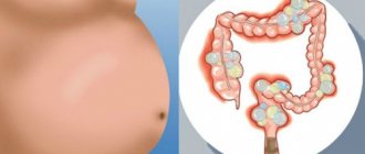
Hepatic encephalopathy - symptoms
The list of symptoms of hepatic encephalopathy includes specific mental and neuromuscular changes, as well as changes in the electroencephalogram.
Considering that the development of PE affects all parts of the brain, the symptoms of PE are quite diverse.
The main manifestations of the pathology are the development in the patient:
- personality disorders (patients are constantly irritable and absent-minded, they have no interest in the family, they behave childishly);
- sleep disorders;
- fixed gaze, lethargy, appearance of apathy;
- speech disorders (monotonous slow speech, short answers to all questions);
- intellectual impairment (difficulty drawing straight lines, counting, memory loss);
- flapping tremors (asterixis) and the appearance of a sweetish-sweet odor from the mouth;
- involuntary urination and bowel movements.
Early signs of this complication include the appearance of personality disorders. This symptomatology of PE is most pronounced in patients with chronic lesions of hepatocyte cells.
The behavior of patients becomes “childish”, they become capricious, irritable, and tearfulness may be observed. There are frequent mood swings. In some patients, depressive symptoms come to the fore.
As PE progresses, neuroses, manic-depressive states, mania, and delirium may appear.
Symptoms associated with personality disorders may persist even during remission of the disease. Symptoms and the degree of their progression in this case are assessed using special psychometric tests.
For reference. Individuals with minimal hepatic encephalopathies do not have intellectual, speech, or coordination impairments, but they often exhibit pathological distractibility and impaired attention.
Additional symptoms
Patients also have problems recognizing objects, changes in handwriting, and impaired ability to draw complex shapes and draw straight lines.
Intellectual impairments can vary from mild inhibition and slow reactions to complete inappropriate behavior of the patient.
Speech disorders in hepatic encephalopathies are manifested by slow speech, disruption of the logical structure of sentences, monotony of speech, and slurred voice.
With the development of cerebral edema against the background of PE, patients are concerned about constant drowsiness, lethargy, apathy, lethargy, depression, depression.
For reference. A frequent manifestation of PE is the specific smell of the patient’s breath (sweetish-sweet aroma).
With the progression of symptoms of damage to the nervous system, the appearance of asterixis is noted (a person is unable to fix and maintain any body position), disorientation in space, and a pronounced impairment of motor skills and coordination of movements.
With the development of coma against the background of this disease, there is a sharp decrease in reflexes, lethargy, fogged consciousness, fainting, the disappearance of pathological tremor of the limbs, the disappearance of the reaction to stimuli, and persistent dilation of the pupils.
Symptoms according to the severity of PE
With unexpressed lesions of the central nervous system in PE, the appearance of pathological absent-mindedness and distractibility, sleep disturbances, constant irritability and emotional instability, inability to concentrate, the appearance of constant tremor of the limbs, and slight unsteadiness of gait are noted.
In moderately severe cases of PE, constant drowsiness, the appearance of lethargy and indifference to others, disturbance of spatial and temporal orientation, increased activity of reflexes, the appearance of pathological reflexes, the development of asterixis, the appearance of significant personality disorders and intellectual impairments, monotony of voice, and the appearance of speech disorders are noted.
For reference. The severe course of PE, accompanied by the development of coma, is manifested by complete spatial and temporal, as well as personal disorientation, severe lethargy, loss of consciousness, the appearance of convulsive symptoms, involuntary defecation and urination, the appearance of pathological reflexes, muscle rigidity, the appearance of aggressiveness, delirium, pronounced “hepatic » aroma.
In fulminant, acute liver failure, the progression of HE is accompanied by the development of true (endogenous) hepatic coma. In patients with terminal forms of hepatic pathologies, the development of false, shunt or portacaval comas is noted.
Symptoms and first signs
Clinical manifestations distinguish between neurological disorders and mental disorders. Characteristic manifestations of encephalopathy include asterixis - large-scale twitching of the limbs. Appear due to tonic muscle tension against the background of dysfunction of the nervous system.
General signs of hepatic encephalopathy:
- fixation of gaze;
- spatial disorientation;
- insomnia;
- monotony of speech;
- forgetfulness;
- anxiety;
- liver tremor;
- decreased intelligence;
- indifference;
- speech and writing disorders;
- decreased body temperature;
- fainting;
- lack of sexual desire.
Lethargy, drowsiness, insomnia are the first signs of pathology. Due to metabolic disorders, bad breath appears. At the decompensated stage, patients become aggressive or apathetic and react inadequately to the environment.
At the penultimate (terminal) stage, depression of the central nervous system occurs. Patients do not respond to irritating factors other than pain. In a comatose state, severe convulsions are common, which are provoked by muscle hypertonicity. At the coma stage, 9 out of 10 patients die without regaining consciousness.
Diagnosis of hepatic encephalopathies
The main goal of diagnostic measures is to confirm the presence of the disease, determine its causes, characteristics and stage.
In a conversation with the patient, the doctor collects information about the phenomena that bother him, his history of liver diseases, and the medications he is taking. The specialist prescribes a consultation with a gastroenterologist and neurologist, a general and biochemical blood test, a coagulogram, and a test for hepatitis markers.
The studies carried out will reveal the presence of inflammation, determine the level of blood clotting, urea, alkaline phosphatase, creatinine, and other elements.
The diagnosis is based on:
- anamnestic data (chronic, progressive liver diseases, acute liver dysfunction, alcoholic cirrhosis);
- results of assessment of neurological status, motor skills and psychometric tests for hepatic encephalopathy;
- electroencephalogram data (slowdown of a-rhythms, appearance of low amplitudes, prolongation of the latent period);
- magnetic resonance spectroscopy results (glutamine peaks, low levels of myoinositols/creatinines);
- assessment of specific symptoms;
- identifying progressive personality disorders.
The degree of organ damage is determined by ultrasound diagnostics of the gallbladder and liver, MRI and CT. Studies demonstrate changes in organ size, areas of death and atrophy.
The cause of the pathology is discovered by performing a biopsy. It is usually performed under ultrasound guidance, since the procedure for cirrhosis can be dangerous.
For reference. An electroencephalogram will assess how damaged the brain is. In some cases, a cerebrospinal fluid tap is taken to check protein levels.
Also, this diagnosis should be suspected in patients with drowsiness, inhibited reaction, speech disorders, inappropriate behavior if they have specific signs of chronic liver damage:
- liver stars on the skin;
- jaundice;
- hair loss;
- exhaustion
- bloating and constant nausea;
- reduction of the liver in size and enlargement of the spleen;
- decreased libido;
- the appearance of a “jellyfish head”;
- identification of specific biochemical abnormalities in blood tests.
According to indications (usually for acute PE), tests are carried out for poisons and toxins, and a test for alcohol content in the blood.
Also, it is mandatory to perform general and biochemical diagnostics, ultrasound examination of the liver, and liver biopsy (if indicated).
A frequently used test for hepatic encephalopathy, which allows assessing the degree of progression of the pathology, is the Reitan test, based on connecting numbers.
Complications
Other diseases may occur against the background of hepatic encephalopathy:
- Brain swelling. With hepatic encephalopathy, the water balance in the body is disturbed, fluid accumulates in the tissues of organs, including the brain.
- Bleeding. With encephalopathy, the functioning of the gastrointestinal tract and the production of its own enzymes are disrupted. This can lead to exacerbation of chronic diseases, stomach or intestinal bleeding, which must be stopped urgently. Stomach bleeding often manifests itself in the form of vomiting like coffee grounds and black feces.
- Infections. With hepatic encephalopathy, the body weakens, immunity decreases, so there is a high probability of various infections that aggravate the course of the disease.
- Aspiration pneumonia. If the patient is in serious condition and lies for a long time, sometimes the contents of the stomach enter the lungs, which causes an inflammatory process.
- Acute pancreatitis. Pancreatitis with hepatic encephalopathy is common. Inflammation in the tissues of the gland develops very quickly and leads to severe abdominal pain, nausea and prolonged vomiting of bile, fever, and jaundice.
At the stage of complications, the patient is hospitalized. Typically, all of the above consequences appear at stages 3-4 of the disease, when the damage to the brain and other organs is significant.
First aid
Emergency care should be provided to the patient when symptoms of hepatic coma appear. Need to:
- record the onset of an attack and report it to the doctor;
- lay the person on his side;
- ventilate the room;
- clear your mouth of vomit;
- provide clean water at all times.
In case of excessive excitement, an injection of one percent Diphenhydramine in a volume of 2 ml is indicated. To normalize cardiovascular activity, an injection of Cordiamin or Metazon is given.
Medicines
| Name | Action | Admission rules | Price |
| Normaze | A laxative that activates the intestines, allowing the body to remove accumulated feces and toxins. | For children under 12 years old – 15 ml. means 1 time per day, for adults – 40 ml. in a day. Course – 5 days. | 280 rub. for 1 bottle. |
| Kanamycin | The drug is in powder form for the preparation of solutions (used for intravenous or intramuscular administration). It has an antimicrobial effect, reduces the activity of pathogenic microflora (viruses, fungi, anaerobic forms). | The daily dose is 2-3 grams, diluted in 1 liter. water. Take once every 6 hours. The course is determined individually. | 15 rub. for 1 g. |
| Gepa - Merz | Sachet for preparing the solution. Helps protect liver cells and remove toxins from liver tissue. | Daily dose 20-40 g. facilities. Use in equal doses 4 times a day. Duration – up to 10 days. | 750 rub. for 10 sachets of 5 g. |
| Gordoks | The drug in the form of ampoules is used when there is a threat of internal bleeding, as well as during surgical operations. | The product is intended for intravenous administration. The solution is administered in an amount of 5-10 ml. in a minute. The duration of therapy is determined individually. | About 4900 rub. |
Treatment of hepatic encephalopathy
All medications for hepatic encephalopathy should be prescribed exclusively by a doctor. Self-medication and the use of traditional therapy methods are unacceptable.
For reference. Diet for hepatic encephalopathy is one of the most important stages of treatment.
Patients are given a protein-free (during periods of exacerbation) or low-protein diet. However, with minimal manifestations of the disease, the diet is expanded. This is due to the fact that excessively reduced consumption of protein foods leads to exhaustion of the patient and the development of a negative nitrogen balance.
In this regard, the degree of protein restriction directly depends on the severity of the manifestations of PE.
When the neurological status is normalized, a gradual expansion of the diet is carried out in order to prevent the development of exhaustion and anorexia.
It is necessary to take into account that in the diet preference is given to proteins of plant origin, since plant foods contain a lot of fiber and fiber. Also, plant foods practically do not contain aromatic amino acids.
Drug treatment will necessarily include the prescription of lactulose (Duphalac). This substance promotes acidification of intestinal contents and actively suppresses the growth of pathogenic and opportunistic intestinal microflora, which leads to a decrease in the synthesis of ammonia compounds.
Dosages are selected individually. The prescribed dosages are considered adequate if the patient has semi-liquid stools two to four times a day. If diarrhea, abdominal pain, or bloating occurs, reduce the dosage.
For patients in serious condition, lactulose is prescribed through nasogastric tubes.
For reference. Also, in order to suppress the proliferation of pathogenic and opportunistic intestinal microflora, antibiotics are recommended. The most commonly used drugs are neomycin, metronidazole, and vancomycin.
The most effective drug is the rifampicin derivative Rifaximin (an antibacterial agent from the ansamycin class, characterized by a wide spectrum of antimicrobial effects).
Dosages of antibacterial agents and frequency of administration are selected individually.
The use of antimicrobial drugs helps to suppress the overgrowth of intestinal microflora and reduce ammonia synthesis.
In order to remove ammonia compounds from the bloodstream, preparations of L-ornithines - L-aspartates are used. Also, in order to suppress the entry of false neurotransmitter substances into the brain, amino acids with branched chains can be used (the use of leucines, valines, and isoleucines is recommended).
Surgical intervention
The main methods of surgical treatment are:
- Portovacal shunting. A shunt is installed to provide bypass blood flow. As a result, blood pressure in the portal vein decreases, the likelihood of hypertension in local vessels decreases, and a stable blood supply is ensured.
- Organ transplant. It is performed if the liver pathology is irreversible and drug treatment is ineffective.
Why does the disease develop?
Scientists still cannot say exactly why hepatic encephalopathy develops. There are three theories explaining why brain activity is disrupted when liver tissue dies, and each of them has convincing evidence:
- False neurotransmitter theory. She says that with liver failure, protein decay in the large intestine increases. The body tries to use some amino acids - those with a branched chain structure (leucine, valine, isoleucine) - as energy. As a result, aromatic amino acids (phenylalanine, tyrosine, tryptophan) enter the blood, which should normally undergo metabolism in the liver. They reach the brain and stimulate the formation of transmitting substances that should not be there (false neurotransmitters). This inhibits the enzyme system that should convert tyrosine into dioxyphenylalanine (from which dopamine and norepinephrine are obtained). Phenylethylamine, octopannine, and thyronine, which inhibit its functioning, also accumulate in the brain. This dictates the need to stop the intake of protein nutrition, replacing it with the introduction of only balanced mixtures consisting of the right amino acids.
- Toxic (ammonia) theory. She says that brain function deteriorates due to toxins (primarily ammonia and protein decay products), formed largely in the large intestine, as well as in the muscles, in the small intestine and in the liver - when proteins are broken down in it . The body always tries to maintain a balance between the formation and neutralization of ammonia, but if this does not happen, the toxins flow through the portal vein to the liver. There, ammonia would have to enter a cycle of reactions called the ornithine cycle to form urea. But since the liver cells are damaged, the rate of neutralization of toxins and ammonia is greatly slowed down. They, together with those substances that bypassed the liver and went directly into the inferior vena cava, are toxic substances to the brain. The situation can be improved by the “correct” amino acids, which neutralize ammonia “bypassing” the ornithine cycle - and along the path of the formation of glutamine from it. These are arginine, glutamic acid, ornithine and aspartate. They convert ammonia into glutamine, which is non-toxic to the brain.
- The neuroglia hypothesis (this is the name of the tissue that is auxiliary in the brain). She says that toxins that appear during liver failure, as well as an imbalance of amino acids, lead to swelling of neuroglia and disruption of its functioning. But if in cirrhosis or fibrosis of the liver compounds toxic to the brain are formed in the intestines, then in acute hepatitis caused by various viruses they are products of liver destruction.
Risk factors
Acute or chronic liver damage, leading to damage to brain tissue, is provoked by the following factors:
- liver failure;
- viral, alcoholic hepatitis;
- drug abuse;
- cirrhosis of the liver;
- hepatocellular carcinoma;
- intestinal dysbiosis;
- abuse of protein foods;
- liver fibrosis;
- alcohol addiction;
- frequent constipation;
- helminthic infestations;
- cholelithiasis;
- autoimmune disorders;
- peritonitis against the background of abdominal dropsy;
- postoperative complications;
- infectious diseases;
- bleeding from the stomach.
Encephalopathy appears in pathological conditions that are accompanied by hepatocytolysis - mass death of liver cells (hepatocytes).
Prevention
Preventive measures will help prevent hepatic encephalopathy and reduce the severity of its course. These include:
- a rational diet with limited protein intake;
- refusal of uncontrolled use of medications;
- timely treatment of liver diseases;
- stopping drinking alcohol;
- using phosphate enemas to cleanse the intestines;
- timely treatment of constipation.
Hepatic encephalopathy has a negative prognosis if the patient ignores its symptoms and refuses timely diagnosis and treatment.
It influences all areas of a person’s personality and life. Properly prescribed therapy and compliance with preventive measures will help improve the prognosis and quality of life of the patient.
Risk factors
The main reasons for the development of a dangerous condition are factors such as:
- Hepatitis of various types (most often these are hepatitis B and D, less often - C and A, during pregnancy the hepatitis E virus has an effect);
- Other infectious liver diseases occurring in acute form (herpes, chickenpox);
- Fungal infections affecting organ tissue;
- The presence of abscesses in the liver tissue;
- Purulent diseases of the liver and gall bladder;
- Food poisoning (for example, when eating poisonous mushrooms, such as toadstool, fly agaric);
- Long-term negative effects of toxic substances (for example, ethyl alcohol, chlorine compounds);
- Uncontrolled use of medications (antibiotics, hormonal agents, drugs based on acetylsalicylic acid, fluorotane anesthetics);
- Disorders of metabolic processes leading to the accumulation of copper in organ tissues;
- Damage to large vessels of the liver;
- Oncological diseases and the use of potent chemicals for their therapy;
- Systematic consumption of alcohol;
- Autoimmune pathologies.
Regarding treatment methods and diet, we recommend that you read the comments of the doctor of the State Medical Center named after. A.I. Burnazyan FMBA for a specific diagnosis of a patient (male 37 years old): https://health.mail.ru/consultation/531687/.
How to prevent the disease
Encephalopathy is provoked by many factors, so there is no specific prevention of the disease. To reduce the risk of brain damage, it is recommended:
- eat rationally;
- to live an active lifestyle;
- refuse hepatotoxic drugs;
- treat liver diseases in a timely manner.
Quitting alcohol and eating a balanced diet reduces the load on the liver. Prevention of mass death of liver cells reduces the risk of disruption of the organ’s neutralizing function.
Hepatic encephalopathy (HE), or portosystemic encephalopathy, is a potentially reversible syndrome of brain dysfunction in patients with progressive liver failure. However, P.E. is not a single category and may reflect the clinical manifestations of reversible metabolic encephalopathy, brain atrophy due to hepatocerebral dystrophy, cerebral edema, or any combination of these conditions. The mechanisms that impair brain function in liver failure are still not fully understood, but it is clear that they are directly related to liver failure and impaired ammonia metabolism. If the underlying liver disease is not curable, PE is associated with poor survival and a high risk of relapse [1, 2]. Even in its mildest form, PE reduces health-related quality of life and is a risk factor for episodes of severe PE [3, 4].
Pathogenesis
Despite more than 100 years of research, the pathogenesis of PE is still not entirely clear. This is mainly due to the difficulty of studying the brains of patients with PE in vivo
. Most published data come from experimental models of PE, which are far from perfect. The most common hypotheses for pathogenesis reflect the role of neurotoxins, impaired neurotransmission due to metabolic disturbances in liver failure, changes in brain energy metabolism, amino acid imbalance, systemic inflammatory response and disturbances in the permeability of the blood-brain barrier [5–7]. Various hypotheses for the pathogenesis of PE are not mutually exclusive. It is likely that many of the described disorders may act simultaneously and ultimately lead to the development of PE.
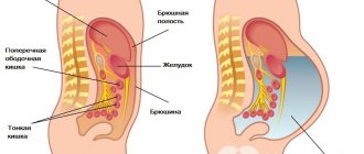
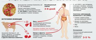
Neurotoxins.
The most studied neurotoxin associated with HE is ammonia, which is produced primarily in the gastrointestinal tract and travels through the portal vein to the liver. A healthy liver detoxifies incoming ammonia by converting it into glutamine, thereby preventing it from entering the systemic circulation. With progressive liver diseases, an increase in the concentration of ammonia in the blood occurs both as a result of a violation of its conversion by the liver into glutamine, and as a result of porto-systemic shunting, in which blood, bypassing the liver, enters the systemic circulation [3].
When the concentration of ammonia in the blood increases, it penetrates the blood-brain barrier and has a neurotoxic effect primarily on astrocytes, the most numerous cells in the brain, closely associated with the functioning of neurons. A key mechanism for the development of PE is astrocyte swelling due to hyperammonemia [8–11].
Excess ammonia leads to the accumulation of glutamine formed in astrocytes, which is accompanied by an increase in intracellular osmolarity and in high concentrations causes brain edema [12].
Impaired neurotransmission.
Impaired functioning of cerebral cortex neurotransmitters and their receptors plays an important role in the pathogenesis of PE. Abnormalities in several neurotransmitter systems have been studied in experimental models of liver failure. Most of these studies describe changes in the GABAergic neurotransmitter system, dopaminergic, serotonergic and glutamatergic neurotransmitter systems [13, 14]. In particular, the role of GABAergic influences in the development of PE may be associated with the activation of benzodiazepine receptors in the brain and the associated increase in the synthesis of neurosteroids [15].
Amino acid imbalance.
An important role in the pathogenesis of PE is played by amino acid imbalance in the form of an increase in the level of aromatic amino acids (tyrosine, phenylalanine, tryptophan), which are precursors of false neurotransmitters, and a decrease in the content of branched-chain amino acids (valine, leucine, isoleucine). Under these conditions, there is an excessive supply of aromatic amino acids to the brain, which serve as the starting product for the synthesis of false neurotransmitters. These changes in amino acid composition are also accompanied by a decrease in dopamine synthesis, which also contributes to the formation of false neurotransmitters [16].
Clinic
The clinical picture of PE is a wide range of nonspecific neurological and mental disorders [17]. Signs of encephalopathy in patients with chronic liver diseases depend on the etiology of the underlying disease, the nature and severity of pathogenic factors.
The severity of PE varies from latent (stage 0) and mild (stage I) to coma (stage IV). With minimal PE, it is manifested mainly by disturbances in abstract thinking and a general mild decline in cognitive functions without impairments of memory, intelligence, speech, and learning ability, which remain intact for a long time [18, 19]. In some patients with liver failure, for a number of years only disorders of higher brain functions are detected without any other neurological symptoms [20].
As PE progresses, personality changes increase, such as apathy, irritability and decreased behavioral control, as well as obvious changes in consciousness and motor function [21]. Sleep-wake cycle disturbances with excessive daytime sleepiness are common, although complete sleep-wake cycle disruption is usually absent [22–24]. With increasing liver failure, progressive disorientation in time and space, behavioral disturbances develop, episodes of confusion with agitation or drowsiness, stupor and, finally, coma occur [25]. The consensus of the International Society for Hepatic Encephalopathy and Nitrogen Metabolism (ISHEN) considers the appearance of disorientation or asterixis (“flapping” tremor) as the initial signs of overt HE [26].
In noncomatose patients with PE, motor dysfunction such as muscle hypertonia, hyperreflexia, and a positive Babinski reflex is observed. As coma progresses, deep tendon reflexes usually decrease [27] and even disappear, although pyramidal signs may persist. Occasionally, focal neurological deficits may occur [28]. Convulsions in PE are very rare [29–31].
Frequent manifestations of the disease are extrapyramidal disorders in the form of facial hypomia, muscle rigidity, bradykinesia, hypokinesia, monotony and slow speech, Parkinson-like tremor and dyskinesia with limitation of voluntary movements [27].
Asterixis (“flapping tremor”) is often noted in the early and middle stages of PE, which precedes stupor or coma. It is not actually a tremor but a negative myoclonus and is caused by hyperextension of the patient's wrist with the fingers spread apart. It is noteworthy that mental and motor disorders in PE may be mild or do not progress equally in different patients, which leads to difficulties in determining the stage of PE.
Classification
According to the practical recommendations of the American Association for the Study of Liver Diseases and the European Association for the Study of the Liver [32], PE is classified according to four criteria: depending on the reasons that led to its development; by duration and nature of the flow; according to the severity of the course (stages) and depending on the presence of provoking (trigger) factors.
A - hepatic encephalopathy as a result of acute liver failure;
B — portosystemic shunting in the absence of liver cirrhosis (LC);
C - hepatic encephalopathy in patients suffering from cirrhosis.
Classification of PE by duration and nature of the course:
1) episodic PE;
2) recurrent PE - these are attacks of PE that occur with a time interval of 6 months or less;
3) persistent PE represents behavioral disturbances that are constantly present and interspersed with relapses of overt PE.
Classification of the severity of PE by stages (West Haven scale)
I. Minimal (latent) stage: there are no clear clinical symptoms, but there is a violation of standardized psychomotor tests (number connection test, line test).
II. The first (mild) stage: apathy, excitement, irritability, anxiety, euphoria, fatigue, disturbances in the rhythm of sleep and wakefulness, mild tremor, impaired coordination of movements, asterixis.
III. Second (middle) stage: drowsiness, lethargy, disorientation in time and space, inappropriate behavior, asterixis, dysarthria, ataxia.
IV. Third (severe) stage: stupor, severe disorientation, unclear speech, hyperreflexia, the presence of pathological reflexes (Gordon, Zhukovsky), myoclonus, hyperventilation.
V. Stage four: hepatic coma, decerebrate rigidity, oculocephalic phenomenon, lack of response to any stimuli.
Depending on the presence of provoking (trigger) factors, PE is divided into unprovoked and provoked (in this case, the provoking factor must be indicated). It is advisable to identify precipitating factors (infections, gastrointestinal bleeding, diuretic overdose, electrolyte disturbances, constipation) for all episodes of type C HE and eliminate them if present.
Diagnostics
Diagnosis of obvious PE
The diagnosis of overt PE is based on an objective clinical examination. It involves assessing the symptoms of HE, as well as excluding other causes of brain dysfunction. In the clinical picture, patients with obvious PE have symptoms of diseases of the hepatobiliary system, for example, in most patients the development of PE is preceded by jaundice. A liver odor and hyperventilation can often be detected in patients with encephalopathy [33]. Also, the diagnosis of PE is confirmed by identifying provoking factors (infections, bleeding and constipation, etc.).
The “gold standard” for diagnosing overt PE is the West-Haven criteria, however, they have limited diagnostic value due to the subjectivity of the assessments, especially in stage I PE, since mild hypokinesia, psychomotor retardation and absent-mindedness can be missed during clinical examination. The leading diagnostic symptoms of obvious PE are the presence of disorientation and asterixis in the patient [32].
To describe the condition of patients with significantly altered consciousness, it is recommended to use the Glasgow Coma Scale (GCS). The Glasgow scale involves calculating points that reflect the severity of the patient's reaction to stimuli. The ability to open the eyes, the nature of motor and verbal reactions to simple stimuli (voice and pain) are determined. Coma is preceded by less profound forms of depression of consciousness: confusion, stupor and stupor.
Diagnosis of minimal PE
Minimal HE is defined as abnormal brain function detected by tests in patients with chronic liver disease who are not confused or have not developed asterixis. The examination of such patients may include two main types of tests: psychometric and neurophysiological. ISHEN recommends the use of at least two tests, depending on their availability and local conditions. The importance of testing for minimal HE is that it predicts the development of overt HE.
To diagnose minimal PE, it is recommended to carry out neurophysiological and psychometric tests, among which the most important are both simple ones performed on paper with a pen (psychometric scale of PE) and computerized ones (reaction time delay test, Stroop test, inhibitory control test and SCAN test ) and neurophysiological (critical flicker fusion frequency test) tests [32]. Electroencephalography can detect changes in cortical brain activity in PE, but this method is not specific enough, since it can be influenced by concomitant metabolic disorders and medications.
Laboratory tests.
In patients with PE, serum-biochemical liver syndromes of varying severity are detected, depending on the predominant direction of pathological processes in the liver [34].
Cytolysis syndrome, or hepatocyte damage syndrome (violation of integrity, necrosis of hepatocytes), is characterized primarily by an increase in aminotransferases (aspartate and alanine aminotransferase), as well as other enzymes - lactate dehydrogenase-5, aldolase, ornithine carbamyltransferase, sorbitol dehydrogenase, glutamate dehydrogenase.
Cholestasis syndrome is characterized by an increase in bilirubin, mainly conjugated (direct), alkaline phosphatase, gamma-glutamyl transpeptidase, 5-nucleotidase, cholesterol and bile acids.
Inflammation syndrome is characterized by increased levels of various globulin fractions, dysproteinemia, and increased levels of serum immunoglobulins.
The AASLD/EASL guidelines [32] note that in patients with chronic liver disease, only high blood ammonia levels have diagnostic or prognostic value. However, if ammonia levels are within normal limits, the diagnosis of PE is questionable. Repeated ammonia measurements may be used to assess the effectiveness of treatment when patients are taking medications that lower ammonia levels.
Computed tomography or magnetic resonance imaging and other imaging methods do not provide complete diagnostic information and are of limited value. They are mainly used to exclude structural damage to the brain in patients with cirrhosis.
Differential diagnosis
AASLD/EASL guidelines indicate the need to exclude other diseases that may resemble PE [32]:
1) diabetes mellitus (hypoglycemic, hyperglycemic and hyperosmolar coma, lactic acidosis);
2) alcohol abuse (intoxication, withdrawal syndrome, Wernicke syndrome);
3) overdose of drugs (benzodiazepines, antipsychotics, opioids);
4) neuroinfections and electrolyte disorders (hyponatremia, hypercalcemia);
5) non-convulsive epilepsy, mental illness;
6) intracerebral hemorrhage and ischemic stroke;
7) dementia (primary and secondary);
 brain damage (traumatic, neoplastic, normal pressure hydrocephalus, obstructive sleep apnea syndrome).
Treatment
Treatment of PE includes elimination of precipitating factors, dietary measures and drug therapy. Elimination of precipitating factors is the primary goal in the treatment of overt PE, since in 90% of patients this is sufficient to improve the condition [32].
Diet
Correction of disorders of protein-nitrogen metabolism is crucial in the treatment of all degrees of PE, since 75% of patients with PE have moderate or severe protein-calorie deficiency, accompanied by loss of muscle mass. Long-term restriction of protein intake is harmful for patients with PE, since the protein requirement in these patients is relatively higher than in healthy individuals.
Therefore, according to the AASLD/EASL recommendations, daily energy intake should be maintained at 35–40 kcal per 1 kg of body weight, and daily protein intake should be within the range of 1.2–1.5 g/kg [32]. Limiting protein intake is recommended only during the first few days after the development of PE, but then this measure should be abandoned. Animal proteins should be replaced with milk and plant proteins, and foods enriched with branched chain amino acids should be consumed.
Drug therapy
Drug therapy is an important part of the treatment of overt P.E. With minimal PE in the absence of its clinical manifestations, pharmacotherapy, as a rule, is not used. At the same time, it can be prescribed to this category of patients in cases of a noticeable impact of minimal PE on the quality of life. To correct HE, various drugs can be used (non-absorbable disaccharides, antibiotics, branched chain amino acids, L-ornithine-L-aspartate) with varying levels of evidence of effectiveness.
Non-absorbable disaccharides.
Lactulose is a synthetic disaccharide that prevents the formation of ammonia. Lactulose is the drug of first choice in the treatment of overt P.E. Lactulose syrup is prescribed 25 ml every 1-2 hours until at least 2 bowel movements with soft or unformed stools occur during the day. Subsequently, the dose of the drug is titrated individually to maintain 2- or 3-fold bowel movements during the day [35]. Taking excessively high doses of lactulose can lead to complications such as aspiration, dehydration, hypernatremia, irritation of the perianal skin, and in some cases even worsen the course of PE [36].
Antibiotics.
Rifaximin is a non-absorbable antibiotic that inhibits ammoniogenic proteolytic bacterial microflora of the intestine [37]. The results of a number of studies have shown the effectiveness of rifaximin in the treatment of P.E. According to AASLD/EASL guidelines [32], rifaximin is an effective addition to lactulose for the prevention of recurrence of overt PE. In comparative studies, 3-6 months of rifaximin therapy improved cognitive function and reduced ammonemia in patients with PE [38]. Rifaximin is prescribed at a dose of 200-400 mg 2-3 times/day (1200 mg/day) for 5-7 (up to 14) days.
Among other antibiotics, neomycin is still used for the treatment of PE; metronidazole can be used for short-term therapy, however, due to side effects, the long-term use of these drugs is limited [39, 40].
Branched chain amino acids.
A number of studies have shown the effectiveness of therapeutic nutrition with the consumption of food enriched with branched chain amino acids [15]. Due to the preferential absorption of these amino acids, the relative content of aromatic amino acids, which serve as precursors of false neurotransmitters, decreases. In addition, branched chain amino acids help increase muscle mass, resulting in increased ammonia detoxification, which partially occurs in skeletal muscle. The AASLD/EASL guidelines only recommend oral administration of branched-chain amino acids as an alternative or adjunctive treatment for patients who have not responded to conventional therapy [41, 42].
L-ornithine-L-aspartate (LOLA).
Since ornithine and aspartate play a major role in the conversion of ammonia to urea, taking LOLA helps reduce the clinical manifestations of PE. Over the past 10 years, several randomized studies have been conducted, the results of which have shown the high efficacy and safety of this drug in the treatment of PE [43]. LOLA is available as a solution for intravenous infusion and as a granule for oral administration. The standard regimen of use involves intravenous drip administration of 20-30 g of the drug for 7-14 days, followed by transition to oral administration of 9-18 g/day. To achieve a faster and more lasting result, a combination of intravenous and oral administration is possible.
Flumazenil
- an antagonist of benzodiazepine receptors, directly affecting the functions of the central nervous system. Flumazenil is used quite rarely, since giving a short-term effect of restoring consciousness, it promotes only a transient improvement in mental state, but does not affect recovery and survival rates [15].
The effectiveness of the therapy is determined by the reverse development of clinical symptoms.
In case of acute liver failure with the development of hepatic coma and cerebral edema, treatment is carried out in the intensive care unit using the same medications as for chronic liver failure, but in higher doses.
Liver transplantation for cirrhosis with recurrent PE, difficult to respond to conservative therapy, can increase the life expectancy of patients. The average 5-year survival rate of patients after transplantation exceeds 72% [15].
The authors declare no conflict of interest.