Bruise is one of the most common types of injuries. With such damage, soft tissues suffer, but the integrity of the skin is not compromised. Such injuries often occur when struck with a blunt object, falls from a height onto a flat surface and can be combined with fractures, dislocations, scratches, and abrasions.
In addition to bruises of individual parts of the body, there are also bruises of internal organs.
As a rule, special treatment for bruises is not required. However, if you have pain for a long time after the injury, for example, a sore elbow or back pain, you should consult a doctor.
1
Treatment of bruises and hematomas
2 Treatment of bruises and hematomas
3 Treatment of bruises and hematomas
Causes
The main reason for the formation of hematomas is traumatic effects on soft tissues: bruise, compression, blow, pinching, stretching. The mechanism for the appearance of bruises lies in the rupture of blood vessels. The size and severity depend on their size, location and number of damaged areas. Certain medications can also cause vascular rupture, for example, anticoagulants and acetylsalicylic acid. Non-mechanical causes also include some diseases, such as:
- Mallory-Weiss syndrome - cracks in the upper part of the stomach or lower esophagus, caused by vomiting due to excessive overeating or drinking alcohol.
- Leukemia is a malignant neoplastic disease of the hematopoietic system.
- Atherosclerosis is the formation of cholesterol plaques in the lumen of blood vessels.
- Hemorrhagic vasculitis is a lesion of the smallest vessels in the body: capillaries, arterioles and venules.
Intramuscular hematoma can be associated with injections given into the buttock. Cephalohematomas are sometimes observed in newborns. Their cause is the discrepancy between the baby's head and the woman's narrow birth canal. Intracerebral trauma in newborns is also associated with difficult births. A separate group consists of postoperative hematomas. In pregnant women, they can occur after cesarean section . The list of causes of postoperative hematomas also includes the following:
- decreased blood clotting;
- vascular pathologies;
- increased fragility of vascular walls;
- worsening blood clotting;
- high blood pressure after surgery;
- increased permeability of blood vessels, which causes their rupture.
What is a bruise
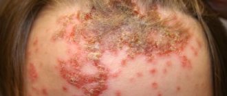
Almost every one of us first becomes acquainted with these formations on the body in childhood, because there is not a single child in the world who has never fallen or hit something. Therefore, everyone knows what a bruise is. This is a bluish (hence the name) spot that appears at the site of the bruise. If bruises form accidentally in children, then in older people, especially among representatives of the stronger half of humanity, they can “bloom” on the face or other parts of the body as a result of a fight. Among the peaceful part of the adult population, bruises appear from accidental bruises, after injections, intravenous injections, squeezing the skin with great force, pinches or bites, for example, from a pet. In all these cases, small blood vessels rupture in the subcutaneous layer, and blood flows out of the wounds into the resulting cavity. That's what a bruise is.
In the cavity, the blood can be in a liquid state, thickened or already completely coagulated. If an injury causes a rupture of not only blood vessels, but also the skin, the blood flowing from the wound can be seen visually. If the top layer of skin remains intact, it will appear as a bluish or purple bruise.
What does a hematoma look like?
When blood leaks into the subcutaneous tissue, a dense and painful swelling forms at the site of injury. Externally, a bruise looks like a spot on the skin, the color of which can determine the age of the injury. At the initial stage, the skin turns red, and then gradually becomes purple-bluish. After 2-3 days the bruise becomes yellowish, and after 4-5 days it becomes greenish. During this time, hemoglobin decomposes. In 4-5 days, the spot may slide down a little.
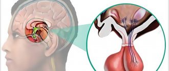
Diagnosis of hematoma in brain structures
Neuroimaging is of clinical value in making a diagnosis. A computed tomography scan is prescribed as an initial examination of the brain. This inexpensive method allows you to quickly and easily determine the presence of blood in the GM substance, the location and volume of the clot. The information value of CT is highest when 2-3 weeks have passed since the hematoma appeared (maximum 5 weeks). Until this time, the VMH area has the highest density, which facilitates diagnosis and can be limited to one CT scan.
MRI.
As the lesion ages (on average, after 14-21 days), the density of the hemorrhagic mass decreases, it becomes isodense, that is, close to normal brain tissue. During this period and later, purely magnetic resonance imaging can provide qualitative information about the intracerebral hematoma and the state of the brain.
Additionally, the patient is often recommended to undergo cerebral angiography. This technique does not have the necessary potential for verification of VMG. But angiography of cerebral vessels determines the intensity and distribution of vasospasm and allows one to exclude or confirm the involvement of a vascular malformation or arterial aneurysm in the development of ICH.
Symptoms
The main symptom is a change in skin color in the wound area to crimson-red, burgundy, blue-violet. The general clinical picture of the development of hematoma depends on the degree of its severity. In total, according to the main classification, there are three of them: light, medium and severe. They are determined by the size and depth of the damage. The characteristic differences of each stage are as follows:
- Easy. Develops within 24 hours after injury. The damage is accompanied by moderate pain. Edema and swelling are not observed, motor activity is maintained at a normal level. With timely assistance, the formation quickly regenerates.
- Average. Forms 3-5 hours after injury. The tissue damage is deeper, which causes pain. Against this background, swelling of the skin and sometimes limitation of motor activity are noted.
- Heavy. It is noted in the first couple of hours after injury. The patient experiences a general and local increase in temperature, impaired movement of the injured limb, and severe pain.
When a brain injury occurs, nausea, vomiting, dizziness, and headache occur. Intracranial hematomas are accompanied by a number of other symptoms, such as:
- psychomotor agitation, turning into epileptic seizures;
- bradycardia;
- hemiparesis on the side of injury;
- visual impairment;
- pyramidal signs;
- loss of consciousness up to coma;
- anisocoria – different pupil sizes in the right and left eyes.
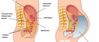
In case of hemorrhage into the abdominal cavity, peritonitis develops, which is indicated by sharp abdominal pain and a rise in temperature. Additionally, the victim experiences nausea and vomiting, cough, and shortness of breath. The retrochorial type of hematoma, which can occur in the first trimester of pregnancy, is accompanied by the following symptoms:
- brownish or bloody vaginal discharge;
- pain in the lower abdomen and lower back;
- red vaginal discharge.
Folk remedies
Treatment with herbs, infusions and decoctions from them has proven its effectiveness for many centuries. In addition to the fact that these remedies are effective, with a reasonable approach they are absolutely safe, especially when it comes to external use, as is the case with superficial hematomas. More than one good remedy for bruises has been created based on natural ingredients:
- herbal decoction of herbs such as wild rosemary and St. John's wort, in the form of compresses 2-3 times a day;
- a gruel of finely chopped wormwood and castor oil - apply the mixture to the bruise for a short time (10-15 minutes) in the morning and evening;
- compresses from white cabbage leaves – relieve swelling from damaged tissues;
- juice from aloe leaves mixed with honey is an excellent anti-inflammatory agent; it is good to use as the basis of a compress, additionally wrapping the bruise in polyethylene.
A little-known, but certainly useful substance that helps fight bruises is dark laundry soap. You need to make a concentrated solution from it or just lather it on the bruise immediately after the blow. After such a procedure, even a subcutaneous forehead hematoma (bump) will disappear in just a few hours.
Kinds
A bruise and a hematoma need to be distinguished from each other. The latter is a more serious injury that often requires urgent treatment. A bruise is a mild form when only small capillaries are damaged . It is characterized by slight swelling and moderate pain. No increase in temperature is observed.
There are many classifications. One of the criteria for distinguishing their types is the degree of severity: mild, moderate or severe - their symptoms were described above. Depending on the clinical signs, hematoma can be of the following types:
- Limited to the periphery. In the center they have a softened structure, and at the edges they have a dense contour.
- Ensembled. They dissolve on their own only in small sizes. An encysted hematoma is characterized by the accumulation of a large amount of fluid inside.
- Diffuse. They increase very quickly, so they require immediate opening to detect a bleeding vessel.
If there is a bruise in the eyeball, a paraorbital hematoma is diagnosed. Because of it, a person can lose his sight. A periorbital hematoma is a bruise around the eyes associated with head trauma. These are two different disorders that need to be distinguished. Taking into account the state of the spilled blood, hematomas are divided into coagulated and non-coagulated (fresh), uninfected and suppurating. The criterion for another classification is the appearance of the hemorrhage. It depends on the type of blood shed. Taking this factor into account, the following are distinguished:
- Arterial. When arterial blood spills into the cavity, the color of the injury site is bright red, and its area is often more extensive compared to other types.
- Venous. If the integrity of the vein is broken or the vein is compressed, venous blood leaks into the cavity, causing a bluish-violet spot to appear on the skin.
- Mixed. A more common case in which both arterial and venous blood enters the cavity. The color of the bruise is mixed.
Taking into account the relationship to the vessel, pulsating and non-pulsating hematomas are distinguished. Ripple occurs when all layers of the vascular wall are damaged, causing blood pressure to be transferred to the liquid contents at the site of hemorrhage. The most extensive classification divides hematomas by location into the following types:
- Subcutaneous. The most common ones are formed in different areas of the skin after injury or as a result of vascular pathologies.
- Submucosal. They are localized not in the subcutaneous tissue, but in the mucous membrane.
- Intramuscular. They are formed as a result of more serious injuries and are localized in muscle tissue.
- Subserous. Affects internal organs, most often the abdominal cavity or lungs.
- Subfascial. The fascia, the connective tissue membrane covering the vessels, nerves and organs, is injured.
- Retrochorial. Hematomas that occur during pregnancy due to detachment of the fertilized egg from the chorion.
The most dangerous are hematomas in the brain area. They differ from other types in the complexity of treatment and more dangerous consequences for the patient. They are also classified into several types:
- Intracerebral. They are rare and are accompanied by the accumulation of blood inside the brain tissue.
- Subarachnoid. Blood accumulates between the arachnoid and inner layers of the dura mater.
- Intraventricular. Blood enters the cerebral ventricles.
- Epidural. An accumulation of blood between the inner surface of the skull and the outer layer of the dura mater, which often accompanies fractures or injuries in the temporal and parietal bones.
- Subdural. Blood contents accumulate under the dura mater of the brain. They are divided into acute, subacute and chronic.
Articles on the topic
- Classification of cerebral strokes by degree - ischemic, hemorrhagic and apoplectic
- Spinal stroke - manifestations, treatment with medications and folk remedies, massage, physiotherapy
- Intracranial hypertension: signs and treatment
Separation of concepts
When the skin is damaged, intra- or subcutaneous hemorrhages may occur, which are called differently, often considered an analogy. But they are all different. The injury is called a bruise, a hematoma, and a bump, but these are not synonyms. For example, hematoma and bruise: the only reason they have in common is an external or internal influence or disease. But with a hematoma, there is always a cavity into which blood flows and the tissue structure is damaged. It appears due to tissue delamination.
If there is no cavity, this is not a hematoma; it can occur not only in the skin, but also inside organs. For example, in the cranial cavity, according to localization, they distinguish:
- epidural (extradural) hematoma - between the bones of the skull and the dura mater;
- subdural hematoma (under the dura mater);
- subarachnoid hematoma (under the pia mater);
- intracerebral, or parenchymal in the substance of the brain, subcutaneous hemorrhage on the head is often called a lump - in adults.
A bruise is also a hemorrhage into soft tissue, but the structure here is not disturbed and a cavity does not occur. In common parlance this is called a bruise.
Bruising is only a colloquial term, not a medical one. It is not used in official documents. Some people like to show off medical terminology and call a bruise a hematoma, although this is completely wrong. To be precise, this is hemorrhagic penetration of the skin.
Why is it so important to separate these concepts? Because they have different effects, treatments and severity. The severity of the injury may determine the appearance of a bruise or hematoma.
Why is a hematoma dangerous?
Only mild soft tissue hematomas occur without serious consequences. Complications develop after severe and extensive hemorrhages. Without opening by a surgeon, they can lead to the formation of scar tissue. This is at best, but at worst the cavity into which the blood has spilled becomes infected and suppurates. When it accumulates in the joint, the following diseases may develop:
- Chronic synovitis is an inflammation of the synovium of the joint, in which effusion accumulates in the cavity lining it.
- Hemarthrosis is hemorrhage inside the joint that occurs due to bruises and intra-articular injuries.
- Bursitis is an inflammation of the synovium of a joint due to injury.
With extensive hemorrhages in the cavities of internal organs, irritation of nerve receptors occurs. This condition is called paresis. It affects the motor pathways of the nervous system, causing incomplete paralysis. When this condition resolves, the spilled blood begins to decompose, i.e. hemoglobin breaks down. This process causes endotoxicosis - the accumulation of toxins in the tissues of the body that poison it.
Intracranial hemorrhages do not go away without consequences. After a head injury, amnesia, disturbances in reaction and attention, and increased anxiety may develop. Serious consequences include epileptic seizures. They appear not only immediately after brain damage, but also after a long period of time. After recovery from intracranial injury, many patients complain of the following symptoms:
- headache;
- attacks of dizziness;
- fatigue;
- increased weakness;
- severe anxiety;
- mental disabilities;
- frequent mood changes;
- headaches.
Diagnostics
A doctor can diagnose superficial injuries visually by examining the site of the injury. To clarify the nature of the disease, the specialist performs palpation, which helps determine the degree of pain, possible local hyperemia and swelling . More serious hemorrhages require detailed diagnosis, which includes the following measures:
- X-ray. Necessary for identifying subserous forms of hemorrhages. An X-ray of the head is taken in two projections to clarify the location of the spilled blood and the extent of damage.
- Computed tomography (CT) and magnetic resonance imaging (MRI). Mandatory for any intracranial injuries. These procedures pinpoint the location of the damage and help study its nature.
- Ultrasound. Helps evaluate the location and structure of any internal bleeding.
- Echoencephalogram. Evaluates intracranial pressure, which is necessary to identify brain pathologies.
- Lumbar puncture. Prescribed in doubtful cases for the purpose of studying cerebrospinal fluid. This procedure helps identify subarachnoid bleeding in brain injuries.
Treatment of hematoma
Superficial subcutaneous hematoma tends to resolve on its own. Immediately after receiving an injury, it is recommended to apply cold to the injury site. This will help prevent blood from entering the tissue and causing swelling. Then the limb can be tightly bandaged, leaving the bandage on for 1-2 hours. In the future, ointments based on bodyaga, heparin or hirudin are used to speed up the resorption of blood . 6-8 hours after injury, the following drugs can be used:
- Voltaren gel;
- Dexpanthenol ointment;
- Lyoton gel;
- Finalgon ointment;
- Troxevasin gel;
- Gepatrombin gel.
For bruises on the face, it is recommended to use Bruise-OFF ointment. It is based on hirudin, a leech extract. In addition to resorption, this drug helps to tone the injury site. The ointment smells pleasant, so using it on the face does not cause discomfort. Since a bruise is a local problem, topical medications are more effective at treating it. In some cases, the use of systemic means is allowed. They are indicated for those who bruise very often. The following drugs help cope with this:
- Ascorutin – contains vitamin P and ascorbic acid. Prescribed for vascular pathologies and some types of hemophilia. The dosage is 1 tablet up to 2-3 times per day. The course of treatment lasts 3-5 weeks.
- Capilar is a plant-based drug. Its action is to strengthen the walls of capillaries. Capilar is especially often prescribed to elderly people suffering from the initial form of hypertension.
More extensive intermuscular hematoma requires surgical opening. During this procedure, the doctor opens the site of injury, removes blood from there and treats it with antiseptics. If there is pulsation, hyperemia and swelling in the area of injury, you should definitely consult a doctor, as bleeding may continue. In this case, the doctor also performs an autopsy, but additionally finds the source of the hemorrhage and eliminates it.
For intracranial hemorrhages, conservative treatment is indicated only if their volume is up to 40 ml, there are no symptoms of brain dislocation and slight depression of consciousness. Otherwise, the patient is hospitalized for craniotomy. The procedure for this operation is as follows:
- Under general anesthesia, the neurosurgeon bleeds the scalp using a cutting suture.
- Next, the specialist cuts out a flap of soft tissue and opens the skull.
- The neurosurgeon then removes the blood using an aspirator, after which the cavity is washed and the source of bleeding is eliminated.
- The bone flap is returned to its place and fixed with thick threads. The tissues are sutured in reverse order, sometimes leaving drainage.
Opening
The procedure for opening and cleansing the cavity into which blood has leaked is indicated for extensive hematomas. This is especially true for injuries to internal organs. For small formations, the autopsy is performed on an outpatient basis, otherwise the patient is hospitalized. The course of the operation includes several stages:
- The patient is provided with local anesthesia.
- The area of spilled blood is cut and cleared of contents.
- Next, it is washed with antiseptics and sutured. If the patient has a festering hematoma, then the area of damage is infected. For this reason, full suturing is not carried out, but the cavity is drained to ensure that the pus escapes out.
- A tight bandage is applied on top.
- After 10 days, the stitches are removed. During this time, the patient takes antibiotics prescribed by the doctor.
Folk remedies
Traditional medicine methods can be used for minor injuries that are not accompanied by severe pain, hyperemia and swelling, i.e. for superficial injuries. Most remedies only help relieve mild pain and speed up the resorption of accumulated blood. The following recipes receive positive reviews:
- Collect a couple of fresh cabbage leaves, not from the surface of the head, but from 2-3 layers of greens. Crush one of them a little and then apply it to the sore spot. Wrap the sheet with a bandage and leave the compress for the whole day.
- Peel raw potatoes, rinse, cut in half. Apply one half to the bruise and hold for 15-20 minutes. Repeat several times throughout the day.
- Wash a couple of plantain leaves and grind them to a paste. Apply it to the sore spot and wrap it with a bandage. Keep the compress all day.
- Dissolve 2 tbsp in 100 ml of water. l. salt. Moisten a clean natural cloth in the resulting solution. Place it on the sore spot and wrap it with a bandage. Leave for the whole day.
First aid for hematomas: what to do?
If the injury is superficial, then you can treat the hematoma at home yourself. First, you need to apply an ice pack to the bruise. This is done no later than two days after receiving the damage. With the help of cold, blood circulation in the tissues slows down and bleeding stops. To do this, put ice in a bag and apply it to the bruised area for 10 minutes. This action should be repeated 5 more times.
With the help of cold, swelling is reduced and the location of the hematoma is reduced, and the risk of cell destruction from lack of oxygen is also reduced. Warming ointments help get rid of residual bruising.
Forecast
A favorable prognosis is typical for soft tissue hematomas. The correct treatment regimen ensures the patient’s full recovery without any serious consequences. The same applies to retrochorial hematomas that develop in the first trimester of pregnancy. With timely diagnosis and adequate therapy, childbirth is carried out naturally. The cesarean section method is used only when a hematoma forms late in pregnancy - at 38 weeks of pregnancy. This does not have any dangerous consequences for the mother and child.
An unfavorable prognosis is observed only with brain injuries. This is especially true for subdural and epidural hematomas. Only mild and moderate injuries are completely cured, but the process can take several years. Among the consequences of such injuries, the following are especially common:
- problems with urination, bowel movements, or swallowing;
- post-traumatic convulsions;
- speech disorders;
- behavioral disorders;
- perception disorders (inability to reproduce what you see);
- disturbances of mental activity and memory.
Prevention of hematomas and bruises
It is impossible to completely protect yourself from environmental influences and prevent potential injuries. But it is very important to increase the level of your immunity so that the body is more protected from the negative effects of the environment. The very frequent appearance of bruises on the skin may indicate a lack of vitamin C or K in the body. Therefore, it is worth adding fresh vegetables and fruits, as well as vitamins, to your daily diet.
It is not possible to completely protect yourself from injuries and damage to the skin. But, despite this, you should know how to provide first aid for bruises, in what ways you can cure hematomas on the body. If there are no results from the treatment, you should immediately contact a traumatologist.
Prevention measures
The main preventative measure is to minimize injuries. Young children often hit furniture or doors. You can reduce the risk of childhood injuries and generally reduce the likelihood of developing hematomas as follows:
- arrange furniture, observing minimal passages between objects;
- in a house with small children there should be a minimum of sharp corners;
- teenagers when cycling or roller skating, skating, etc. are recommended to use protective equipment such as knee pads, elbow covers and a helmet;
- take anticoagulants and Aspirin with caution;
- Athletes should warm up before each workout.
Etiology of bruises
To the above explanations of how a hematoma differs from a bruise, we can add the reasons why a person develops such formations. First, let's look at where bruises come from. Since they always form in the subcutaneous layer, their appearance can be caused by any injury that disrupts the integrity of small and medium-sized blood vessels located in the subcutaneous fat. These are:
- injury;
- hit;
- compression, squeezing;
- pinch;
- injection (bruises are observed with intramuscular injections, because the syringe needle touched and injured small vessels);
- intravenous injections (bruises are observed because blood has leaked from the punctured vein into the resulting space).
Bruises can appear on the legs due to varicose veins and phlebitis, and on the face after medical procedures, such as tooth extraction.
The weaker the walls of a person’s blood vessels, the more often and in greater numbers bruises “bloom” on his body.