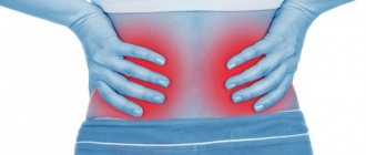
The appearance of unbearable pain in the lower back, which does not give the patient peace, makes him rush about in bed and does not allow him to lie or sit quietly, is almost always a manifestation of acute renal colic. This is not a disease, but only a symptom of some disease.
But, as with all emergencies, the first step is to get rid of this pain in order to alleviate the person’s condition. Treatment of the pathology itself should, in most cases, come second.
In order to detect an attack of colic, provide proper assistance and eliminate the cause of its occurrence, it is necessary to have reliable information about this condition. You can get acquainted with it in this article.
Definition of the concept
Renal colic is a common symptom complex characterized by intense pain in the lower back, which is caused by a violation of the outflow of urine or spasm of the muscular structures of the ureter. It is formed on the basis of existing pathologies of the urinary tract and kidneys, for example, urolithiasis.
Usually appears when the urinary tract is blocked by stones at the level of the ureters or renal pelvis. The condition is observed in absolutely any age group, but most often between the ages of 20 and 55 years. At an older age, colic diagnosed for the first time is considered a very rare occurrence.
As a rule, renal colic is unilateral.
It is a serious condition, in some cases life-threatening for the patient, and therefore requires immediate hospitalization and emergency care.
general description
An attack of severe pain, caused by a sudden disruption of urine output, as well as renal ischemia, is called renal colic.
This is a fairly common pathology that is observed in people in many countries around the world. Most often, the disease is registered in residents of developed countries who abuse fast food and other junk food.
The disease manifests itself as severe pain in the lower back that spreads downwards. In addition, the patient experiences frequent urination, severe nervous agitation, and vomiting.
Treatment is carried out by applying heat to the lesion, introducing strong analgesic and anti-spasm drugs, and novocaine blockades. After the attack is relieved, diagnostics are carried out aimed at identifying the causes of its occurrence.
Interesting facts about renal colic
- One of the first mentions of this condition are the records of the greatest Roman healer Galen, who lived in the second century AD.
- The oldest stone that could contribute to the development of pathology was identified in a mummy; according to scientists, its age was more than seven thousand years.
- The most common location of renal colic is on the right side; in only 10% of cases both kidneys are involved in the process.
- The risk of developing kidney stones increases depending on a person's wealth. The richer a person is, the higher he is.
- Recurrence of renal colic occurs in more than a third of cases.
Complications
With timely assistance, the prognosis for this condition is favorable. Complications arise only in case of delayed treatment or erroneous tactics. The severity of these conditions may vary, depending on the duration of stagnation of urine and the condition of the patient. The most common of them include:
- Nephrosclerosis or kidney atrophy;
- Pyelonephritis – purulent inflammation of organ tissues;
- Persistent narrowing of the ureter;
- Sepsis;
- Shock.
In clinical practice, there are even cases of death after attempts at long-term self-medication at home. All these complications are extremely difficult to treat (with the exception of pyelonephritis), but it is quite easy to prevent - all you need to do is seek medical help.
Briefly about the structure of the urinary system
It is very problematic to understand the mechanism of development of pain without knowing the basic structures of the urinary system and the process of urination. Secondary urine is formed in the kidney tissue in a volume of about 1.5 - 2 liters per day, then it enters the pelvis - specific formations located at the exit from the kidneys.
Their diameter is quite narrow (up to several millimeters), so stones most often get stuck in this section. Next comes another organ related to the urinary system - the ureter. It consists of two hollow tubes with a diameter of up to 8 mm, through which the kidneys and bladder communicate. This organ is also often the target of stones. After entering the bladder, urine moves through the urethra into the environment. In this area, stone obturation occurs least rarely.
Causes of renal colic
This condition is not an independent disease, but only a complication of a number of diseases, such as:
- urolithiasis disease;
- kidney inflammation
- kidney injuries;
- tuberculosis;
- congenital features;
- benign or malignant neoplasms;
- allergic reactions accompanied by swelling of the ureters;
- Ormond's disease.
Due to unfavorable factors, there is a disruption in the movement of urine from the renal tissue, leading to filling of the renal pelvis, an increase in pressure inside them, irritation of pain receptors located at the gates of the kidneys and in their fibrous membrane, which ultimately provokes an attack of renal colic.
Urolithiasis disease
It provokes an attack of the disease in 9 cases out of 10. A calculus that enters the lumen of the ureter or renal pelvis clogs it, disrupting the movement of urine and additionally injuring the walls of these formations, which initiates colic. Stone formations can form due to a violation of salt metabolism, leading to an imbalance of substances that contribute to the formation of stones and maintaining urine in a liquid form.
The first include:
- creatinine;
- urea;
- citric acid salts;
- hippuric acid, etc.
Elements that promote stone formation include:
- calcium salts (calcinates);
- uric acid (urates);
- oxalic acid (oxalates);
- phosphorus (phosphates), etc.
When urine is oversaturated with these substances, a crystallization nucleus is formed, which is an accumulation of stable microscopic crystals. Gradually, an increasing amount of salts are deposited on this core, which leads to the appearance of a full-fledged stone of various diameters and shapes. The development of urolithiasis is influenced by the following factors:
- excessive consumption of water enriched with salts and minerals;
- food ration, including sorrel, parsley, chocolate, pickles, smoked meats;
- warm southern climate;
- dehydration as a result of insufficient fluid intake, persistent vomiting or diarrhea;
- prolonged lying down, for example, after extensive surgery;
- chronic infections of the urinary system;
- lack of vitamins A and D.
For a long time, urolithiasis develops secretly and makes itself felt only when the stone clogs the lumen of the urinary tract.
Pyelonephritis
The most common infectious disease of the renal pelvicaliceal system. It is usually provoked by Pseudomonas aeruginosa or Haemophilus influenzae, Proteus, and staphylococcus. Formed during hypothermia, diabetes, reduced immune status, pregnancy and other conditions.
The pathology is characterized by desquamation of the renal epithelium, suppuration and fibrin deposition (a specific post-inflammatory protein formation), which can contribute to occlusion (blockage of the lumen) of the ureter, especially in places of its physiological narrowing, the diameter of which is about 5 mm. As a result, renal colic is formed, characterized by acute pain in the lumbar region.
Kidney injuries
Injury (fall from a height, blows to the lumbar region, etc.) of the kidney tissue leads to bleeding, which can result in the formation of hematomas of various sizes - limited accumulations of blood clots that block the lumen of the urinary tract. In addition, the outcome of any injury can be increased formation of scar tissue at the site of injury, which significantly blocks the lumen of organs.
Tuberculosis
Damage to the kidneys by Mycobacterium tuberculosis (bacillus, Koch's bacillus) is accompanied by the formation of specific tuberculous granuloma formations, as well as purulent masses and desquamation of the epithelium of the renal tissue, which together leads to difficulty in the outflow of urine and the occurrence of renal colic. Kidney damage by mycobacteria is not so common; pulmonary localization of the process occupies the first place.
Congenital characteristics
These include:
- prolapse of the kidneys (nephroptosis);
- incorrect attachment of the ureters to the bladder (ectopic orifices);
- incorrect location of organs (dystopia);
- congenital narrowing or absence of the lumen of the ureter (stenosis and atresia);
- the formation of additional vessels that compress the ureters.
Inadequate congenital location and functioning of the organs of the urinary system leads to incorrect urodynamics, and under the influence of unfavorable factors (damage, infections), a violation of the outflow of urine and the formation of an acute process are possible.
Tumors
Tumor formations (adenoma, lipoma, lymphangioma, fibroma, sarcoma, fibroangiosarcoma) can grow into the cavity of the genitourinary system or compress it from the outside, being nearby and reaching a significant size, ultimately leading to an attack of renal colic.
Allergic reactions accompanied by swelling of the ureters
They are relatively rare. Observed when taking certain drugs, especially iodine-containing drugs and antibiotics (for example, Ampicillin, Levofloxacin). The release of biologically active substances (bradykinin, histamine, serotonin, etc.) is accompanied by severe tissue edema, which can block the lumen of the ureter.
Ormond's disease or retroperitoneal fibrosis
This condition is rarely diagnosed and is associated with increased formation of coarse fibrous tissue behind the peritoneum, compressing the ureters from the outside. It is believed that Ormond's disease occurs due to chronic infections, inflammatory and autoimmune diseases (tuberculosis, systemic lupus erythematosus, scleroderma, etc.), and malignant neoplasms.
In the event of an attack of renal colic, the presence or absence of stones in the lumen of the ureter or pelvis is initially determined, only then other factors are excluded.
Causes
Various diseases can lead to the development of colic, but they are united by one feature - blockage (synonym - obstruction) of the passages for urine excretion. Each of them disrupts the outflow of this fluid from the body, which leads to the occurrence of all symptoms. Closure of the lumen of the urinary tract can occur at various levels (in the pelvis, ureter and even bladder), but the manifestations of the disease practically do not change.
What pathologies can lead to obstruction? Currently, the most common reasons are:
| Disease | Blockage mechanism |
| Urolithiasis disease | In the vast majority (more than 92%), the cause of colic is a stone blocking the pelvis or ureter. When characteristic symptoms occur, doctors first rule out this particular pathology. |
| Pyelonephritis | Infection in the kidney, as a rule, occurs under the influence of microbes: E. coli, staphylococci and streptococci, influenza bacilli, etc. The inflammatory process is often accompanied by the formation of pus, fibrin and desquamation of the inner wall of the organ (epithelium), which exits through the urinary tract. If they are in excess, they can close the lumen of the ureter, the diameter of which in places of narrowing is less than 5 mm. It should be noted that pyelonephritis often occurs due to the presence of a stone. |
| Congenital structural features | This group of causes includes conditions such as prolapse (nephroptosis) or abnormal position (dystopia) of the kidneys, abnormalities of the attachment of the ureters to the bladder, and others. As a rule, these features do not bother the patient in any way and often go unnoticed throughout life. However, under the influence of provoking factors, such as injury or an infectious process, patients may experience impaired urine flow and an acute condition. |
| Injury | Mechanical damage to the urinary organs can lead to compression by hematomas (collection of blood) or the formation of blood clots in the lumen of the organs. |
| Kidney tuberculosis | According to modern statistics, in almost 30% of patients in TB hospitals, tuberculosis is located outside the lungs. Kidney tissue is one of the “favorite” locations for mycobacteria that cause this disease. Therefore, if colic occurs in a patient who has confirmed tuberculosis or its typical signs (persistent cough, significant weight loss, prolonged fever of 37-38°C), kidney damage from this infection should be excluded. |
| Tumor (benign or malignant) | Pathological tissues can compress the ureter or pelvis in two cases: if it grows from these organs or if it is located next to them (in the retroperitoneal space). |
One important point should be emphasized - when symptoms of renal colic appear after first aid, you should first determine the presence/absence of a stone in the lumen of the ureter or pelvis. Only after this can other diseases be excluded.
Renal colic. Symptoms
Renal colic symptoms in women are the same as in the stronger sex.
They are:
- soreness;
- altered urine parameters;
- disorder of the digestive tract in the form of nausea, vomiting, delayed passage of intestinal gases;
- change in heart rate;
- high blood pressure;
- chills.
Acute pain syndrome
The leading symptom of renal colic. Soreness appears due to an increase in pressure affecting the renal pelvis and the fibrous capsule of the organ, which causes irritation of pain nerve endings, from which signals are transmitted through the sympathetic fibers through the celiac ganglion (solar plexus) to the spinal cord at the level of the upper lumbar and lower thoracic segments.
Where the pain will radiate depends on the location of the stone. Typically the following zones are distinguished:
- outer thighs and groin area. Characteristic of blockage at the level of intersection of important iliac vessels with the ureter;
- in the navel area and one of the lateral surfaces of the abdomen with occlusion of the junction of the renal pelvis and the ureter - one of the physiological narrowings;
- into the head of the penis in men or into the area of the vestibule of the vagina and clitoris in women with occlusion of the prevesical zone of the ureter.
Acute pain in renal colic is permanent, which distinguishes it from intestinal or hepatic colic, which are characterized by a wave-like course of the process. An increase in pressure inside the ureters or pelvic system is a progressive phenomenon (can only decrease with severe damage to the organ or the passage of a stone into the bladder), so changing the position does not bring any relief. Because of this, the victim is very worried, literally tossing about in bed. The duration of pain will depend on the speed at which the stone moves through the urinary tract.
Unpleasant sensations last from a couple of hours to several days.
Impaired outflow of urine from the kidney tissue for more than 3-5 days leads to irreversible damage to the organ, and up to 24 hours - to reversible damage.
Changed urine parameters
Changed quality of urine (suppuration, blood discharge, change in composition) can be identified only after removing the obstacle that prevents the outflow of urine. In turn, quantitative characteristics may also be present in colic.
Characteristic:
- increased urination (pollakiuria). Increased urge is typical when the stone is located low, as a result of which the nerve endings are irritated and the bladder reflexively contracts;
- decreased volume or complete absence of urination (oligo- and anuria). It occurs rarely, since the intact kidney fully compensates for the situation in which the outflow in one of the organs is impaired. But this circumstance can occur in the presence of one kidney (developmental anomaly, condition after surgical removal) or bilateral obstruction;
- the feeling of pain when urinating is a consequence of a reflex spasm of the urinary tract or injury to a calculus in the walls of the organs;
- disorder of the digestive tract in the form of nausea, vomiting, delayed intestinal gases.
It is a reflex process and is associated with the close location of the nerve plexuses innervating the gastrointestinal tract - celiac and perinephric. Due to their irritation, a vomiting state is formed, which is not associated with food intake, and it also does not bring any relief. Peristalsis is disrupted, flatulence appears (increased gas formation, bloating), intestinal gases are delayed.
Change in heart rate
Due to excessive pain, a number of neurovegetative reactions are activated in the brain, which contributes to both a slowing of the pulse - bradycardia (observed most often) and an acceleration of the pulse - tachycardia (usually found at elevated body temperature).
Increased blood pressure
Since the kidneys are involved in regulating blood pressure (which is necessary for an adequate level of blood filtration and removal of toxic substances from it), any functional changes in them lead to a slight increase. This condition is short-lived. Also, pressure numbers may change due to neurovegetative reactions that occur during pain.
Chills
Pressure in the calyx-pelvis system can provoke pyelovenous reflux, which consists of regurgitation (reverse movement) of urine and blood, containing a significant amount of decay products, from the calyces and pelvis into the network of veins. A similar symptom may be accompanied by fever (up to 37.5 °C) and chills.
When the narrowing of the lumen of the ureter is eliminated, the symptoms of renal colic gradually disappear, the pain decreases, the pain becomes aching, and a large volume of urine is released, possibly mixed with pus, blood or sand.
The process of the release of individual small-sized stones is called “birth of a stone.” Their movement along the urinary canal is accompanied by severe pain.
Symptoms
In order to diagnose this condition in a patient, only one symptom is sufficient - this is characteristic pain. In addition, it may be accompanied by two more signs: vomiting and changes in urination. They are not obligatory manifestations of colic, but are quite often observed in patients.
Pain
This is the main complaint of any patients with this condition, which must be present in the clinical picture. What pain occurs with renal colic? They are very intense, sharp cutting in nature, patients describe it as “unbearable”. Unpleasant sensations do not allow one to lie/sit quietly; patients are very agitated and cannot find a place to rest.
The pain is located in the lumbar region and usually spreads to:
- Anterior thigh;
- In the crotch;
- Colic in men “radiates” to the testicle, scrotum and head of the penis;
- Renal colic in women radiates to the labia and vagina.
This symptom intensifies when tapping the lower back and palpating the abdomen at certain points (3-5 cm away from the navel). The second sign is optional and is observed only when the ureter is damaged.
Urinary problems (dysuria)
Blockage of one of the urinary tracts almost always leads to this symptom. The patient is often “pulled” to the toilet due to the occurrence of false urges, but at the same time a small amount of urine is released. The act of urination itself is very unpleasant, it leads to cutting pain in the perineum and lower back. Due to injury to the walls of organs and slight bleeding, urine often acquires a reddish or pink tint.
Can urine remain normal? Yes, but only if it comes from a healthy kidney. Unfortunately, it is not possible to determine the outflow path at home, so this symptom has only additional significance.
Vomit
Two mechanisms play a role in the occurrence of this symptom of the disease. The first is severe pain that the brain cannot cope with on its own. His unsuccessful attempts are manifested by various (vegetative) disorders: vomiting, nausea, increased sweating, general weakness, etc. The second is local disruption of the solar plexus nerves, which leads to malfunction of most of the digestive system.
As a rule, vomiting is repeated, has nothing to do with ingestion of food or water, and occurs spontaneously. Taking various sorbents (activated carbon, neosmectin, smecta) does not help cope with attacks.
Can all symptoms suddenly subside? Yes they can. The reason for this spontaneous improvement is a change in the position of the stone and restoration of normal urine flow. With its small size (up to 3-5 mm), it can come out on its own, which will also lead to the disappearance of all signs of the disease. Unfortunately, self-recovery occurs quite rarely, which forces patients to seek medical help.
Renal colic during pregnancy
About 1% of pregnant women experience urolithiasis, and a simple conclusion can be drawn that the problem of renal colic in this category is not so rare.
Specifically, bearing a child is not considered a predisposing factor for the formation of stones.
The remaining causes of the condition make up a very small percentage of cases. Most often, colic occurs in the third trimester of pregnancy. Often, expectant mothers are brought to the hospital due to renal colic, mistaking it for contractions. There are cases when an attack of the disease is provoked by early labor. This is why it is extremely important to be careful when diagnosing pregnant women.
But how can you relieve an attack of renal colic in a pregnant woman? After all, the use of many medications negatively affects the formation and development of the fetus. First aid should be aimed at preventing negative consequences and relieving pain.
For this purpose the following can be used:
- antispasmodics - Papaverine hydrochloride, No-shpu, Platiphylline hydrotartrate;
- antispasmodics that affect only the smooth muscles of the ureters - Avisan, Cystenal in the form of drops and tablet forms.
The use of thermal procedures - heating or taking a bath - is contraindicated for expectant mothers.
An attack of renal colic in this category of women must be stopped in a hospital setting.
Negative consequences of renal colic
The possibility of their occurrence depends on:
- provoking factor;
- stages of narrowing of the urinary tract;
- general condition of the body;
- correctness and timeliness of medical care.
The most common undesirable consequences are:
- acute obstructive pyelonephritis. Develops in one out of four cases of renal colic. It is formed when various infectious agents (staphylococcus, Proteus, Pseudomonas aeruginosa and others) are introduced into the affected kidney through the bloodstream. Characterized by a rise in body temperature to 38°C, pain in the lumbar region, painful urination;
- pyonephrosis - purulent inflammation of the kidney tissue (terminal, final stage of obstructive pyelonephritis). Characteristic: severe weakness, general malaise, headaches, pallor of the skin, severe pain in the lumbar region, discharge of cloudy urine with a significant amount of flakes and an admixture of pus (pyuria) up to a quarter of the total volume;
- urinoma is a urinary pseudocyst surrounded by a dense fibrous capsule (“urinary dropsy”). It can form when the renal calyx ruptures. It is a round formation similar to a tumor. Large urinomas can compress the kidney tissue, leading to failure or complete dysfunction of the organ;
- acute renal failure is a condition requiring immediate medical intervention that occurs with prolonged complete blockage of the ureter. It consists of a decrease in kidney function, mainly in the elimination of substances toxic to the body;
- hydronephrosis. It is a progressive expansion of the pyelocaliceal system, resulting from a violation of the outflow of urine, followed by atrophy of the renal parenchyma;
- ureteral rupture requiring immediate surgical intervention;
- formation of the structure of the ureter - connective tissue adhesions in the area of its walls. They interfere with the normal outflow of urine, which creates the preconditions for the development of chronic infectious processes in the kidneys;
- urosepsis. It is characterized by the spread of infection (usually Pseudomonas aeruginosa) through the bloodstream throughout the body. The patient's condition worsens significantly, body temperature rises, accompanied by chills, and signs of intoxication reach their peak. This condition is often fatal.
To prevent the development of such serious complications, it is necessary to consult a doctor in a timely manner.
Diagnostics
If renal colic develops, you should contact a urologist, surgeon, nephrologist, but it would be best to call a medical team that will take the patient to the required department.
Start diagnostics:
- from the collection of characteristic complaints (pain in the lower back, nausea, vomiting, change in the nature of urination);
- from the medical history (whether the patient suffers from urolithiasis or another disease of the urinary system, whether there have been injuries or operations in this area, whether he has any allergic reactions to anything);
- with a clinical examination, which can help the doctor identify the presence of renal colic, and in some cases even determine the cause of the development of this condition. To do this, light tapping (percussion) is performed with the edge of the palm in the lumbar region, approximately at the level of the twelfth rib. The appearance of pain on one side or the other (a positive symptom of effleurage) indirectly indicates kidney damage. Moreover, the detection of an increased number of red blood cells in a general urine test most reliably indicates a disease of this organ (Pasternatsky’s symptom). Next, to assess the condition of the kidneys, they are palpated (palpated) through the anterior abdominal wall - normally they are not palpable in adults who are not thin, but if this was done, this prompts the specialist to think about a pathological enlargement of the organ. In addition, during manipulation it is possible to detect benign or malignant neoplasms when they reach a significant size;
- To exclude other pathologies with similar symptoms, it is necessary to conduct a gynecological examination, deep palpation of the navel area, and digital examination of the rectum.
To identify risk factors, it is necessary to clarify the following data:
- Lifestyle;
- nature of nutrition;
- bad habits;
- place of work and residence;
- whether surgical operations were performed in the projection of the urinary system;
- the use of any herbal or medicinal products that promote the passage of stones.
It is also necessary to clarify the date of the last menstruation (in order to exclude ectopic pregnancy) and the nature of the stool - for differential diagnosis with intestinal obstruction.
Laboratory tests include:
- general urine analysis. With its help, you can evaluate the volume of urine secreted, the reaction (alkaline or acidic), relative density, the presence of fragments or whole red blood cells, bacterial agents, the level of calcium salts, cysteine, phosphates, oxalates, urates, citrates (stone-forming substances). The most typical changes in renal colic are detected immediately after an attack of the disease;
- blood chemistry. Important for diagnosing acute renal failure. The level of creatinine, urea, residual nitrogen and glomerular filtration rate are determined. An increase in the first three and a decrease in the last indicator indicates its presence.
It is extremely important to determine the chemical composition of the stone, as this directly affects further treatment tactics.
Among the instrumental research methods used:
- ultrasonography. A very informative non-invasive (without damaging the skin) diagnostic method. In case of renal colic, it is carried out without excluding gas-forming products - potatoes, legumes, milk, cabbage, etc., since an urgent diagnostic procedure is required. It is possible to detect the following changes: visualization of dense formations in the ureter or pelvis (calculi), proliferation of the kidneys, expansion of the pyelocaliceal component, the presence of ulcers, changes in blood circulation in the renal vessels;
- plain radiography of the peritoneal organs. Allows you to identify X-ray positive stones - calcium, oxalate, phosphate. Carrying out may be difficult in the presence of obesity or ascites (the presence of free fluid in the peritoneum). In addition, on an x-ray, stones often have to be differentiated from the shadow of stool, calcified lymph nodes, and intestinal foreign bodies;
- excretory urography. A more accurate way to detect renal colic, which can accurately determine the degree of urinary tract obstruction. The essence is the intravenous administration of a contrast agent (Ultravist, Verografin, etc.), which is excreted through the kidneys. It also allows you to assess the state of blood circulation in the kidney tissue. It is carried out only after the condition is stopped, since at its height the renal hemodynamics are impaired, which can affect the removal of the contrast agent. Contraindications are: allergy to iodine, creatinine more than 200 mmol/l;
- CT scan. With its help, you can identify the presence of stones of any nature, anomalies in the development of the kidneys and ureters, various diseases of the urinary tract, malignant or benign neoplasms. But computed tomography is rarely used, since the cost of the study is quite high.
In the case of colic, a plain radiography is first prescribed, since stones are usually radiologically positive.
Diagnostic methods
The first task of doctors is to exclude other diseases with similar symptoms. The diagnosis is clarified using the following methods:
- A general clinical blood test to detect inflammation and toxic products excreted by healthy kidneys.
- A urine test confirming traces of blood.
- Ultrasound of the kidneys, identifying stones and pathology of the organ.
- CT scan of the urinary system for correct detection of the culprit of colic.
- Excretory urography - examination with a contrast agent. An important advantage is that it makes it possible to see and evaluate the substance blocking the ureter.
Conditions similar to renal colic
Among the diseases that can imitate the symptoms of renal colic are:
- acute cholecystitis - an inflammatory process in the gallbladder, which is characterized by intense pain in the right side of the body, nausea, vomiting, but is distinguished by irradiation of pain to the right shoulder blade, cervical and shoulder region, nipple of the right breast; intensified by tapping on the right costal arch, deep palpation of the right hypochondrium (Ortner and Kehr symptoms);
- radiculitis is an inflammation that affects nerve endings located in different parts of the spine, including the lumbar region. It is distinguished by the absence of altered urine parameters, absence of nausea and vomiting;
- ectopic pregnancy. Rejection of a fetus located in the right or left fallopian tube can simulate an attack of renal colic. It is characterized by a long absence of menstruation and other symptoms of pregnancy;
- intestinal obstruction, which is also characterized by nausea, vomiting, diffuse pain in the abdominal area, difficulty passing gas, and lack of bowel movements. Distinctive features can sometimes be determined only after additional research methods - radiography, irrigoscopy, etc.;
- inflammation of the appendix, especially when retrocecal (behind the cecum). Distinguishes the initial appearance of pain in the epigastric region, rigidity of the muscles of the anterior abdominal wall (protective muscle tension - “defense”) and the characteristic symptoms of Obraztsov, Sitkovsky, Razdolsky, Bartomier-Mikhelson, etc.;
- perforation of a gastric or duodenal ulcer. It is characterized by the formation of a through defect in the organs and severe “dagger” pain, which is localized in the navel, sometimes reminiscent of renal colic;
- acute pleurisy - inflammation affecting the pleural membrane. It also manifests itself as intense pain, in which the patient takes a forced position, trying to spare the affected side, excluding it as much as possible from the act of breathing.
Since an attack of renal colic can imitate various dangerous diseases, you should not engage in self-diagnosis and self-medication of such a serious condition.
First aid
An attack of colic requires urgent hospitalization. Before the ambulance arrives, the patient must be put to bed and calmed down, and then, if the nature of the discomfort is beyond doubt, emergency medical measures must be taken.
Emergency care for renal colic includes:
- taking painkillers. It could be Baralgin, No-shpa or Analgin;
- heat on the sore spot - apply a heating pad strictly to the lower back (not on the stomach or between the legs) or take a hot bath. The procedure will help get rid of spasms and ease the attack.
If the pills do not help and the person continues to suffer, the painful symptom can be relieved with injections. Adults and adolescents over 15 years of age are administered Revalgin. Drotaverine (Noshpa) and Ketorolac provide a good analgesic effect.
All medications taken and administered must be written down and then reported to the doctor. Properly performed first aid for renal colic can shorten the hospital period, and in some cases, do without it.
Renal colic. Treatment. How can I help during an attack?
The main goals in treating this condition are:
- elimination of spasm of the urinary tract and pain syndrome;
- restoration of urine outflow;
- eliminating the root cause of renal colic.
First aid consists of the following activities:
- using a hot bath. It allows you to reduce spasms of the smooth muscles of the urinary tract, which helps reduce pain;
- local warming (hot water bottle, heating pad) of the abdomen or lower back, if it is impossible to use the bathroom;
- intramuscular or intravenous administration of antispasmodics (No-shpa, Drotaverine, Papaverine). It is possible to use tableted drugs, for example, No-shpa, in the amount of four pieces at a time;
- use of painkillers. Diclofenac sodium has proven itself to be excellent for renal colic, but it is also possible to administer Ketorolac, Xefocam, Meloxicam, etc. Nonsteroidal anti-inflammatory drugs are used only for left-sided localization of the process, since the occurrence of pain on the right can be associated not only with this condition.
Self-treatment of renal colic is impossible; it can only be provided by a medical team.
Further treatment is carried out in a hospital setting. Even if the stone comes out on its own, dynamic monitoring of the patient is necessary for 1 to 3 days. The following are subject to mandatory hospitalization:
- persons with a transplanted or only functioning kidney;
- patients with a combination of renal colic and an infectious process in the urinary system;
- in the absence of effect from the use of painkillers.
The following medications are used for treatment:
- painkillers. Both non-narcotic (Ketorolac, Diclofenac, Baralgin) and narcotic (Omnopon, Tramadol, Morphine) are used. For the purpose of local nerve blockade, Novocaine and Lidocaine are used to interrupt the transmission of pain impulses;
- antispasmodics to relieve spasm of smooth muscles of the urinary tract (myotropic - Papaverine, Drotaverine; M-anticholinergics - Platyfillin, Atropine, Hyoscine butyl bromide);
- antiemetics - to stop reflex vomiting (Cerucal, Metoclopramide);
- reducing urine production. An antagonist of hypothalamic hormones, Desmopressin, is used. With its help, the pressure in the collecting system is reduced.
It is also possible to use additional groups of drugs - nitrates (Isosorbide dinitrate), calcium channel blockers (Nifedipine), alpha-blockers (Alfuzosin, Prazosin) to eliminate spasm of smooth muscles and relieve pain. And to eliminate uric acid stones, it is possible to use sodium bicarbonate and potassium citrate. They are capable of alkalizing urine.
The combination of Diclofenac, Metoclopramide and No-shpa has proven itself well for relieving an attack of renal colic.
If conservative measures are ineffective, surgical intervention is resorted to. In addition, the indications are:
- the presence of stones of significant diameter that are not capable of self-extrusion;
- hydronephrosis or shrinkage of the kidney;
- complicated urolithiasis (rupture of the kidney, ureter).
The following manipulations are possible:
- remote lithotripsy. Using a high-energy focused beam of ultrasound, it affects the calculus, leading to its destruction. It is carried out without damaging the skin by applying a special device to the lumbar region. For best effectiveness, it is performed under general anesthesia. This method is used when the size of the stones is no more than 20 mm and their location in the middle or upper part of the pelvis. Contraindications are pregnancy, blockage of the ureter, too dense location of stones, blood clotting disorders. The effectiveness of the technique reaches over 95% of positive outcomes;
- contact lithotripsy. A special tube is inserted into the ureter through the urethra (less often through a puncture at the level of the stone) and using physical factors (compressed air, ultrasound, laser) they act directly on the stone, leading to its destruction. This method also allows you to simultaneously extract destroyed fragments;
- percutaneous nephrolithotomy. A specialized instrument is inserted through a small skin incision (approximately 10 mm), with which the stone is removed. Surgical removal of the stone is carried out under careful fluoroscopic or ultrasound guidance;
- endoscopic removal of stones. A special device is inserted through the urethra - an endoscope, which is equipped with a camera and, thanks to the ability to fully visualize the process, the stone is removed without any problems;
- ureteral stenting. It is a highly specialized method for the prevention of renal colic. It consists of installing a cylindrical frame at the site of narrowing of the ureter, which prevents stones from getting stuck;
- open kidney surgery. It is now being carried out less and less, thanks to the introduction of non-traumatic methods for removing stones. It is performed when the organ is severely damaged, there are purulent processes in it, massive stones that are not amenable to lithotripsy and other methods of extraction.
Urate, phosphate and oxalate stones can be treated using traditional methods. The average course of therapy is about two months. To treat urate stones, decoctions of the following medicinal plants are used:
- lingonberries;
- parsley;
- calamus rhizomes;
- knotweed;
- juniper;
- black elderberry flowers;
- rose hips.
For folk therapy of phosphate and oxalate stones, the following decoctions are used:
- birch;
- barberry;
- mint;
- from blue cornflower flowers, bearberry leaves, budra grass, etc.
It is possible to use medicinal herbs only in the absence of allergic reactions to them, and also only during the interictal period of the disease and not as the main therapy for urolithiasis.
Treatment
Help for renal colic should consist of 2 main stages. The first is to relieve the attack. Eliminating discomfort and restoring the flow of urine is very important, both for the patient’s well-being and for the condition of his kidney. Once this goal has been achieved, it is necessary to move on to the next stage. It consists of treating the underlying disease that led to colic. This problem should already be dealt with by doctors of narrow specialties, after the end of the acute period.
First aid
What to do for renal colic at home? First of all, you should call an ambulance. Considering the congestion of the ambulance and the state of traffic, it is unlikely that medical workers will be able to arrive earlier than in 30 minutes. During this time, it is recommended to perform the following measures to alleviate the patient’s condition:
- Warm the lumbar region . The optimal effect will be a hot bath (water temperature 38-40°C), which affects not a limited area, but the entire body. An alternative can be a regular heating pad. However, if kidney tuberculosis is suspected, heating is contraindicated;
- Give the patient a pain reliever . For this purpose, combination drugs combining NSAIDs and antispasmodics are most suitable. Together, they have an anti-inflammatory and relaxing effect on the urinary organs. Examples of these drugs: Revalgin, Spazmalgon, Baralgin. An alternative is the usual NSAIDs - Diclofenac, Ketorolac, Paracetamol, Citramon.
These steps should be performed simultaneously, since the effect of the tablets occurs only after 30 minutes. The combined effect of first aid allows the patient to feel better before the doctor or paramedic arrives.
How to relieve pain if the measures taken are ineffective? In this case, the patient needs to perform a blockade (local anesthesia of the nerve) and promptly restore the outflow of urine. But assistance of this level is provided only in a hospital setting.
Who should be hospitalized?
Emergency doctors almost always suggest continuing treatment for renal colic in a hospital setting. Unfortunately, some patients refuse to go to the hospital for some personal reasons. This can lead to a lack of adequate therapy and relapse of the attack.
However, there is a group of patients for whom hospitalization is vital. Even if the acute period of the disease begins to subside, you should seek inpatient care if the following conditions exist:
- The patient has only one kidney;
- If pain occurs on both sides;
- The appearance of signs of severe complications: an increase in temperature above 38 ° C, impaired consciousness, a decrease in pressure less than 100/70 mmHg. and others.
If patients with these problems do not regain urinary function within a few hours, the result can be irreversible organ damage and even death.
Restoring urine flow
The standard algorithm for renal colic that is resistant to conventional therapy involves surgery. In modern conditions, doctors perform all interventions through the opening of the urethral canal or through 1 opening on the skin. The following options for restoring urine flow are possible:
- Endoscopic stone removal is an operation performed through the external urethra. Allows you to restore urination in minimal time and with minor trauma;
- Ureteral stenting is another type of endoscopic surgery in which doctors install a special drain (tube) into the pelvis. This method allows you to create a bypass for urine and eliminate the symptoms of the disease;
- Percutaneous nephrostomy is usually used as an emergency treatment when endoscopic techniques are ineffective or the surgeon is unable to use them. Its principle is to introduce drainage into the pelvis through a puncture in the skin.
Only after normalization of urination does it make sense to begin treatment of the underlying disease. If the patient is hospitalized, all necessary diagnostics are usually carried out in a hospital setting. In case of outpatient medical care, the patient is referred to a specialist by a local physician.
What should you do to avoid an attack of renal colic?
To prevent this condition it is important:
- maintain sufficient drinking regime (at least 1 - 1.5 liters of water per day);
- eat foods rich in calcium and vitamin A (cottage cheese, carrots, spinach, cheese, almonds, leafy vegetables);
- spend more time in the fresh air (sunbathing stimulates the production of vitamin D);
- daily, at least 30 minutes, devote to physical exercise (in the absence of existing stones), because thanks to them, the risk of stone formation is significantly reduced;
- promptly treat chronic diseases of the urinary system and pelvis (pyelonephritis, cystitis, prostatitis - inflammatory pathologies of the collecting system, bladder and prostate gland).
Must be avoided:
- lumbar region injuries;
- hypothermia;
- sedentary lifestyle;
- dehydration.
If you have urolithiasis, you must follow a specialized diet:
- for oxalate stones, you need to exclude potatoes, sorrel, spinach, lettuce, cheese, chocolate, tea;
- phosphate - dairy products, vegetables, cheese;
- urate - meat and meat products, alcohol, legumes, smoked meats, coffee;
- cysteine - eggs, chicken, peanuts, beans, corn.
Compliance with these recommendations not only protects against a recurrence of an attack of renal colic, but also prevents the formation of stones.
Frequently asked questions about renal colic
- What can trigger an attack of renal colic? Most often it occurs spontaneously, without previous actions. But in some cases, an attack is provoked by a long trip by car or train; taking herbal or other drugs that promote the release of stones; with a strong blow to the lumbar region. There are also several known cases of renal colic occurring after prolonged abstinence from drinking fluids, and then taking too much of it.
- Can a stone reach the bladder but not come out? This condition is relatively rare, for example, with strictures of the urethra or prostate adenoma. In these conditions, the urethra narrows, which prevents the stone from passing out. But in healthy people, the diameter of the urethra is much larger than that of the ureter, which affects the unimpeded passage of the stone.
- Why does the bladder feel full even though very little urine is produced? The reason lies in the structure of the human nervous system. When a calculus passes through the lower third of the ureter, irritation of receptors occurs, provoking a false urge to urinate.
- How long can a stone pass during litholytic therapy? Usually, after 1 - 2 days, fragments of stones begin to come out, but if after this time they do not come out, an additional examination is carried out with a possible change in treatment tactics, for example, they resort to endoscopic removal of stones; in severe cases, open kidney surgery is performed.
- What to do if the pain was relieved, but the stone never came out? Stones must be removed for any outcome of renal colic, as they severely injure the walls of the organs of the urinary system. And disruption of the outflow of urine leads to serious complications - hydronephrosis, obstructive pyelonephritis, urosepsis and others.