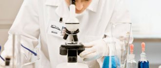
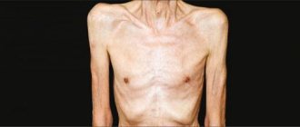
The human intestine is part of the digestive tract. A huge number of processes take place in it that directly affect the entire functioning of the human body. The main, but not the only function of the intestine is the digestion of food, the absorption of all useful elements, and the removal of processed foods from the body. Disturbances in the structure or functioning of this part of the digestive tract are immediately manifested by weight loss, decreased immunity, general malaise, and deterioration of the condition of the skin, nails, and hair. One of the endoscopic methods for studying the human body is sigmoidoscopy (rectoscopy), a visual diagnostic method that allows the doctor to directly examine the mucous walls of the rectum, and, if necessary, the cavity of the distal sigmoid colon.
Indications and Contraindications
Indications:
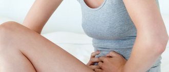
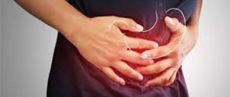
complaints of constipation, diarrhea; discharge from the rectum of mucus, pus, blood; pain in the lower abdomen and rectum; suspicion of the presence of an inflammatory process or neoplasm of the colon; assessment of the results of treatment of certain diseases of the rectum and sigmoid (sigmoid colon, T.) intestines; performing a number of manipulations and operations (taking scrapings and biopsies, removing polyps, foreign bodies, etc.). R., carried out for preventive purposes, is becoming increasingly widespread.
Contraindications:
pronounced acute inflammatory diseases of the rectal wall and surrounding tissues; low-lying stenotic tumors of the rectum, Ch. arr. anal canal; chemical and thermal burns in the acute stage, state of decompensation in cardiovascular pathology, psychosis.
What to take with you
To make the procedure comfortable, it is necessary not only to prepare the intestines, but also to collect the necessary things in advance. Pack your bag the day before and take with you:
- slippers and sheets, disposable;
- socks if you are afraid that your feet will freeze;
- all medical documents: insurance policy, test results, medical record, etc. This should be discussed with your doctor in advance;
- sometimes older people need to have an electrocardiogram done first;
- passport;
- wet wipes and toilet paper - just in case. Moreover, if the procedure is performed in a clinic and not in a private medical institution.
Disposable panties or shorts and shoe covers must be purchased in advance at the pharmacy.
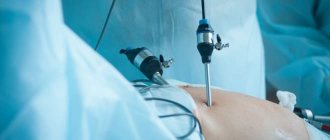
Equipment
Sigmoidoscopy is performed using special endoscopic devices - sigmoidoscopes (rectoscopes). A modern rectoscope includes a set of hollow tubes of various diameters and lengths, obturators for them and certain other instruments (for example, a telescopic magnifier or optical tube). Lighting is typically provided from a tabletop illuminator using a fiber-optic lighting cable or fiber light guide located inside the tube along its entire length or sometimes only in its proximal part. Rectoscopes with illumination from a miniature incandescent lamp mounted inside the tube at its distal end are also used. Among them, rectoscopes from the Leningrad Production Association (LPO) “Krasnogvardeets” (model 185, intended for adults, and model 170, for children) have become widespread.
Since 1973, LPO "Krasnogvardeets" has been producing rectoscopes with optical fibers, developed at the All-Union Research Institute of Medical Instrumentation, including two models of rectoscopes for adults - Re-VS-3 (large set) and Re-VS-3-1 ( small set) and two models of children's rectoscopes - Re-VS-5 (large set) and Re-VS-5-1 (small set). Rectoscopes are equipped with illuminators for equipment with optical fibers, for example, type OS-100.
Rice. 1. Rectoscope with fiber light guide Re-VS-3: 1 — tube; 2 — eyepiece; 3 — fiber light guide; 4 - light source.
Each tube contains a fiberglass light guide. The set of the Re-VS-3 device (Fig. 1) includes rectoscopic tubes 11, 20, 25 and 30 cm long, dia. 15, 20 mm, as well as proctoscope and anoscope. The Re-VS-5 rectoscope is equipped with tubes 15, 20, 25 and 20 cm long, dia. respectively 10, 15, 20 and 20 mm. All components and units of rectoscopes are unified. Each tube has an obturator. The kit includes a nozzle, a protective cover, a telescopic magnifier, a handle for holding tubes, collet-type cotton holders, a rubber balloon and a set of biopsy forceps - serrated and spoon-shaped. For photography through rectoscopes, a device is produced consisting of an optical tube for photocopying and a photo attachment for endoscopes.
Rice. 2. RMS-1 rectomicroscope: 1 - light source for inspection; 2 - light source for photography; 3 - tube; 4 — docking device for connecting a camera; 5 — camera; 6 - fiber light guide.
A type of rectoscope is a rectomicroscope (Fig. 2), which is used for intravital examination of the rectum and adjacent areas of the sigmoid colon using a contact method at high magnification (up to 220 X) for the purpose of diagnosing early forms of diseases.
For the study of the rectum and sigmoid colon, LPO Krasnogvardeets also produces flexible endoscopes - sigmoid colonoscopes (see Colonoscopy).
Additional recommendations
Below are some precautions that are recommended for every patient to observe when diagnosing with a proctoscope:
- It is not recommended to drive for several hours after the diagnosis.
- During pregnancy, a woman must inform the endoscopist, nurse and attending physician about her situation before the start of manipulations.
- For two hours after rectoscopy, it is better to refrain from consuming food and liquid.
- If there are any suspicious symptoms or painful sensations after the examination, it is imperative to report them to your doctor.
When prescribing a study called sigmoidoscopy, the patient needs to discuss in advance with the proctologist the method of preparation for the diagnostic procedure. The doctor will prescribe the most appropriate examination option for a particular person, taking into account the characteristics of the patient and his problem.
VIDEO “HOW TO PREPARE FOR RECTOSCOPY OF THE INTESTINE”
Preparing the patient
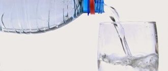
Preparing a patient for sigmoidoscopy depends on the nature of the disease, the goals and objectives of the examination. To identify tumors and chronic diseases when performing various manipulations and operations, careful cleaning of the colon from contents is necessary. For this purpose, before the study, patients are prescribed a low-residue diet (the day before breakfast and lunch without bread, for dinner - sweet tea). The study is carried out on an empty stomach. The evening before the examination, a cleansing enema is given (see). In the morning on the day of the study, the enema is repeated no later than 1.5-2 hours in advance so that the irritating effect of the enema on the mucous membrane of the colon passes.
In cases where long-term preparation for R. is impossible, the use of special microenemas is recommended - microlax, micro-cleist, aerosol microenema. In acute inflammatory diseases (dysentery, acute stage of ulcerative colitis), R. can be performed after the next bowel movement without prior preparation. Special preparation is not recommended in case of increased bleeding of the rectal mucosa.
Reviews
Sigmoidoscopy causes a lot of fear in patients, and even positive reviews do not particularly reassure them.
Valentina, 28 years old: I, like many, am more afraid of a proctologist than a gynecologist or dentist. I really don’t like preparing for the procedure - doing enemas - it’s so tiring. But I was lucky with the structure of my intestines; there were no bends, so the examination was painless, with only mild discomfort.
Victoria, 34 years old: I had read that this was a completely painless procedure and went through it quite calmly. But I wouldn’t describe my feelings as simple discomfort—I was in a lot of pain.
Kirill, 36 years old: The procedure, of course, is not the most pleasant, but without it it is impossible to make an accurate diagnosis. I walked for 10 days in depression while I waited for my “fateful” hour, and everything passed very quickly with moderate pain. More psychological discomfort than physical.
Sigmoidoscopy is a diagnostic that is accessible to everyone, moderately uncomfortable, and allows many dangerous pathological conditions to be identified at an early stage. For a high-quality examination in this way, a good psychological attitude and high-quality preparation are required.
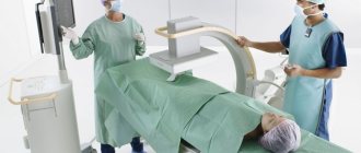
Research methodology
The most common positions of the patient during sigmoidoscopy are the knee-elbow position, and the knee-shoulder position is also used. In this case, the abdominal organs shift towards the diaphragm, the small pelvis is freed from the loops of the small intestine, and the physiological bends of the distal colon are straightened to the maximum. This creates favorable conditions for the advancement of a rigid metal tube and allows for better examination of the mucous membrane of the colon along the entire length of the apparatus tube. In cases where such a position is intolerable for the patient (severe weakness, shortness of breath, high blood pressure) or impossible (joint damage, absence of a limb), you can use the position on the left side with the pelvis raised and the thighs pressed to the stomach.
Rice. 3. Schematic representation of the stages of insertion of a rectoscope during sigmoidoscopy in the patient’s knee-elbow position: a - beginning of insertion; b - subsequent insertion with simultaneous lowering of the tube downwards according to the bend of the rectum; c — lifting the tube upward; d — installation of the tube parallel to the axis of the body after passage of the rectum; The arrows indicate the direction of movement of the proctoscope tube.
First, the doctor must carefully examine the anal area and perineal area, and then perform a digital examination of the rectum (see Rectal examination) to exclude situations where R. may be dangerous or impossible (low stenosing tumors, stricture, sharp fissures). Sigmoidoscopy is usually performed without anesthesia, although it is possible to lubricate the anal canal with anesthetic or xylocaine ointment. The rectoscope tube, lubricated with petroleum jelly and closed with an obturator, is inserted through the anal canal into the rectum (Fig. 3, a). After inserting the device to a depth of 4-5 cm, the obturator is removed and the tube is closed with an eyepiece or magnifying glass. Further use of the rectoscope must be carried out under visual control. The advancement of the instrument should be smooth, without brute force; the direction of movement of the apparatus is determined by the location of the intestinal lumen (Fig. 3, b, c, d). Inserting a proctoscope into the sigmoid colon requires great care. As a rule, the entrance is located asymmetrically, the intestinal walls are in a collapsed state, blocking the lumen, which is not always easy to determine behind the folds of the mucous membrane. You have to give the instrument different positions, moving apart and straightening these folds one by one.
In this case, a slight blowing of air is of great help. The movements of the instrument and the introduction of air should not cause pain to the person being examined. You should avoid resting the proctoscope tube against the intestinal wall, and especially pressing it against the sacrum and pubic symphysis. If slight movement of the distal end of the device and the introduction of air not only cause pain, but also do not allow the lumen to be determined, the study should be stopped. A similar situation may be due to fixed kinks of the sigmoid colon, in which rough manipulations can lead to perforation. After the proctoscope tube has been inserted to its full length, it is slowly withdrawn out. At this point, the mucous membrane is re-examined. Before removing the device from the rectum, it is necessary to remove excess introduced air. This is achieved by removing the eyepiece. In some cases (for example, with large tumors or rectal stenosis), the device can be inserted only 15-17 cm, i.e., the study is limited to rectoscopy.
Features of nutrition before rectoscopy
A few days before the procedure, you need to switch to a slag-free diet. Below are recommendations regarding nutrition during preparation for a diagnostic examination of the intestines:
- the day before the procedure, exclude cereals, baked goods, flour, legumes, fruits and vegetables from the diet;
- eliminate toxins as much as possible;
- remove from the diet foods that cause flatulence and excessive bloating;
- saturate the menu with stewed or steamed foods (lean meat, fish);
- add semolina and rice porridge, soft cheeses, low-fat meat broth to the menu.
The day before the procedure, you must refuse dinner; you can only drink weak tea without sugar. And the next morning it is permissible to eat a small amount of low-fat cottage cheese, but it is better to endure and limit yourself to one glass of tea.
Foods prohibited for several days before the procedure include fatty fish, potatoes, lamb, pork, duck, strong teas, spices, fermented milk products, pasta, coffee, chocolate, beer, kvass, carbonated drinks and any alcohol. It is better to boil or steam all meals in the days before the examination.
Normal endoscopic appearance
In the rectum (see) there is a number of transverse folds, the lower of which (coccygeal) is located at a distance of 5-7 cm from the outer edge of the anal canal. Above it, 2-3 cm, the lower sacral fold is visible, intersecting with the coccygeal fold at an acute angle. Another 2-3 cm higher is the upper sacral fold, usually smaller. At a distance of 13-14 cm from the anus (see), there is a terminal fold that defines the transition to the distal part of the sigmoid colon (see). The mucous membrane of the rectum is moist, shiny, pink (see color table to the article Rectum, Fig. 4), submucosal vessels are sometimes visible in the lower ampullary section, and there are no circular folds. In the sigmoid colon, the mucous membrane forms many semilunar folds, which are easily straightened by the movement of the apparatus or the introduction of air. The lumen of the sigmoid colon is much narrower than the lumen of the rectum, the mucous membrane is more juicy and dark pink in color.
Endoscopic picture of the main types of pathology. Nonspecific ulcerative colitis - changes in the mucous membrane from slight swelling with hyperemia and lack of vascular pattern to the formation of ulcers covered with fibrinous-purulent plaque.
Crohn's disease of the colon is characterized by rigidity of the rectal wall and the formation of multiple longitudinal cracks in the mucous membrane, which usually have fibrinous-purulent deposits. Multiple cracks and swelling of the mucous membrane create a characteristic endoscopic picture, often compared to a “cobblestone street.”
Colon polyps are tumor-like formations with a smooth or villous surface on a narrow base - a pedicle (see color table for the article Rectum, Fig. 7) or a wide base. These formations can be single or multiple.
Colon cancer. Based on the endoscopic picture, two main forms of cancer are distinguished: exophytic and endophytic. The exophytic form is a formation in the form of a node, polyp or villous tumor with growths along the periphery, reminiscent of cauliflower (see color table to the article Rectum, Fig. 6). Endophytic forms have the appearance of a crater-shaped ulcer with infiltration of the entire thickness of the intestinal wall. The bottom of the ulcer is usually covered with fibrin or gray necrotic masses.
See also color. table to Art. Proctitis.
Approximate diet
3 days before the procedure:
- Breakfast: 200 ml tea, oatmeal, 1 piece of toast.
- Second breakfast: 250 ml low-fat fermented baked milk.
- Lunch: chicken broth, 2 steamed meatballs, cucumber salad.
- Afternoon snack: cottage cheese casserole and compote.
- Dinner: biscuits and 250 ml of low-fat kefir.
In 2 days:
- Breakfast: hibiscus, buckwheat, 1 slice of white bread.
- Second breakfast: 250 ml of dairy products.
- Lunch: steamed sea fish, rice porridge and juice.
- Afternoon snack: low-fat yogurt.
- Dinner: tea and semolina porridge.
For 1 day:
- Breakfast: 1 soft-boiled egg, toast and fruit compote.
- Lunch: green tea, broth and low-calorie cookies.
Due to the fact that therapeutic nutrition is designed solely for cleansing the body, it is recommended to reduce physical activity. The diet will help stabilize intestinal activity and reduce the formation of gases.
Complications
If the indications and technique are followed, sigmoidoscopy is a relatively safe examination method. Rough manipulations and excessive distention of the intestine can lead to perforation of its wall. With certain diseases, such as hemangiomas, large villous tumors, bleeding is possible (see Gastrointestinal bleeding). This complication can also occur when performing a biopsy or removing polyps. Bleeding is stopped using electrocoagulation of the bleeding area. In case of perforation, an emergency operation is necessary, the nature of the cut depends on the location of the perforation hole and the period that has passed since the perforation.
Consequences
The most dangerous complication after the procedure is perforation of the intestinal wall. If the procedure is carried out carelessly, the instrument can damage the intestinal walls, and this will cause its contents to enter the abdominal cavity. But as a rule, this happens extremely rarely, if you choose wisely the specialist who will carry out the procedure.
If perforation cannot be avoided, the patient is urgently hospitalized and undergoes surgery. More often, after sigmoidoscopy, patients have the following complaints:
- increased gas formation;
- abdominal cramps;
- slight nausea.
If the patient begins to develop a fever or finds streaks of blood in the stool, then this must be immediately reported to the proctologist who performs sigmoidoscopy.
Sigmoidoscopy in children
Sigmoidoscopy in children is usually used as an addition to digital rectal examination. Indications for it: bleeding through the anus, constipation, unstable stool, suspicion of polyps, ulcers, tumors, damage to the mucous membrane, portal hypertension. R. is performed to identify varicose veins in places of anastomosis of the upper rectal (hemorrhoidal), middle and lower rectal veins, if necessary, to monitor the dynamics of the pathological process, the progress of restoration of the colon mucosa in nonspecific ulcerative colitis, acute and chronic dysentery. Contraindications are the same as for adults.
Rice. 4. Position of the child and the doctor during sigmoidoscopy. The child's knees are pulled up to the stomach (the direction of pull-up is indicated by the arrow).
The night before, the child is given a cleansing enema and is not given dinner. In the morning, 1.5 - 2 hours before the study, an enema is given again, and 20-30 minutes before the test. a gas tube is inserted. The success of the study largely depends on the child’s calm behavior during the procedure. The purpose and necessity of the study are explained to the child in advance, and they are warned about possible subjective sensations. During the research process, the child's attention is distracted by conversation or a toy. Usually R. is performed without anesthesia and only in small and restless children, and also in cases of surgical manipulation there is a need for short-term anesthesia. In young children, it is preferable to conduct the study in the supine position (Fig. 4), in older children (10-14 years) - in the knee-elbow position or on the left side.
Rice. 5. Schematic representation of the stages of insertion of a rectoscope during sigmoidoscopy in a child lying on his back: a - beginning of insertion; b - subsequent insertion with simultaneous lifting of the tube upward according to the bend of the rectum; c — lowering the tube; d — installation of the tube parallel to the axis of the body after passage of the rectum; The arrows indicate the direction of movement of the proctoscope tube.
The introduction of a rectoscopic tube and examination of the mucous membrane is carried out in stages (Fig. 5). The depth at which it is possible to insert the rectoscope tube depends on the age of the child and the position of the pelvic segment of the sigmoid colon. On average, 15-20 cm of the intestine is available for inspection, but the use of devices with fiber optics makes it possible to significantly expand these boundaries up to the examination of the entire colon (see Colonoscopy).
See also Endoscopy.
Bibliography:
Aminev A. M. Guide to proctology, vol. 1, p. 141, Kuibyshev, 1965; Lenyushkin A.I. Proctology of childhood, p. 53, M., 1976; Lukomsky G.I. and Berezov Yu.E. Endoscopic techniques in surgery, M., 1967; Fedorov S.P. Rectoscopy, Surgery, vol. 1, no. 6, p. 516, 1897; Tsepelev Yu. A. and Gorokhov L. I. Endoscopic devices for gastroenterology, p. 47, M., 1976; Chulkov P. S. Sigmoidoscopy, Leningrad, 1952; Endoscopy for diseases of the rectum and colon, Atlas, ed. V. D. Fedorova, M., 1978; Yanchev V. G. Sigmoidoscopy, Sofia, 1959 (in Bulgarian); Bensaude R. Rectoscopie, Sigmoidoscopie, P., 1956; Eisenberg SW Proctosigmoidoscopy, J. int. Coll. Surg., y. 36, p. 243, 1961; Freeman G.C. Twenty-five hundred proctoscopic examinations, Amer. Surg., v. 26, p. 431, 1960.
G. I. Vorobiev; A. I. Lenyushkin (det. surgeon), Yu. A. Tsepelev (techn.).
Which doctor should I contact, what is the cost of the procedure?
For any complaints related to the rectal area, you need to visit a proctologist. Or if there are stool problems or bloating, you can contact a gastroenterologist, and if necessary, he will refer you to a proctologist. The examination is carried out by an endoscopist specializing in intestines. If such a specialist is not available in the hospital, the procedure is performed by a proctologist or surgeon.
The test can be done in any public hospital, as well as in special diagnostic centers, of which there are about 60 in Moscow. The cost of the procedure varies from 1,200 to 6,000 rubles, it depends on the technology used. The highest price is computerized video sigmoidoscopy. The examination can be done free of charge for persons with a compulsory medical insurance policy; to do this, they need to have a doctor’s referral and sign up for a waiting list.
Reviews from patients who have undergone sigmoidoscopy indicate that the procedure is very quick, painless, and easily tolerated.
Photo gallery
The photo shows laxatives that are used to cleanse the large intestine.
Lavacol, 197 rub.
Duphalac, from 300 rub.
Fleet phospho-soda, 659 rub.
Forlax, from 150 rub.
Fortrans, 549 rub.
Microlax, from 357 rub.
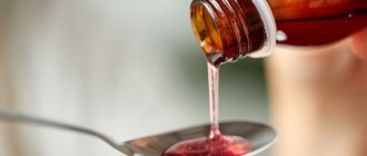
Use of laxatives
The advantages of this cleaning method include ease of use and no risk of injury to the walls of the rectum. A modern laxative allows you to cleanse the intestines delicately and thoroughly without disturbing its microflora. You can use one of the laxatives, after consulting with your doctor in advance and strictly following the recommended dosage regimen.
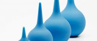
Microlax
The drug, produced in the USA, is produced in the form of microenemas that have a mild effect on the intestinal mucosa. Contraindications include only intolerance to the components of the laxative.
To administer the drug you need:
- Clean the area of the body near the anus.
- Break off the seal covering the tip of the microenema.
- Squeeze the container slightly until drops of the contents appear and lubricate the tip with the preparation.
- Squat down and insert the tip into the anus, holding the enema vertically.
- Press firmly on the container.
- Without unclenching your fingers, remove the enema.
Glycerol, which is part of the drug, irritates the intestinal mucosa and improves its peristalsis, due to which the urge to defecate appears 5-20 minutes after administration of the drug. When preparing for rectoscopy, it is recommended to use two enemas in the evening with an interval of 10 minutes. In the morning, 2-6 hours before the start of the examination, the procedure is repeated.
Fortrans
When used correctly, it is an absolutely safe product that will cleanse the intestines without disturbing its microflora, without causing painful spasms and without changing the natural rhythm of physiological processes occurring in the body. Among the disadvantages is the possibility of an allergic reaction in patients sensitive to the components of the drug.
Another disadvantage is the need for large water consumption, since the action of the product is based on the dilution of feces and slag deposits located in the folds of the intestines, with their subsequent removal. For this reason, the drug is not prescribed to people with kidney disease, heart disease, or intestinal obstruction.
The laxative is a powder packaged in sachets. The intake rate is calculated at the rate of one sachet for every 20 kg of weight. The contents of the sachet are diluted in a liter of water and drunk gradually, in small sips, over an hour.
A few drops of lemon juice will help you overcome attacks of nausea caused by the unpleasant taste of the product. It is permissible to use antiemetic drugs: Domperidone or Motilak. Do not reduce the volume of liquid, as this will lead to a decrease in the effectiveness of the drug!
Depending on the time of rectoscopy, Fortrans is used:
- If the examination is in the morning, then the day before, at 14-15 hours, take 2 sachets, take an hour break and take the rest.
- If it is afternoon, taking the first 2 sachets is postponed until 18:00, and the remaining portions are taken from 7:00 am.
Duphalac
Can be used by patients of any age, including children. The action of Duphalac is based on the ability of the components of the product to soften stool and stimulate contractions of the intestinal walls. The medicine is available in the form of a suspension, packaged in portioned vials.
In preparation for rectoscopy, scheduled for the morning, it is recommended to start taking the drug on the day preceding the procedure, 2 hours after a light lunch (at 14-15 hours). To do this, one bottle should be dissolved in 2 liters of clean water and drunk in small portions for 2-3 hours.
You can wash the product down with a few sips of tea or juice without pulp.
Complete bowel movement should be expected in the evening, approximately 3 hours after taking the last portion of the laxative. If RRS is scheduled for the afternoon, Duphalac should be taken in the evening, take a break 2 hours before bedtime and continue cleansing from 6 am.
Fleet phospho-soda
The drug can reduce the absorption of fluid, due to which it accumulates in the intestines, softening its contents. Thanks to the action of Fleet phosphosoda, feces and waste accumulated in the folds are removed quickly, without causing painful spasms during bowel movements.
The drug is contraindicated for patients with kidney pathologies, heart failure, or narrowing of the intestines, as well as for those who are forced to limit salt intake. Sigmoidoscopy of the intestines, the preparation of which is associated with the use of Flit phosphosoda, will also be interconnected in terms of the timing of the drug dosage regimen.
For example:
- During the morning examination: on the day preceding it, after breakfast, take 1 bottle, the contents of which must be diluted in 120 grams of water and washed down with another glass of water. The second dose is taken in the evening, after drinking a glass of liquid.
- During the daytime examination, the first dose is transferred to lunchtime, and the second is taken in the morning, on the day of the procedure. Doses are the same as in the first case.
Lavacol
The drug acts similarly to those described above, having similar contraindications. Sold in a package containing 15 sachets, each of which must be diluted in a glass of water. To mask the unpleasant taste, you can add syrup or jam.
Depending on the time of rectoscopy:
- Morning examination – start of taking the drug – 16:00 the previous day. The time interval between servings is 20 minutes. It is necessary to drink the entire package, alternating taking Levkol with weak tea and weak broth.
- Examination in the afternoon - the first sachet is taken at 18:00, then, with a break of 20 minutes, another 10 sachets are drunk. The remaining 3 or 4 are consumed in the morning (from 6 o’clock) on an empty stomach according to the same scheme.