What is ultrasound examination of the peritoneum?
Abdominal ultrasound is a highly informative ultrasound diagnostic technique. It is painless, based on the ability of waves to penetrate tissue and be reflected from them. Radiation passes freely through internal organs that are filled with air. Ultrasonic waves are reflected from all dense structures.
This signal is recorded by the sensor and the data, after processing in the computer, is transmitted to the monitor. The image shows organs, their shape, density and deviations from the norm. At the same time, the retroperitoneal space, pelvic area or kidneys can be examined.
Contraindications
There are no restrictions on how often an ultrasound of internal organs can be performed on an adult or child. Ultrasound is not dangerous to the body; the examination is equally safe for newborns, pregnant women, and the elderly.
Relative contraindications are:
- alcohol or drug intoxication of a person;
- psychosis;
- wounds or inflammation of the skin at the examination site.
If emergency diagnosis is necessary, even these contraindications are canceled.
How does ultrasound work?
Abdominal ultrasound uses the ability of ultrasound waves to illuminate tissue. They have a high frequency, penetrate the skin and are fully or partially reflected from tissues. The information is transmitted to the machine, which is both a highly sensitive sensor and receives data.
They are transmitted to a computer on which a special program is installed to create a 2 or 3 dimensional image. For a better picture, the air gap is removed between the skin and the highly sensitive device using gel. For scanning, devices operating at 1.8-7.6 MHz are most often used.
CT scan of the abdomen with contrast
In order to more accurately determine the pathological focus, its location, size and extent of spread, specialists resort to the use of computed tomography of the abdominal cavity with contrast. A special substance is introduced into the body orally or intravenously. Contrast is applied to the area of interest and a series of images are taken. The resulting volumetric image reflects an increased accumulation of contrast agent or, conversely, a lack of it. In this way, high accuracy of diagnostic testing is achieved. The question of the need for a statement is decided by the attending physician.
What organs are checked?
During ultrasound diagnostics, all vessels and aortas that are located in the peritoneum are checked. The kidneys, sections of the ureters, and cellular spaces are examined. Internal organs partially or completely hidden in the peritoneum (and in the space behind and in front of it) are checked:
- The spleen is checked for enlargement, ruptures and internal bleeding. The area of the organ, its thickness, length and width are measured.
- The gallbladder's contractility, bile density, and the absence or presence of stones are determined.
- The dimensions and volumes of the pancreas are measured (its body, tail, and head are examined separately), and the pancreatic duct is checked.
- When examining the bladder, the presence or absence of stones is noted, whether there are wall tumors, reflux, or fistulas.
- In the liver, bile ducts, parenchymal structures, veins and large vessels are checked. The dimensions of the organ, its fragments and lobes, and edges are measured.
- In the kidneys, the structure of the parenchyma is scanned, the size and volume of the organ, the condition of the cups and pelvis are measured. Its contractility is recorded.
- Stomach.
During the abdominal ultrasound procedure, a partial examination of the intestines is possible. However, due to the fact that it contains semi-liquid contents and air, it is impossible to completely check the organ. However, ultrasound can detect some dense intestinal fragments (foreign bodies, tumors).
Structure
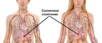
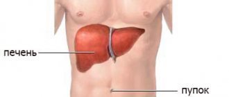
The human abdominal cavity (anatomy in the pictures is presented below) is the largest container of organs. It is surrounded by soft tissue on all sides. Bone protection is represented by the spine, ilium and chest bones.
The abdominal cavity contains not only the gastrointestinal tract organs, but also organs related to the genitourinary system. All organs are covered by peritoneum differently. Accordingly, they are divided into those directly related to the abdominal cavity, and those located in the retroperitoneal space.
Although the abdominal cavity appears as one unit, it is anatomically divided into two parts:
- The first section is that part of the abdomen that is lined with peritoneum inside and occupied by organs:
- stomach,
- large intestine and small intestine;
- spleen;
- liver;
- pancreas.
- The other part of the abdominal cavity is the retroperitoneum, which is located behind. Here pass the inferior vena cava with its tributaries and nerves, the aorta and its branches. Near the posterior wall of the peritoneum you can see:
- kidneys;
- ureters;
- adrenal glands
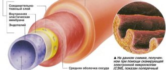
All organs are fixed and function among themselves. The abdominal aorta is the main vessel supplying the abdominal cavity. Its large unpaired branches supply blood to the cavity and paired arteries of the kidneys, adrenal glands, diaphragm, ovaries and lumbar region.
Structure of the human abdominal cavity
The vessels of the genitourinary system, on the other hand, are supplied by the inferior vena cava. Together with the splenic vein, the azygos veins of the digestive tract form the portal vein, which extends to the liver.
What does the study show?
What is included in an abdominal ultrasound? All organs located in this area are examined. This method helps identify tumors, but ultrasound cannot determine whether they are malignant or benign. Ultrasound also shows:
- fibrosis;
- cysts;
- appendicitis;
- changes in the kidney parenchyma;
- echinococcal parasitic cysts;
- glomerulonephritis;
- amyloidosis;
- jaundice;
- subcapsular rupture of the spleen;
- splenomegaly;
- Hydnephrosis;
- pyeelectasis;
- gastritis;
- stones in the kidneys;
- fatty hepatosis;
- cirrhosis of the liver;
- pyelonephritis;
- adrenal gland diseases;
- non- and calculous cholecystitis;
- renal agnosis;
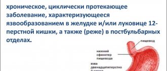
- stomach ulcer;
- calicopyelectasia;
- alcoholic, infectious and toxic hepatitis;
- organ ruptures.
Ultrasound of the abdominal cavity reveals swelling and changes in organ volumes. The presence or absence of neoplasms behind the peritoneum is determined. Diagnostics shows some signs of ureteral reflux, vesicouterine or rectal fistulas.
Indications for ultrasound
The reasons for performing an ultrasound scan of the pancreas are deviations from normal values in a blood test and girdle pain. A bladder scan is done when urination is painful and difficult, and sediment and salts appear in the urine. The examination is carried out when:
- painful constant sensations in the lumbar region;
- hypertension;
- painful urination;
- chronic form of pancreatitis;
- damage to abdominal organs;
- all types of hepatitis;
- suspected Addisson's disease;
- the appearance of cysts in the peritoneum;
- swelling;
- an increase in the size of the spleen;
- jaundice;
- suspected Itsenko-Cushing's disease;
- abdominal injury;
- after car accidents;
An abdominal ultrasound may be done if signs appear:
- throbbing and abdominal pain;
- excessive gas formation;
- belly growth;
- stable elevated temperature that is not associated with colds;
- heaviness and discomfort under the ribs on the right;
- dyspeptic syndrome, accompanied by nausea, vomiting, heartburn, loss of appetite;
- hypersalivation;
- distension of the stomach, nagging pain after meals;
- bitterness in the mouth.
An abdominal ultrasound is prescribed to assess damage to the genitourinary system after inflammation and changes during pregnancy. Ultrasound scanning is performed when tumors are suspected or to monitor already detected diseases. As a preventative measure, it is advisable to do an ultrasound of the abdominal organs twice a year.
Further actions
After diagnosis, the results are given to the patient or local physician. The doctor writes out a conclusion, which, however, is not a thorough diagnosis. Depending on the changes detected on ultrasound in certain organs, the doctor prescribes further treatment or, if necessary, additional diagnostics.
Although ultrasound is an accurate and highly informative diagnostic method, the doctor may still prescribe additional diagnostics. He refers the patient to the following additional types of diagnostics:
- FGDS.
- Biopsy of the gastric mucosa.
- Colonoscopy or sigmoidoscopy.
- X-ray examination, including the use of a radiopaque contrast agent.
- Capsule endoscopy.
- CT scan.
- Magnetic resonance imaging.
Depending on the final diagnosis, the necessary treatment is prescribed.
Preparing for an ultrasound
If a person is taking medication, this should be reported to the doctor before the procedure. Before it, the intestines are cleansed. Four days before the ultrasound, they begin to drink medications and herbs that reduce gas formation and improve food digestion.
During the preparatory period, you can drink Espumisan, activated carbon, or other drugs prescribed by your doctor. Take 2 tablets three times a day, before meals. For persistent constipation, an enema is performed two days before the procedure. It can be recommended by your doctor on the day of the ultrasound. You cannot undergo a colonoscopy before the examination.
For three days before the ultrasound, you should adhere to a strict diet. You need to eat 5-6 times a day, but in small portions. The diet should include barley and oatmeal, buckwheat. Before the examination, all foods that cause flatulence are excluded. Accumulated gases can distort the results of the study. Excluded from the menu:
- peas;
- dairy products;
- fresh juices;
- black bread;
- chocolate and other sweets;
- White cabbage;
- milk and yoghurts;
- fried food;
- fat meat;
- raw fruits and vegetables;
- excessively salty foods;
- flour and confectionery masterpieces;
- spicy food;
- alcohol;
- soda.
Avoid smoking; chewing gum and hard candies can cause stomach cramps. Food should be steamed or boiled. Adding salt to dishes is kept to a minimum. Allowed:
- lean fish;
- oatmeal, buckwheat, millet, boiled in water;
- low-fat soups (for example, chicken or vegetable);
- hard cheeses;
- chicken meat;
- unsweetened tea;
- beef;
- hard boiled eggs.
The last time you can eat before the ultrasound is twenty hours. On the day of the procedure, eating is allowed only after the examination. You can drink during the day, but stop drinking any liquids a couple of hours before the ultrasound.
If the kidneys and bladder are examined, then the patient, on the contrary, should drink a lot. Immediately before scanning, drink 1.5-2 liters of water. You need to drink very slowly so that as little air as possible gets into the stomach with the liquid. It can provoke distortion of the results.
Prevention
Certain steps will help avoid many diseases and complications:
- Reduce alcohol consumption, quit smoking.
- Maintaining a healthy weight
- Restriction on the amount of salt in the diet, consumption of harmful foods.
- Switching to a healthy eating regimen.
- Protecting the body from pathogenic bacteria.
- Keeping your body in good physical shape and developing a habit of physical exercise.
Carrying out ultrasound diagnostics
An abdominal ultrasound does not take long. The patient lies down on the couch in the position required by the doctor. If it is the liver, then the patient is placed on his left side or stomach up. A conductive gel is then applied to the skin to improve ultrasound transmission.
The study is carried out with a small sensor. It moves around the body in the desired area. An image appears on the computer monitor. According to it, the doctor evaluates:
- general condition, structure, size of the internal organs of the abdominal cavity;
- presence or absence of neoplasms;
- location of organs.
The examination lasts approximately 15-20 minutes. The duration of the scan depends on the quality of preparation and the individual characteristics of the body. If necessary, attention is focused on suspicious points that require more careful study.
Interpretation of ultrasound results of OBP
For each organ or system there are certain standards - size, density, etc. Their deviations may indicate specific diseases.
Liver
Increased echo density of the liver (presence of small lesions) indicates fatty hepatosis. The edges of the organ are rounded. Due to its compaction in the final stages of the disease, the portal vessels become completely invisible.
Cirrhosis of the liver is determined by enlargement of the organ and dilation of the veins. In this case, uneven contours are visible, the lower edge is rounded. A large focal increase in density is noted. Fluid accumulates in the abdomen.
Enlargement of the organ, rounding of its edges and dilation of veins may indicate pulmonary diseases or heart pathologies. If the echo structure is disturbed, this may be a sign of neoplasms, cysts, abscesses.
Gallbladder
Thickening of the gallbladder wall indicates acute cholecystitis. The size of the organ is any. The wall may have a “double” contour. Peritonitis is indicated by fluid spilling around the bladder.
The walls of the organ also appear thickened in the chronic form of cholecystitis. At the same time, the contours maintain density and clarity. Acoustic shadow, thickening of the gallbladder wall, uneven contours - everything speaks of calculous cholecystitis. An increase in the width of the ducts indicates the presence of stones.
Pancreas
A decrease in the echo density of the pancreas is observed in acute pancreatitis. Its chronic form or oncology is indicated by an increase and expansion of the duct. Additional signs include “ragged” edges of the contours, pits on the surface of the organ, compression of the aorta or displacement of the vena cava.
Spleen
Diseases of the blood and liver are reflected in the spleen in the form of its increase in size. Tissue compactions indicate the death of some area. The image clearly shows organ ruptures due to trauma.
Lymphatic structures
If the lymphatic structures are normal, they are not visible during an ultrasound. Enlarged nodes indicate infection, malignant tumors, and metastases.
After the abdominal ultrasound procedure, a conclusion is made that describes in detail what diseases the echo signs resemble. If a specific organ is examined and no deviations from the norm were observed, it is written that the echo symptom did not reveal any pathology.
Ultrasound is an effective and accurate method for determining changes in internal organs and systems. The procedure helps determine the development of oncology and other serious diseases. The method is painless and has virtually no contraindications.
- Ultrasound of the adrenal glands
- Normal kidney sizes according to ultrasound in adults and children
- Ultrasound of the pancreas
- Ultrasound of the liver. Preparation for the procedure
- Ultrasound of the uterus and appendages. How does it go and what will it show?