Placenta: concept and its importance
The organ that arises during pregnancy in the uterus and connects the mother and the fetus is called the placenta (baby place). Its significance is very great. The organ is responsible for the biological processes due to which the baby develops normally. The life of a child depends on the placenta. Deviations and pathologies associated with it can lead to his death.
The following functions of the placenta can be distinguished:
- Gas exchange . The baby in the womb needs oxygen: it enters the fetal blood from the maternal blood through the placenta. It also transfers carbon dioxide from the baby to the mother. A small placental abruption can disrupt gas exchange.
- Trophic and excretory . For the normal development of a baby, a whole range of nutrients is required. He receives all of them through the placenta. Through it, waste products are removed.
- Hormonal . The placenta produces important hormones (chorionic gonadotropin, placental lactogen, prolactin progesterone, etc.), without which a normal pregnancy is impossible.
- Protective . The placenta provides the fetus with immunological protection. Mother's antibodies passing through the baby's place protect the baby from various diseases.
- Barrier . Between the blood of the fetus and the blood of the mother there is a hematoplacental or fetoplacental barrier with selective permeability. The blood-placental barrier protects the baby's body from contact with potentially harmful metabolites and chemical compounds that may be in the mother's blood.
Bottom line
Premature placental abruption causes oxygen starvation in the baby, as it plays the role of lungs during pregnancy. Hypoxia (oxygen deficiency) can result in irreversible processes and cause the death of an unborn child. Therefore, at the first signs of placental rejection (bleeding, pain in the groin area, poor health), you need to urgently seek help. Timely obstetric care can save the baby and prevent complications in the mother.
Placental abruption: what is it, what does it look like and what happens?
Placental abruption is its separation (partial or complete) from the lining of the uterus. In this case, blood accumulates between the baby's place and the wall of the uterus, which pushes the placenta away.
The placenta should not be expelled during pregnancy. Its separation from the uterus should occur in the third labor period. However, there are cases when the placenta leaves prematurely.
What are the risks of placental abruption during childbirth? This is extremely dangerous for the baby, as it can deprive him of oxygen and nutrients.
The importance of the placenta for the child
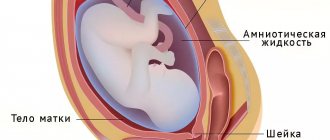
The placenta (baby place) is a special organ that is formed during pregnancy and provides a connection between the child and the mother. The life, development and nutrition of the fetus depend on its work.
Nutrients, oxygen, and maternal antibodies are supplied to the baby through the placenta.
The formation of the placenta begins at 2 weeks of pregnancy. Its growth and development continue until the 12th week, after which it begins to fully function.
The placenta is attached to the anterior, posterior wall or fundus of the uterus. On one side it is attached to the wall of the uterus, on the other it is connected to the fetus through the umbilical cord.
Causes of premature abruption of a normally located placenta
Primipara women experience premature expulsion of the placenta in 0.4–1.4% of cases. After 3 births, the risk of premature detachment increases to 15–20%. Detachment can occur both during pregnancy and during childbirth - in the first or second period. Why does placental abruption occur? The reasons for this process are various.
The separation of a child's seat can be caused by disorders in the vascular system . The capillaries of the uterus and placenta may become more fragile and brittle. Because of this, blood patency may be impaired. Similar changes in the female body can occur during gestosis.
They are also observed in the presence of certain diseases: cardiovascular pathology, hypertension, kidney disease, obesity, diabetes, etc.
The threat of placental abruption can come from inflammatory, degenerative and other pathological processes occurring in the baby's place and uterus. Disturbances are observed with uterine fibroids, malformations, and postmaturity.
Other provoking factors include polyhydramnios, multiple pregnancies, a short umbilical cord, increased density of the walls of the amniotic sac, abortion and a history of spontaneous termination of pregnancy.
Bad habits predispose to premature expulsion of the placenta : drinking alcohol, addiction to cigarettes, drugs. The situation may worsen with anemia (anemia, decreased number of red blood cells, low hemoglobin).
Most often, symptoms of placental abruption in early or later stages of pregnancy are observed in women for whom the upcoming birth is not the first . The reason for this lies in changes in the uterine mucosa.
There are rare cases of placental abruption due to autoimmune conditions , in which the female body produces antibodies to its own cells. This can be observed with a disease such as lupus erythematosus.
Allergy to drug therapy is another cause of placental abruption in late or early stages. Typically, pregnant women experience an allergic reaction when transfusion of donor blood and its components, or administration of protein solutions.
Complications can result from abdominal trauma caused by a fall, blow or accident. Placental abruption can also be facilitated by sudden changes in blood pressure that occur during stress and other neuropsychic influences.
Causes
Experts cannot say for sure why this condition occurs.
In their opinion, placental abruption occurs due to the presence of one or more risk factors in a pregnant woman:
- complicated obstetric and gynecological history;
- high blood pressure;
- bad habits (smoking, taking drugs);
- the presence of endocrine diseases;
- autoimmune pathologies;
- blood diseases accompanied by the formation of blood clots;
- previous intrauterine infection;
- injuries (falls on the stomach).
According to research, the occurrence of placental abruption is possible after IVF even in women who do not have the risk factors described above.
Who's at risk
Symptoms of placental abruption
In early and late pregnancy, symptoms of placental abruption may be as follows:
- bleeding;
- uterine tension and pain during placental abruption;
- cardiac dysfunction in a baby.
Bleeding can be external (visible), internal (hidden) or mixed. External bleeding is easy to notice, since scarlet or brown discharge . It is observed when the edges of the placenta are detached.
If the baby's place is disconnected from the uterus in the center, and the edges remain attached to its wall, then the bleeding will be internal. Blood will accumulate between the uterus and placenta.
When a child's place is detached, uterine tension . When it is palpated, pain . It can be dull and paroxysmal. Sometimes the pain radiates to the hip and pubic area, as well as to the lumbar region. It is felt most strongly with internal bleeding.
In the fetus with premature abruption, cardiac dysfunction . When listening to the fetal heartbeat, a sharp increase in heart rate (tachycardia) is observed, followed by depression of cardiac activity and bradycardia.
Signs begin to appear when 1/4 of the placenta is separated. If 1/3 of it leaves, the child begins to experience severe oxygen deficiency. His death occurs when 1/3–1/2 of the placenta is detached.
How is it diagnosed?
The characteristic signs of placental abruption usually leave no doubt about the diagnosis.
If the clinical symptoms are mild (no bleeding or pain), a vaginal examination and ultrasound are performed to exclude other pathologies.
To determine the uterine nature of the bleeding, the doctor examines the vagina and cervix.
This allows you to assess whether the bleeding is caused by infectious changes, polyps, or dilatation of the cervix.
The ultrasound procedure allows you to unambiguously determine the presence, size and location of the detachment.
A small abruption may not be detected by ultrasound, but in this case blood clots are visualized behind the placenta.
This allows us to exclude another common cause of bleeding - placenta previa.
Placental abruption at different stages of pregnancy
The separation of the baby's place from the uterus manifests itself differently depending on the stage of pregnancy. Doctors often encounter premature placental abruption in the first trimester .
With timely diagnosis and proper treatment, the consequences can be avoided - pregnancy continues, there is no discharge.
Placental abruption in the second trimester is characterized by such signs as high muscle tone and tension. Treatment tactics directly depend on the stage of pregnancy.
No less dangerous is the separation of a child's place in the 3rd trimester . Signs of placental abruption in the later stages are typical: abdominal pain, tension and soreness of the uterus, bleeding, fetal suffering.
The only way out is delivery. Natural birth is possible only if there is partial detachment of the placenta, it does not progress and the birth canal is completely ready. In all other cases, a caesarean section is performed.
Premature placental abruption during childbirth is a fairly common occurrence. Ideally this should happen in the third stage. However, it also happens that detachment occurs at the first or second stage. In such a situation, a caesarean section is started.
Consequences
Detachment affects not only the fetus, but also the pregnant woman. The severity of the consequences directly depends on the degree of detachment, as well as the time of initiation of treatment measures.[5]
For mother
The main complications for a pregnant woman are:
- Kuveler's uterus (a condition accompanied by saturation of the uterus with blood due to massive hemorrhages);
- DIC syndrome (disruption of the coagulation system, against which a large number of blood clots form);
- hemorrhagic shock.
If left untreated, death may occur due to the development of shock.
There are consequences for mother and child
Bleeding
Abundant effusion, in addition to general weakness, leads to progressive anemia and the development of dangerous DIC syndrome.
Anemic condition
Excessive uterine hemorrhages provoke anemic conditions. The woman experiences weakness, often loses consciousness and faints, and her blood pressure decreases.
For a child
The most common complication in the fetus is hypoxia. In this condition, there is oxygen starvation of the fetus against the background of impaired transplacental circulation. In severe cases, fetal death occurs.
Intrauterine fetal hypoxia
If the uteroplacental blood flow is disrupted, the fetus suffers from acute oxygen starvation. The result is intrauterine growth retardation, congenital defects and diseases. In the absence of timely treatment, the embryo may even die from attacks of suffocation.
Diagnosis of premature placental abruption
If there are pronounced symptoms, it is not difficult to find out that the placenta is detached. If the symptoms are not fully manifested, for example, there is no pain factor, external bleeding is not observed, then the diagnosis is made, excluding the presence of other diseases that can cause similar symptoms.
Ultrasound assists in diagnosing placental abruption. Thanks to it, it is possible to determine the area of the placenta that has moved away from the uterine wall and the size of the retroplacental hematoma.
During the examination, one of three possible diagnoses of the separation of a child’s place can be made:
- non-progressive partial;
- progressive partial;
- total.
The placenta may partially detach from the wall of the uterus in a small area. In such situations, damaged blood vessels often become clogged. The bleeding stops and further detachment does not occur. Pregnancy can proceed without complications, and the child will be born healthy.
However, a non-progressive detachment can begin to progress at any time. Any detachment requires timely and proper treatment.
Progressive partial placental abruption poses a danger to the fetus. The size of the hematoma increases. If most of the placenta leaves the uterine wall, the fetus will die.
In such a situation, the woman carrying the baby also suffers, because she loses a large amount of blood. Blood loss can lead to hemorrhagic shock. This situation can be dealt with through a caesarean section.
Total (complete) detachment of the child's place
is extremely rare The fetus dies almost immediately, as gas exchange between it and the mother stops.
Types of detachment
Placental insufficiency occurs.
- Primary (early). The pathology occurs up to 16 obstetric weeks and occurs against the background of endometriosis, fibroids and other uterine defects. In the absence of timely treatment, placental insufficiency takes on a secondary form.
- Secondary (late). Develops after 16 obstetric weeks. This pathology is preceded by chronic diseases of the female body, infectious and inflammatory processes, and late toxicosis.
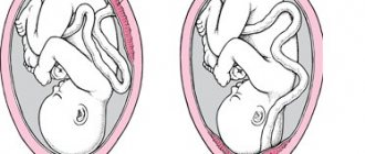
Based on its location, the “children’s place” detachment is classified into:
- Central. The departure of the placenta from the walls of the uterus begins from its central part and moves towards the edges.
- Regional. The edges of the placenta are the first to peel off, as a result of which the blood supply to the embryonic organ is disrupted.
According to the nature of behavior, the following occurs:
- Non-progressive detachment. The disruption of blood flow is insignificant; there is no threat to the health of the mother and child.
- Progressive. The amount of blood loss increases, and the fetus may even die.
- Complete (total). Gas exchange between mother and child is disrupted, and the fetus quickly dies.
What is the danger of partial detachment in a normal location?
This form of pathology does not manifest itself in any way; in the female body it is asymptomatic and does not reduce the quality of life. The child is not suffering. Diagnosis must be accurate, and treatment must be adequate and timely.
Rejection of the placenta is minor, progressive or not. If pathology develops, its consequences negatively affect not only the course of pregnancy, but also natural delivery and the health of the newborn.
To make it clearer
What does marginal detachment look like when the fetus is presented?
A pregnant woman begins to experience heavy uterine bleeding. Urgent hospitalization of the patient is necessary, followed by complex therapy with medications and complete rest.
How does detachment manifest itself in the area of the uterine pharynx?
This problem most often occurs during natural delivery. With partial rejection, the pregnant woman complains of brown vaginal discharge. The abundance of these completely depends on the stage of the pathology.
Treatment of placental abruption
The question of how to treat placental abruption is decided on a strictly individual basis. When diagnosing premature separation of a child's place, the doctor faces a difficult task - to choose a method of gentle and quick delivery. You also need to take additional actions aimed at increasing blood clotting, combating shock and blood loss.
The choice of treatment method for placental abruption in the early stages of pregnancy and later depends on several parameters:
- The moment of detachment (during pregnancy or childbirth).
- The volume of blood loss and the severity of bleeding.
- General condition of the expectant mother and fetus.
Doctors may refuse the option of early delivery if:
- the placenta has separated in a small area, and this condition does not progress;
- pregnancy period is no more than 36 weeks;
- discharge has stopped due to placental abruption and the amount of blood loss is small;
- there are no signs of oxygen starvation in the fetus;
- The pregnant woman feels well and will be in the hospital under the supervision of doctors.
The patient must remain in bed . The condition of the expectant mother and baby should be monitored. It is necessary to regularly undergo ultrasound examination, cardiotocography, Dopplerometry, and monitor blood clotting (it is determined on the basis of a coagulogram).
For placental abruption, the following drugs can be used:
- medicines with a relaxing effect on the uterus;
- antispasmodics;
- hemostatic agents;
- medications to combat anemia.
If there are any concomitant diseases and complications, then appropriate therapy must be carried out.
You will have to abandon wait-and-see tactics if, during your hospital stay, bleeding begins to appear after placental abruption. They may indicate that the detachment is progressing. In such cases, the decision is most often made to perform an emergency caesarean section. Childbirth can also be carried out through natural means, but only if the birth canal is fully prepared and the uterine os is properly dilated.
In any case, childbirth should take place under the close supervision of medical workers over the child’s cardiac activity. If a woman gives birth naturally, then after the baby is born, a manual examination of the uterine cavity is required.
After a cesarean section, the uterus is also examined to assess the condition of its muscle layer. If bleeding has penetrated into it (this condition is called Couveler's uterus), the organ is removed, since in the future it can become a source of bleeding.
Therapy
What should you do if signs of pathology are detected? If there is little time left before the predicted date of birth, you should give birth immediately, even if there is minimal rejection of the placenta from the uterus. The pathological picture can take an unpredictable form and provoke the death of the baby. In obstetric practice, caesarean section is used as an emergency method of delivery. If mother and baby feel comfortable, a natural birth is an option. However, a woman should be under constant supervision of an obstetrician so as not to risk the health of the fetus.
If the fetus is very preterm, the obstetrician weighs the degree of risk of a cesarean section and the possible progression of pathological placental rejection. In this case, the woman is placed in a hospital, where she is under constant supervision of medical staff. If there is a risk of fetal death, a caesarean section is performed, and the newborn is placed in a box for further development.
What measures are taken to prevent the development of a critical condition? Doctors take measures to stop blood loss, then administer medications to activate blood clotting. These are the most important medical measures to normalize the condition of the patient and fetus. The most appropriate method of delivery is then determined. It will depend on the following conditions:
- time of onset of pathology;
- volume of blood loss;
- well-being of the patient and fetus.
Under what circumstances can obstetricians allow vaginal birth? This is possible if:
- placental abruption has stopped, pathology does not develop;
- premature pregnancy (up to 36 weeks);
- gas exchange between the woman and the fetus is not impaired;
- slight blood loss.
To make sure that everything is fine with the fetus, ultrasound and other hardware examinations are regularly performed. They check the heart rate and the dynamics of blood flow in the placenta (Doppler). If a woman is allowed to carry her pregnancy to term and give birth on her own, she must still be in a hospital setting and adhere to bed rest. You should be prepared for this.
During inpatient treatment, drugs for blood clotting and relaxing uterine muscles, antispasmodics such as no-shpa, and iron supplements for anemia are prescribed. Treatment of concomitant diseases is also carried out. If, despite careful treatment and bed rest, blood is still released, a caesarean section is performed immediately.
Pregnancy after placental abruption
Women who suffered placental abruption during a previous pregnancy are interested in the question of whether a similar situation will repeat during the next pregnancy. The likelihood of a child's seat being lost is high. In 20–25% of women the situation repeats itself.
Unfortunately, modern medicine is not yet able to completely eliminate the possibility of placental abruption during pregnancy in subsequent gestations.
You can try to avoid placental abruption. To do this, you need to prevent the occurrence of risk factors in early pregnancy:
- control your blood pressure;
- It is mandatory to attend scheduled examinations;
- periodically undergo ultrasound examination, thanks to which even a small hematoma of placental abruption can be detected;
- maintain a healthy lifestyle (abstain from alcoholic beverages, tobacco products, drugs, junk food);
- protect yourself from injury, always buckle up in the car;
- in case of exacerbation of chronic diseases, the occurrence of inflammatory processes, you should not turn a blind eye to them, but begin treatment;
- prevent the occurrence of allergic reactions.
Premature placental abruption is a serious condition that threatens the life of the child. Any woman can face it.
If the first signs of placental abruption occur (vaginal bleeding or discharge of the same color, uterine pain, pain in the back or lower abdomen, lack of movement of the baby in the womb), you should immediately seek help from a doctor. If the health of the mother and baby is not in danger, the pregnancy will continue in the hospital.
If placental abruption progresses, then urgent treatment and constant medical supervision are required, and in the later stages, a cesarean section.
Reviews
I encountered this situation at 18 weeks. Ultrasound showed the presence of partial placental abruption. Doctors admitted him to the hospital. They gave Dicinon and Duphaston. We were treated for about a month. After the control ultrasound, we were allowed to go home, prohibiting any exertion or stress. She gave birth to a normal healthy baby.
For 2 pregnancies in a row I have been faced with abruption. My previous pregnancy was normal until 28 weeks, when I was diagnosed. We had a caesarean section at 32 weeks. My daughter was born premature, she spent a month in the maternity hospital. Its OK now. This year, already at the 14th week, a moderate abruption was diagnosed, we were hospitalized, and a control ultrasound revealed a frozen fetus.
Comment from an obstetrician-gynecologist
“From the moment a pregnant woman is registered at the antenatal clinic, anamnesis is collected and the degree of risk of changes in the normal location of the placenta is determined. If a woman is at risk, she is advised to undergo a comprehensive examination and constant dynamic monitoring of her condition. Particular attention is paid to patients with severe toxicosis (preeclampsia). Such women are usually hospitalized in a maternity hospital before their due date. The method of delivery and its timing are determined individually, at the stage of hospitalization"
Ilona Ganshina
obstetrician-gynecologist
Photo: ru.freepik.com
Useful video about placental abruption
List of sources:
- Zainulina M.S. On the issue of pathogenetic mechanisms of premature abruption of a normally located placenta. Journal of Obstetrics and Women's Diseases. 2004; LIII (4): 19-25.
- Yakimova N.A. Clinical and immunomorphological indicators of vascular damage in women with premature abruption of a normally located placenta. Author's abstract. diss. ...cand. honey. Sci. Samara. 1997.
- Peretyatko L.P., Storozhenko T.V. Premature abruption of a normally located placenta: predisposing factors, etiology, pathogenesis, clinical and morphological classification. Modern problems of science and education. 2014; 4: 55-63.
- Jobava E.M., Nekrasova K.R., Artizanova D.P., Heidar L.A., Sudakova G.Yu., Danelyan S.Zh., Blinov D.V., Dobrokhotova Yu.E. Endothelial dysfunction and the hemostatic system in risk groups for the development of obstetric pathology. Systematic approach to diagnosis and therapy. Obstetrics, gynecology and reproduction. 2013; 1: 45-53.
Reviewer: Ganshina Ilona Valerievna Author
Editorial board of the portal Mama66.ru
Share