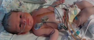
Newborn asphyxia is a pathological condition that occurs in a child in the early neonatal period and is manifested by impaired respiratory function, the development of hypoxic and hypercapnic syndromes.
The condition of asphyxia is observed in approximately 4-6% of newborns and becomes one of the main causes of perinatal mortality.
Asphyxia of newborns is manifested by impaired respiratory function
Definition of newborn asphyxia
Translated from Latin, asphyxia means suffocation, that is, lack of oxygen. Asphyxia of newborns is a pathological condition in which gas exchange in the newborn’s body is disrupted, which is accompanied by a lack of oxygen in the child’s tissues and blood and the accumulation of carbon dioxide.
As a result, a newborn who was born with signs of a live birth either cannot breathe independently in the first minute after birth, or he experiences isolated, superficial, convulsive and irregular respiratory movements against the background of an existing heartbeat. Such children are immediately given resuscitation measures, and the prognosis (possible consequences) for this pathology depends on the severity of asphyxia, the timeliness and quality of resuscitation.
Factors of the uteroplacental circle
But not always the pathological condition of the mother or fetus can cause asphyxia. There are factors that appear during pregnancy for which neither the baby’s parents nor doctors are to blame.
- early placental abruption;
- rapid aging of the placenta;
- post-term fetus;
- repeated entanglement of the fetal neck with the umbilical cord, false and true knots;
- placenta previa;
- pregnancy in which several fetuses develop, that is, multiple pregnancy;
- lack/excess of amniotic fluid;
- incoordination during childbirth; weakness of the flow; rapid labor;
- uterine rupture;
- general anesthesia for the woman in labor;
- C-section.
Factors provoking the development of asphyxia
This pathological condition is not an independent disease, but is only a manifestation of complications during pregnancy, diseases of the woman and the fetus. Causes of asphyxia include:
Fruit factors
- birth injury (traumatic brain injury) in a child;
- Rhesus conflict pregnancy;
- anomalies in the development of organs of the bronchopulmonary system;
- intrauterine infections;
- prematurity;
- intrauterine growth restriction;
- obstruction of the respiratory tract (mucus, amniotic fluid, meconium) or aspiration asphyxia;
- malformations of the heart and brain of the fetus.
Maternal factors
- severe gestosis occurring against the background of high blood pressure and severe edema;
- decompensated extragenital pathology (cardiovascular diseases, diseases of the pulmonary system);
- anemia of pregnant women;
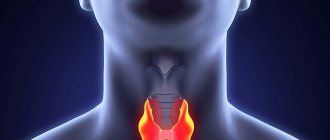
- endocrine pathology (diabetes mellitus, thyroid disease, ovarian dysfunction);
- woman's shock during childbirth;
- disturbed ecology;
- bad habits (smoking, drinking alcohol, taking drugs);
- insufficient and malnutrition;
- taking medications contraindicated during gestation;
- infectious diseases.
Factors contributing to the development of disorders in the uteroplacental circle:
- post-term pregnancy;
- premature aging of the placenta;
- premature placental abruption;
- umbilical cord pathology (umbilical cord entanglement, true and false nodes);
- constant threat of interruption;
- placenta previa and bleeding associated with it;
- multiple pregnancy;
- excess or lack of amniotic fluid;
- anomalies of labor forces (weakness of labor and incoordination, rapid and rapid labor);
- drug administration less than 4 hours before completion of labor;
- general anesthesia for women;
- C-section;
- uterine rupture;
Secondary asphyxia is provoked by the following diseases and pathologies in the newborn:
- impaired cerebral circulation in a child due to residual effects of damage to the brain and lungs during childbirth;
- heart defects that were not identified and did not appear immediately at birth;
- aspiration of milk or formula after a feeding procedure or poor-quality sanitation of the stomach immediately after birth;
- respiratory distress syndrome caused by pneumopathy: the presence of hyaline membranes;
- edematous-hemorrhagic syndrome;
- pulmonary hemorrhages;
- atelectasis in the lungs.
Mother's factors
A healthy mother means her future baby is healthy. What pathologies of a woman can cause asphyxia in a baby?
- gestosis, manifested in the third trimester;
- diseases of the heart, blood vessels or lungs;
- anemia during pregnancy;
- birth shock;
- blood diseases, thyroid gland, diabetes mellitus, ovarian dysfunction;
- poor ecology in the place of residence;
- bad habits during pregnancy (smoking, drinking alcohol and drugs);
- poor or insufficient nutrition (if the expectant mother is a vegetarian, she will have to give up her beliefs for nine months and start eating meat and dairy products (at least in small quantities), which are necessary for the full development of the fetus);
- infections suffered during pregnancy;
- treatment with drugs prohibited during pregnancy.
Mechanism of development of asphyxia
It doesn’t matter what caused the lack of oxygen in the body of a newly born child, in any case, metabolic processes, hemodynamics and microcirculation are rebuilt.
The severity of the pathology depends on how long and intense the hypoxia was. As a result of metabolic and hemodynamic changes, acidosis develops, which is accompanied by a lack of glucose, azotemia and hyperkalemia (later hypokalemia).
In acute hypoxia, the volume of circulating blood increases, and in chronic and subsequent asphyxia, the blood volume decreases. As a result, the blood thickens, its viscosity increases, and the aggregation of platelets and red blood cells increases.
All these processes lead to microcirculation disorders in vital organs (brain, heart, kidneys and adrenal glands, liver). Disturbances in microcirculation cause swelling, hemorrhages and areas of ischemia, which leads to hemodynamic disturbances, disruption of the functioning of the cardiovascular system, and, as a consequence, all other systems and organs.
Causes
The etiology of oxygen starvation is different, depending on the symptoms. The most common causes of intrauterine developmental asphyxia are maternal diseases. Maternal diseases include:
- pathology of the renal system;
- late toxicosis;
- maternal high blood pressure;
- pathology of the heart and lungs;
- acute anemia.
The most dangerous condition is an increase in body temperature. Body temperature in pregnant women may be a consequence of the inflammatory process. The entanglement of the umbilical cord around the child’s neck also plays a role in the etiology of the disease.
The umbilical cord is entangled in the patient's neck due to a difficult pregnancy. And also due to the incorrect position of the fetus in the womb.
The causes of fetal asphyxia are also the physiological characteristics of the mother. For example, a narrow pelvis. With a narrow pelvis, the patient’s cerebral circulation is disrupted during the birth process. The result is asphyxia.
go to top
Clinical picture
The main symptom of asphyxia in newborns is considered to be respiratory failure, which entails a malfunction of the cardiovascular system and hemodynamics, and also impairs neuromuscular conduction and the severity of reflexes.
To assess the severity of the pathology, neonatologists use the Apgar assessment of the newborn, which is carried out in the first and fifth minutes of the child’s life. Each sign is scored 0 – 1 – 2 points. A healthy newborn gains 8–10 Apgar points in the first minute.
Late complications
The consequences of asphyxia in newborns at an older age - from three days of life and later, are manifested in the following:
- hyperexcitability (increased excitability at any age, tachycardia);
- decreased excitability (lethargy, tendency to lethargy, decreased muscle tone, adynamism, low pulse, decreased and sometimes sluggish breathing;
- convulsions;
- hypertensive-hydrocephalic syndrome, in which there are convulsions, strabismus is often noted;
- vegetative-visceral syndrome;
- movement disorders;
- sepsis;
- development of meningitis;
- development of pneumonia;
- late speech development;
- educational lag;
- weak immunity.
In conclusion, I would like to note that this article describes the most serious consequences of newborn asphyxia. In most cases, with proper therapy, complications can be eliminated.
Degrees of newborn asphyxia
Mild asphyxia
With mild asphyxia, the number of Apgar points in a newborn is 6 - 7. The child takes the first breath within the first minute, but there is a weakening of breathing, slight acrocyanosis (cyanosis in the area of the nose and lips) and a decrease in muscle tone.
Moderate asphyxia
The Apgar score is 4 – 5 points. There is a significant weakening of breathing, possible disturbances and irregularity. Heartbeats are rare, less than 100 per minute, cyanosis of the face, hands and feet is observed. Motor activity increases, muscular dystonia develops with a predominance of hypertonicity. Possible tremor of the chin, arms and legs. Reflexes can be either reduced or enhanced.
Severe asphyxia
The condition of the newborn is serious, the number of Apgar scores in the first minute does not exceed 1 - 3. The child does not make breathing movements or takes separate breaths. Heart beats are less than 100 per minute, severe bradycardia, heart sounds are muffled and arrhythmic. The newborn does not cry, muscle tone is significantly reduced or muscle atony is observed. The skin is very pale, the umbilical cord does not pulsate, reflexes are not detectable. Eye symptoms appear: nystagmus and floating eyeballs, possible development of seizures and cerebral edema, DIC syndrome (impaired blood viscosity and increased platelet aggregation). Hemorrhagic syndrome (numerous hemorrhages on the skin) intensifies.
Clinical death
A similar diagnosis is made when all Apgar indicators are assessed at zero points. The condition is extremely serious and requires immediate resuscitation measures.
Symptoms
Clinical signs of asphyxia in children are different. Most often, the symptoms of asphyxia are reduced to some clinical signs. Symptoms of asphyxia in intrauterine development are as follows:
- tachycardia up to one hundred and sixty beats per minute;
- bradycardia up to one hundred beats per minute;
- dullness of heart sounds;
- arrhythmic phenomena.
Also, the symptoms of asphyxia in intrauterine development are associated with the presence of meconium. Meconium is found in intrauterine fluids. Asphyxia of newborns is also accompanied by certain symptomatic complexes.
Symptomatic complexes for this type of asphyxia are different. Most often, certain external signs are observed. In this case, the external characteristics of the newborn are as follows:
- cyanosis of the skin;
- no reaction to irritating factors;
- slowing heartbeat.
These clinical signs mentioned above indicate a mild course of asphyxia. However, with severe asphyxia, the symptomatic signs in newborns are as follows:
- pale skin;
- slow heart rate;
- lack of basic reflexes in newborns;
- muscle relaxation.
All of the above signs indicate severe asphyxia of newborns. This often requires the provision of proper medical care. Otherwise, the child may die.
If you are interested in this information, you can find out more on the website: bolit.info
Consultation with a specialist is required!
go to top
Diagnostics
When making a diagnosis: “Asphyxia of a newborn,” data from the obstetric history, how the birth proceeded, the child’s Apgar assessment at the first and fifth minutes, and clinical and laboratory tests are taken into account.
Determination of laboratory parameters:
- pH level, pO2, pCO2 (test of blood obtained from the umbilical vein);
- definition of base deficiency;
- level of urea and creatinine, diuresis per minute and per day (function of the urinary system);
- level of electrolytes, acid-base status, blood glucose;
- level of ALT, AST, bilirubin and blood clotting factors (liver function).
Additional methods:
- assessment of the functioning of the cardiovascular system (ECG, blood pressure control, pulse, chest x-ray);
- assessment of neurological status and brain (neurosonography, encephalography, CT and NMR).
In adults
Oxygen starvation in adults can be associated with various pathological processes. Most often, asphyxia is regarded as a serious condition with a risk of death. Asphyxia in adults may be associated with certain diseases:
- severe respiratory injuries;
- damage and chronic pathologies of the respiratory system;
- hypertension.
However, these are not the only reasons for the development of asphyxia in adults. Malignant pathological processes can lead to asphyxia in adults. Malignant pathological processes are associated with the development of a tumor. In severe cases, tracheal incubation is required.
Asphyxia in adults develops at different ages. Moreover, gender does not affect the development of the disease. Young people and middle-aged people are equally susceptible to asphyxia.
Asphyxia in adults requires emergency assistance. Moreover, assistance must be provided immediately. Otherwise the outcome will be death. Asphyxia in pregnant women is the most dangerous condition for the fetus.
Asphyxia in pregnant women has a whole range of preventive measures. It is also necessary to follow therapeutic measures. Asphyxia in adults can lead to oxygen starvation of the brain.
go to top
Treatment
All newborns born in a state of asphyxia are given immediate resuscitation measures. The further prognosis depends on the timeliness and adequacy of treatment of asphyxia. Resuscitation of newborns is carried out using the ABC system (developed in America).
Primary care for a newborn
Principle A
- ensure the correct position of the child (lower the head, placing a cushion under the shoulder girdle and tilt it back slightly);
- suck out mucus and amniotic fluid from the mouth and nose, sometimes from the trachea (with aspiration of amniotic fluid);
- intubate the trachea and examine the lower respiratory tract.
Principle B
- carry out tactile stimulation - a slap on the baby’s heels (if there is no cry within 10 - 15 seconds after birth, the newborn is placed on the resuscitation table);
- jet oxygen supply;
- implementation of auxiliary or artificial ventilation (Ambu bag, oxygen mask or endotracheal tube).
Principle C
- performing indirect cardiac massage;
- administration of drugs.
The decision to stop resuscitation measures is made after 15–20 minutes if the newborn does not respond to resuscitation measures (there is no breathing and persistent bradycardia persists). Termination of resuscitation is due to the high probability of brain damage.
Administration of drugs
Cocarboxylase diluted with 10 ml of 15% glucose is injected into the umbilical vein against the background of artificial ventilation (mask or endotracheal tube). Also, 5% sodium bicarbonate is administered intravenously to correct metabolic acidosis, 10% calcium gluconate and hydrocortisone to restore vascular tone. If bradycardia appears, 0.1% atropine sulfate is injected into the umbilical vein.
If the heart rate is less than 80 per minute, indirect cardiac massage is performed with the mandatory continuation of artificial ventilation. 0.01% adrenaline is injected through the endotracheal tube (can be into the umbilical vein). As soon as the heart rate reaches 80 beats, cardiac massage stops, mechanical ventilation is continued until the heart rate reaches 100 beats and spontaneous breathing appears.
Further treatment and observation
After providing primary resuscitation care and restoring cardiac and respiratory activity, the newborn is transferred to the intensive care unit (ICU). In the intensive care unit, further treatment of asphyxia of the acute period is carried out:
Special care and feeding
The child is placed in an incubator, where constant heating is provided. At the same time, craniocerebral hypothermia is performed - the newborn’s head is cooled, which prevents brain swelling. Feeding of children with mild and moderate asphyxia begins no earlier than 16 hours later, and after severe asphyxia, feeding is allowed after 24 hours. The baby is fed through a tube or bottle. Breastfeeding depends on the baby's condition.
Prevention of cerebral edema
Albumin, plasma and cryoplasma, and mannitol are administered intravenously through the umbilical catheter. Drugs are also prescribed to improve blood supply to the brain (Cavinton, cinnarizine, vinpocetine, sermion) and antihypoxants (vitamin E, ascorbic acid, cytochrome C, aevit). Diuretic and hemostatic drugs (dicinone, rutin, vikasol) are prescribed.
Carrying out oxygen therapy
The supply of humidified and warmed oxygen continues.
Symptomatic treatment
Therapy is carried out aimed at preventing seizures and hydrocephalic syndrome. Anticonvulsants are prescribed (GHB, phenobarbital, Relanium).
Correction of metabolic disorders
Intravenous sodium bicarbonate is continued. Infusion therapy with saline solutions (saline and 10% glucose) is carried out.
Newborn monitoring
The child is weighed twice a day, the neurological and somatic status and the presence of positive dynamics are assessed, and the incoming and excreted fluid (diuresis) is monitored. The devices record heart rate, blood pressure, respiratory rate, and central venous pressure. From laboratory tests, a complete blood count with hematocrit and platelets, acid-base status and electrolytes, blood biochemistry (glucose, bilirubin, AST, ALT, urea and creatinine) are determined daily. Blood clotting indicators and blood vessels are also assessed. cultures from the oropharynx and rectum. X-rays of the chest and abdomen, ultrasound of the brain, and ultrasound of the abdominal organs are indicated.
Proper care of a newborn in a medical institution
Newborns who have suffered asphyxia require special care. Measures for asphyxia of a newborn are carried out strictly according to medical protocols. The child should be at constant rest, the baby's head should be in a slightly elevated state. The child is provided with oxygen therapy. If a baby has been diagnosed with mild asphyxia, then he should be in an oxygen ward; the length of stay in it is individual for each little patient. If the degree of asphyxia is moderate or severe, then the child is placed in a special incubator, where there is a constant supply of oxygen, the concentration of which is about 40%; if there is no incubator in the hospital, the child is put on special oxygen masks.
In intensive care wards, babies receive appropriate drug treatment. In newborns after asphyxia, body temperature, intestinal functions, and the volume of urine excreted are constantly monitored. Feeding of newborns with mild asphyxia begins sixteen hours after birth, with severe asphyxia 22-26 hours after birth using a tube. The decision to start breastfeeding is made by the doctor in each case individually.
Consequences
Asphyxia of newborns rarely goes away without consequences. To one degree or another, the lack of oxygen in a child during and after childbirth affects all vital organs and systems. Particularly dangerous is severe asphyxia, which always occurs with multiple organ failure. The baby's life prognosis depends on the Apgar score. If the score increases in the fifth minute of life, the prognosis for the child is favorable. In addition, the severity and frequency of consequences depend on the adequacy and timeliness of resuscitation measures and further therapy, as well as on the severity of asphyxia.
Frequency of complications after hypoxic encephalopathy:
- in case of I degree of encephalopathy after hypoxia/asphyxia of newborns - the child’s development does not differ from the development of a healthy newborn;
- with stage II hypoxic encephalopathy – 25–30% of children subsequently have neurological disorders;
- with stage III hypoxic encephalopathy, half of the children die during the first week of life, and the rest, 75–100%, develop severe neurological complications with convulsions and increased muscle tone (later mental retardation).
After suffering asphyxia during childbirth, the consequences can be early and late.
Early complications
Early complications are said to occur when they appear during the first 24 hours of the baby’s life and, in fact, are manifestations of a difficult course of labor:
- cerebral edema;
- cerebral hemorrhages;
- convulsions;
- increased intracranial pressure and hand tremors (first small, then large);
- attacks of apnea (stopping breathing);
- meconium aspiration syndrome and, as a result, the formation of atelectasis;
- transient pulmonary hypertension;
- due to the development of hypovolemic shock and blood thickening, the formation of polycythemic syndrome (a large number of red blood cells);
- thrombosis (blood clotting disorder, decreased vascular tone);
- hypoglycemia;
- heart rhythm disorders, development of posthypoxic cardiopathy;
- disorders of the urinary system (oliguria, renal vascular thrombosis, swelling of the renal interstitium);
- gastrointestinal disorders (enterocolitis and intestinal paresis, digestive tract dysfunction).
Late complications
Late complications are diagnosed after three days of the child’s life and later. Late complications can be of infectious and neurological origin. The neurological consequences that appeared as a result of cerebral hypoxia and posthypoxic encephalopathy include:
- Hyperexcitability syndrome
The child has signs of increased excitability, pronounced reflexes (hyperreflexia), dilated pupils, and tachycardia. There are no convulsions.
- Reduced excitability syndrome
Reflexes are poorly expressed, the child is lethargic and adynamic, muscle tone is reduced, dilated pupils, a tendency to lethargy, there is a symptom of “doll” eyes, breathing periodically slows down and stops (bradypnea, alternating with apnea), rare pulse, weak sucking reflex.
- Convulsive syndrome
Characterized by tonic (tension and rigidity of the muscles of the body and limbs) and clonic (rhythmic contractions in the form of twitching of individual muscles of the arms and legs, face and eyes) convulsions. Opercular paroxysms also appear in the form of grimaces, gaze spasms, attacks of unmotivated sucking, chewing and tongue protruding, and floating eyeballs. Possible attacks of cyanosis with apnea, rare pulse, increased salivation and sudden pallor.
- Hypertensive-hydrocephalic syndrome
The child throws back his head, the fontanelles bulge, the cranial sutures diverge, the head circumference increases, constant convulsive readiness, loss of function of the cranial nerves (strabismus and nystagmus are noted, smoothness of the nasolabial folds, etc.).
- Syndrome of vegetative-visceral disorders
Characterized by vomiting and constant regurgitation, disorders of intestinal motor function (constipation and diarrhea), marbling of the skin (spasm of blood vessels), bradycardia and rare breathing.
- Movement disorder syndrome
Residual neurological disorders (paresis and paralysis, muscle dystonia) are characteristic.
- Subarachnoid hemorrhage
- Intraventricular hemorrhages and hemorrhages around the ventricles.
Possible infectious complications (due to weakened immunity after multiple organ failure):
- development of pneumonia;
- damage to the dura mater (meningitis);
- development of sepsis;
- intestinal infection (necrotizing colitis).
Early complications
Doctors note early effects in the first 24 hours of a baby’s life, these include:
- cerebral hemorrhage or swelling;
- convulsions;
- increased intracranial pressure, small and large hand tremors;
- respiratory arrest;
- pulmonary hypertension;
- increase in the number of red blood cells;
- thrombosis, decreased blood clotting;
- hypoglycemia;
- posthypoxic cardiopathy, irregular heart rhythm;
- disorders in the urinary system;
- gastric and intestinal disorders (digestive tract dysfunction, intestinal paresis and enterocolitis).
Features of care after asphyxia
A child who has suffered oxygen starvation should regularly visit a pediatrician and neurologist, who will give all the necessary recommendations. Asphyxia of newborns can cause hidden complications, so special gymnastics and massage are mandatory.
It is also recommended to constantly stay in the fresh air so that the baby receives the necessary amount of oxygen. It is important to maintain a normal eating pattern and daily routine. When breastfeeding, it is better to consult a doctor about how to carry out the feeding procedure. After oxygen starvation, a calm atmosphere in the family in which the baby lives is of great importance. He should not experience stress, but only positive emotions.
Resuscitation measures
Regardless of the type and causes of asphyxia, all newborns require immediate assistance. The faster it is provided, the greater the chances of a positive outcome. Therefore, resuscitation occurs right in the delivery room. Primary care is provided in a specific order. At the same time, the nursing process is very important in case of asphyxia of newborns, without which one doctor simply cannot cope.
First of all, with the help of assistants, the baby's airways are cleared of amniotic fluid, meconium and mucus. Then respiratory function is restored and blood circulation is supported. At the same time, during all activities, the newborn’s heartbeat, breathing rate and skin color are continuously monitored.
If meconium is not detected in the amniotic fluid, neonatologists and resuscitators immediately place the baby under an infrared lamp on a changing table. There, everything unnecessary is removed from his respiratory tract by suction, while avoiding touching the posterior pharyngeal wall. After this, the child is blotted dry with a diaper, placed on his back, and a special cushion is placed under the shoulders. This must be done to improve airway patency. To stimulate respiration, the baby is slapped on the heels and massaged along the spine with the palm of the hand.
If there is meconium in the amniotic fluid, treatment of newborn asphyxia requires additional measures. The contents of the trachea are aspirated using an endotracheal tube and the airway is re-cleared. If spontaneous respiration is not observed, and the heart rate is very low, then indirect cardiac massage and artificial ventilation of the lungs using a mask method are used.
If no actions help in the first thirty seconds, then they decide to give medications. Most often it is a light solution of adrenaline. Resuscitation measures continue for no more than twenty minutes, after which death is diagnosed.