Hemangioma occurs in children from the moment of birth, but most often it appears in the first two months of a newborn child’s life, which is extremely frightening for parents. Meanwhile, there is no reason for serious concern: the neoplasm never degenerates into a malignant tumor and does not cause physical pain to the baby. The exception is when the hemangioma is injured (for example, by clothing), which leads to bleeding from the vascular formation and the formation of a wound surface, which can subsequently become infected. This situation requires immediate surgical treatment.
“Hemangioma has one positive feature: in children from one to 2 to 3 years of age, the process of hemangioma involution occurs, that is, it independently decreases in size, until it completely disappears!”
Alexander Genrikhovich Dorofeev
Candidate of Medical Sciences, pediatric surgeon, doctor of the highest category at the Scientific Research Institute of National Chemistry and Technology
General information
This formation appears as a result of pathologies occurring in the blood vessels during the embryonic development stage.
In appearance, hemangioma in children is a flat, convex, homogeneous or multiple red spot (the color can also be crimson or purple) on the skin or subcutaneous surface. Most often observed in newborns immediately after birth (congenital pathology). In this case, there are cases when the formation appears at a later age, for example, during adolescence, when the hormonal background undergoes changes (acquired hemanginoma). According to medical statistics, hemangiomas are recorded in 1–2 percent of newborn infants and in 10 percent of children in the first years of life.
At the same time, hemangioma is one of the most common benign formations that form in children on the skin and mucous membranes - it accounts for approximately 50 percent of all tumor-like processes in soft tissues. At the same time, girls are susceptible to this disease 2 or 3 times more often than boys.
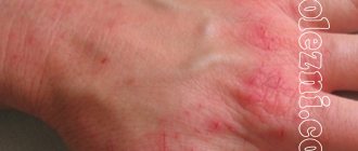
The predominant places where hemanginoma appears are the face (on the lip, nose and mouth, forehead, eyes), back and scalp. In infants, vascular tumors are localized in the area of the lower extremities (on the arm or leg, mainly on the foot and heel), perineum and genitals. Despite the fact that hemangioma in children is benign, it is prone to rapid growth and even bleeding. Due to its growth in width and depth into nearby tissues, hemangioma can lead to problems with vision, hearing and respiratory organs.
Video about the dangers of vascular neoplasms
In the video presented, Dr. Komarovsky talks in detail about what hemangioma is in newborns and what dangers it carries with it. Should you consult a doctor, and what treatment methods are used in modern medicine?
Unfortunately, the birth of a child is not always associated only with joyful moments.
Have you encountered benign vascular neoplasms in newborns? Did you have to seek surgical help, or did the pathology go away on its own? Share your experience with our readers. Perhaps your advice will help save someone’s health.
Leading clinics in Israel
Assuta
Israel, Tel Aviv
Ikhilov
Israel, Tel Aviv
Hadassah
Israel, Jerusalem
What types of hemangiomas in children are known to medicine?
The classification of hemangiomas in children is based on whether it is located on the surface of the skin or inside the skin epidermis and why it appears. In this regard, four types of hemangiosal tumors are distinguished:
- Simple, or as it is also called capillary hemangioma, which is formed from small capillaries and is located mainly on the surface of the skin. This tumor is characterized by a predominantly slow development. If growth becomes rapid, the tumor is removed surgically. The prevalence of this type of hemanginoma is 95 percent of cases;
- Cavernous or cavernous hemangioma in children. In its structure, it consists of several vessels filled with blood and is located under the skin in the form of a nodular tubercle. The consistency is elastic, and the color is bluish. When pressed, this type of hemangioma turns white, and when straining, pushing, strong crying or coughing, it becomes large in size due to blood flow (it may turn blue or red);
- The combined type of hemangioma in newborns is a combination of cavernous and capillary types. It can form both on and under the skin. In this case, the combined type of hemangiomas manifests itself depending on which component predominates in it - simple or cavernous;
- Mixed hemangioma lies in its complex structure, implying the combination in one tumor of several vascular tissue elements, such as connective, nervous, lymphoid and others. Such hemangiomas are hemlymphangioma, angiofibroma, angioneuroma, lymphogemangioma and others. The appearance and structure of mixed hemangioma depend on what tissue it consists of.
Is prevention possible?
There is no specific prevention of the occurrence of vascular formations. The main prevention is to minimize the risk of complications in newborns.
The main danger is the occurrence of a syndrome in which blood clotting is impaired and, in parallel, a tendency to form blood clots develops. This condition requires preventive examination of the blood count and monitoring of its condition.
When the tumor is localized in potentially traumatic areas or in the genital area, there is a high risk of ulcer formation and subsequent infection. As a preventative measure, it is necessary to avoid increased friction in the area of hemangioma and perform timely hygiene in newborns.
Characteristics of the causes of hemangioma in children
Why do hemangiomas appear?
Until today, medicine remains unknown to the clear causes of the occurrence and development of this disease. However, the studies conducted make it possible to identify the probable causes of hemangioma formation in children. Taking into account the fact that such a pathological process is observed in newborns, it is obvious that the reason lies in the abnormal development of vascular tissues in the uterine development of the child. Along with this, the occurrence of hemangioma, regardless of any location, can be caused by:
- The use of a number of drugs by a woman during pregnancy;
- The presence of viruses and infections in the body of the expectant mother;
- Residence of a pregnant woman in environmentally unfavorable areas;
- Hormonal disorders;
- Late pregnancy (over 40 years);
- The appearance of various pathological processes and complications while a woman is carrying a child;
- Pregnancy with twins or triplets;
- Premature birth;
- Hereditary.
Attention ! According to medical research, in adolescence, the appearance of hemangioma on the face, body and head may be a consequence of the development of liver disease.
Symptoms in children
The symptoms of hemangioma are not difficult to recognize. First of all, because this type of tumor has pronounced external signs. Thus, the formation has clearly defined boundaries, red-brown, strawberry or purple color, and is also marked by growth.
Attention ! Sometimes it is difficult to distinguish a hemangiosal tumor from other formations and rashes on the skin. It is often confused with a mole.
The most common hemangiomas in children appear:
- On the scalp, especially on the back of the head and in the neck area;
- On the front part, in the area of the eyes, nose (on the bridge of the nose), lips, eyelid, forehead and cheek;
- In the lower extremities, on the butt, in the abdomen and on the back;
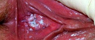
- On the mucous membranes - tongue, lips and genitals;
- In the tissues of the spine, bones and skull;
- On organs located deep inside (for example, in the liver).
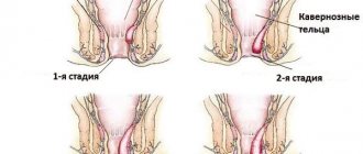
Hemangioma in children is characterized by two stages of development:
- Proliferative (productive) stage – when tumor growth occurs within 6 months or up to a year;
- The involutionary or regressive stage is the reverse development of education, in which in 70 percent of cases it passes when the child reaches 5 or 6 years of age. By the age of 9 years, almost 90 percent of children have completed this stage.
A sign of tumor involution is its blanching from the center to the edges and compaction. In this case, often, the site of the previous localization of the hemangioma subsequently differs from the healthy color of the skin, and is marked by the presence of small scars. When regression of the formation occurs before the age of four, no scars remain on the skin, but if this occurs at seven years or older, then the likelihood of a scar will be high.
What does a hemangioma look like? Hemangiosal tumors can be raised (with a swelling above the skin surface) or flat spots. Their size ranges from 0.1 to fifteen centimeters. The temperature with hemangioma has its own characteristics - the location of the tumor is hot compared to other parts of the body.
Important to know : When a hemangioma is located next to vital organs, this often leads to disruption of their functions. In cases where the tumor is localized on the surface of internal organs, the disease may not manifest itself in any way for many years. Symptoms become severe when the tumor begins to grow and put pressure on the nerve endings.
Treatment
For hemangioma in children, treatment is selected based on the size of the tumor, its growth rate, and the degree of germination into surrounding tissues. For rapidly growing tumors, or those located near important organs, removal of the tumor is indicated. Removal methods can be different:
- cryodestruction;
- electrocoagulation;
- laser removal;
- surgical excision;
- sclerosis.
The type of surgery depends on the age of the child and the type of hemangioma. Surgical methods are used when the tumor is localized in the head (mainly on the face), on the genitals, inside the eye, in the oral cavity, and also when the child’s life may be in danger due to the rapid growth of the tumor and its complicated course.
For small tumors that do not grow, the most effective treatment tactic is observation. There is a high chance that such a tumor will resolve on its own. For slow-growing formations, drug treatment is used. Traditional medicine can be used as a supplement. In a normal course, treatment of hemangioma in children under seven years of age, as a rule, is not carried out.
Cryodestruction
If the tumor has not affected the face and there is slow blood flow, freezing the tumor is used. This method works well for vascular tumors located on the back.
We recommend reading Osteosarcoma of the bone - signs, manifestations, diagnosis, treatment
Sclerosis of hemangioma
During treatment with this method, a substance is injected into the tumor, causing an inflammatory process and blockage of the main vessel feeding the hemangioma. This method is suitable for the cavernous type of disease. Capillary hemangiomas cannot be treated with sclerotherapy.
Electrocoagulation
This treatment involves cauterizing the tumor with a low-frequency electric current. Such removal is not used for extensive lesions, as well as for tumors located on visible parts of the body, since it can leave scars
Laser removal
Laser removal of hemangioma helps with small formations. You can immediately treat several hemangiomas with laser. After treatment, a crust remains at the site of the tumors, which falls off after a week.
Surgical excision
If the hemangioma is located over a large area, it is removed through surgery. In this case, surgery is preferable to laser treatment, since when using the latter, the child will be left with rough scars that have a cosmetic defect, as well as restricting the baby’s movements.
Drug treatment
Hemangiomas located in hard-to-reach places are treated with medications. Therapy can be carried out in two types:
- Acceleration of regression with the help of hormonal drugs injected directly into the vessel or for internal use;
- Use of the adrenergic blocker Propranolol in hospital settings.
Antibacterial or anti-inflammatory drugs cannot cure hemangioma.
Treatment with folk remedies
Doctors have proven that traditional medicine, as an independent treatment, is ineffective for hemangiomas, but many use it as an additional therapy. To do this, drink linden blossom tea, make compresses from cabbage leaves, and apply kombucha.
How is it treated?
In recent years, modern medicine has changed the approach to the treatment of childhood hemangioma. If the tumor had not previously grown and did not threaten the child’s health, observational therapy was applied to it. However, an analysis of recent studies has shown that early diagnosis of the disease and timely initiation of treatment can avoid a number of serious complications.
How to treat? Removal of hemangiosal tumors in children is carried out by:
- Cryodestruction . Freezing with liquid nitrogen tumors located at a shallow distance and small in size (no more than two centimeters in diameter). As a result, the formation is rejected and dies. This procedure is not performed for facial tumors or large tumors because the risk of wide scarring is very high. The advantage of this method is the targeted destruction of tumor tissue, painless nature, minimal risk of damage to healthy tissue, and rapid rehabilitation. The cryotherapy process takes a little time, and to achieve maximum effect it is necessary to repeat this session two or three more times with a break of 5 days. After the procedure, the site of the hemangioma must be smeared with brilliant green until a crust forms in the form of a sore. The healing period is up to 1 month;
- Laser irradiation. The most effective, gentle and relatively cheap method of removing hemangiosal formations in children (in Moscow, the average cost is from 1.5 to 2 thousand rubles). It is used to treat deep-lying hemangiomas (more than two centimeters). This method is effective because: it destroys tumor tissue, prevents the possibility of bleeding and scarring;
- Sclerosis . Used to remove large hemangiomas located both on the skin surface and on internal organs. The method involves cauterizing the tumor with some elements and further destroying it. The procedure is repeated with a break of a week or ten days. The hemangioma completely disappears within two years after treatment;
- Electrocoagulation method . Destruction of the tumor through the application of high-frequency current, as a result of which the formation is charred and rejected. The advantage of using electrocoagulation is that it minimizes the chance of bleeding.
- Methods of close-focus radiotherapy . Exposure to X-rays leads to the destruction of formation capillaries. This method is mainly used as an additional method - on the eve of surgery in order to reduce the size of the tumor. However, X-rays are not advisable for the child, since this may lead to the development of malignant neoplasia;
- Surgical elimination of a tumor is an operation to completely remove it and send it for histological analysis.
Giving help
To ensure that the foot is higher than the body, you need to put a pillow under your feet.
IT IS IMPORTANT TO KNOW! Even “neglected” joints can be treated at home, without surgery or hospitals. Just read what Valentin Dikul says read the recommendation...
- Provide complete rest to the limbs.
- The victim should be seated or laid down so that the foot is in an elevated position.
- Place a cushion or pillow on the ankle area.
- Apply ice to the bruised area on the top and outside of the foot, transferring the ice to a clean rag.
- Apply a fixing bandage to the foot.
- Relieve severe pain with analgesics.
- Deliver to a medical facility.
If you did not notice any serious signs when examining your heel, then you can treat your foot at home.
How to treat:
- Use drugs with external treatment. These can be cold compresses. When you apply them to a bruised area, in our case to a damaged heel, you can slightly reduce inflammation and relieve swelling.
- Physiotherapy treatment helps to quickly reduce foot pain and restore the soft tissue of the heel. But for this you need to contact a knowledgeable specialist.
- Medicines are used for treatment. They are administered internally because they help reduce pain levels and help the heel heal quickly.
Complications and prognosis conclusions
Despite the apparent harmlessness of the disease in question, many people wonder how a hemangioma can be dangerous for a baby? If this disease was not detected at the initial stage, or in the case when the treatment was incorrect, hemangioma can lead to a number of the following complications:
- Ingrowth and damage to adjacent organs;
- Damage to bone, muscle and spinal tissues;
- Development of paralysis;
- Damage to internal organs such as kidneys, liver and others;
- The appearance of ulcers and infections;
- Malignancy;
- Progressive anemia;
- Development of hemangiectasia;
- Scars.
With a disease such as hemangioma, the prognosis for recovery and minimization of harm depend on:
- The original location of the formation;
- Growth rate;
- Time of tumor detection;
- The correctness of the chosen treatment method.
Diagnostic methods
Diagnosis of hemangioma begins with a physical examination, during which the doctor determines the size of the tumor. After the initial examination, additional studies are prescribed:
- general blood test (other tests for hemangioma are not very informative);
- Ultrasound of the affected area (to assess the depth of penetration and structural features of the formation);
- angiography (reveals the characteristics of the blood supply to the hemangioma);
- X-ray (performed when the skeletal system is affected).