Causes of pulmonary atelectasis
Atelectasis is a pathological condition that develops as a result of collapse of the lung or part of it, followed by compaction and a decrease in air content. Atelectasis usually develops against the background of an underlying disease; its occurrence has certain prerequisites. The causes of pulmonary atelectasis include:
- bronchial obstruction - blockage of the bronchus with subsequent resorption of air just below the blocked area;
- the formation of extensive pleural effusion, which compresses the lung tissue from the outside;
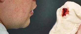
- the formation of a blood clot in the lungs due to internal bleeding, in which the patient cannot cough up blood;
- formation of viscous secretion in the bronchi due to pneumonia, chronic bronchitis, pulmonary infarction and similar diseases;
- compression of the lung tissue due to the impact of a tumor, an enlarged lymph node, or penetration of a foreign object into the mediastinum;
- impaired ventilation of the lungs and accumulation of thick mucus in the bronchi due to the use of general anesthesia during surgery;
- long-term adherence to bed rest, in which the patient does not change body position.
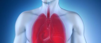
Atelectasis of the lung is accompanied by pronounced symptoms that cannot be ignored. These are chest pain, shortness of breath, cyanosis, hypotension and tachycardia, weakened voice and breathing, increased body temperature if atelectasis is complicated by a bacterial infection. During the physical examination, a dull percussion sound and retraction of the affected area of the chest during breathing are observed.
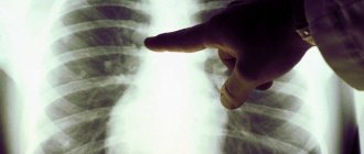
The results of radiography show a high position of the diaphragm, a decrease in the transparency of the affected area of the lung, and a displacement of the mediastinal organs to one side. In case of atelectasis, it is appropriate to conduct bronchography and computed tomography as part of a more in-depth study of the causes of the pathology.
Signs and symptoms
Signs and symptoms may be absent or include:
- cough, but not noticeable;
- chest pain (not common);
- difficulty breathing (fast and shallow);
- low oxygen saturation;
- pleural effusion (transudative type);
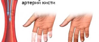
- cyanosis (late sign);
- increased heart rate.
It is a common misconception and pure speculation that atelectasis causes fever. A study of 100 postoperative patients followed by serial chest x-rays and temperature measurements showed that the incidence of fever decreased as the incidence of atelectasis increased. A recent review article summarizing the available published data on the association between atelectasis and postoperative fever concluded that there is no clinical evidence to support this suggestion.
How to treat pulmonary atelectasis?
Treatment of atelectasis is aimed at eliminating the factor that provokes it, restoring bronchial patency and preventing infection. When the bronchi are obstructed by foreign bodies or sputum, bronchoscopy is used. In uncomplicated cases, sputum can be removed by coughing, as well as by using a catheter inserted into the bronchus. If atelectasis is caused by compression of the lung by neoplasms, surgical intervention is indicated. Pleural punctures and drainage of the pleural cavity with aspiration of fluid and air are indicated for compression atelectasis.
To prevent the collapse of the lung from being accompanied by an infectious process, the patient is prescribed antibacterial drugs. To stimulate weakened breathing, etimozol may be used. At the same time, courses of breathing exercises are prescribed, and for active coughing up of thick and viscous sputum, thinning and bronchodilator drugs are prescribed.
Prevention of atelectasis for patients with pneumonia and other potentially dangerous diseases of the respiratory system consists of improving pulmonary drainage, the same is recommended for postoperative patients.
Causes and risk factors
The most common cause is postoperative atelectasis, characterized by splinting, that is, restriction of breathing after abdominal surgery.
Another common cause is pulmonary tuberculosis. Smokers and older people are also at increased risk. Outside of this context, atelectasis implies some obstruction of a bronchiole or bronchus, which may be internal to the airway (foreign body, mucus plug), from a wall (tumor, usually squamous cell carcinoma), or compressed externally (tumor, lymph node, tubercle). Another reason is poor surfactant distribution during inspiration, causing surface tension that tends to collapse the smaller alveoli. Atelectasis can also occur during sanitation of the respiratory tract, since air is removed from the lungs along with sputum. There are several types of atelectasis depending on the underlying mechanisms or extent of alveolar collapse; resorption (obstructive), compression, microatelectasis and contraction atelectasis. Relaxing atelectasis (also known as passive atelectasis) is when pleural effusion or pneumothorax disrupts the contact between the parietal and visceral pleura.
Treatment of other diseases starting with the letter - a
| Treatment of lung abscess |
| Treatment of brain abscess |
| Treatment of liver abscess |
| Treatment of splenic abscess |
| Treatment of overuse headaches |
| Treatment of pituitary adenoma |
| Treatment of adnexitis |
| Treatment of acromegaly |
| Treatment of alcoholism |
| Treatment of alcoholic hepatitis |
| Treatment of alcoholic liver disease |
| Treatment of allergic dermatitis |
| Treatment of alopecia |
| Treatment of alveolitis |
| Treatment of amoebiasis |
| Treatment of liver amyloidosis |
| Treatment of renal amyloidosis |
| Treatment of sore throat |
| Treatment of aneurysm |
| Treatment of ankylosing spondylitis |
| Treatment of anuria |
| Treatment of kidney aplasia |
| Treatment of aplastic anemia |
| Treatment of ovarian apoplexy |
| Treatment of appendicitis |
| Treatment of arthrosis of the knee joint (gonarthrosis) |
| Treatment of ascariasis |
| Treatment of ascites |
| Treatment of atherosclerosis |
| Treatment of atypical pneumonia |
| Treatment of autoimmune hepatitis |
The information is for educational purposes only. Do not self-medicate; For all questions regarding the definition of the disease and methods of its treatment, consult your doctor. EUROLAB is not responsible for the consequences caused by the use of information posted on the portal.
Prevention
Smokers can reduce the risk of developing atelectasis postoperatively by quitting smoking, if possible, 6 to 8 weeks before surgery. After surgery, patients are encouraged to breathe deeply, cough regularly, and begin moving as early as possible. The use of devices that stimulate voluntary deep breathing (stimulus spirometry) and certain exercises, including changes in body position to enhance the removal of mucus and other secretions from the lungs, may help prevent atelectasis.
Prevention of atelectasis is also achieved by deep breathing. If possible, conditions that cause shallow breathing over a long period of time should be treated.
Complications
Atelectasis in the lung can cause complications:
- respiratory failure;
- pneumonia;
- abscesses.
With rapid development (as well as in the absence of timely treatment), the disease can be fatal.
Treatment
Actually, the treatment of pulmonary atelectasis in the first stages has the same tasks as prevention; the main concern is to facilitate the access of air to the atelectatic parts. In congenital atelectasis, this is achieved by carefully examining the newborn's mouth and nose and, if necessary, cleaning them. To further improve breathing (if necessary), artificial respiration is performed.
In case of atelectasis due to bronchial obstruction, the root cause should be eliminated. Thus, in children with bronchitis and infectious diseases, expectorants and, if necessary, antibiotics should be prescribed when signs of atelectasis appear.
With atelectasis due to pressure on the lung, it is also necessary to pay attention to the underlying disease, with improvement of which atelectasis often disappears.
Etimizol is used to stimulate breathing during atelectasis, and pulmozyme is used to clean mucus plugs.
Measures to confirm the diagnosis
Reliable data can be obtained from the results of a chest x-ray or CT scan. X-rays are taken in frontal and lateral projections. The photographs show an area of the “fallen out” lung. Further research may involve performing bronchography (with the help of such a study, you can see and even remove a foreign body from a blocked bronchus).
Methods with contrasting (staining) of vessels and bronchi are also used to assess the overall picture (for small atelectasis).
Symptoms
The degree of intensity of the symptoms of the clinical picture will directly depend on the volumes of the lung involved in the pathological process. For example, microatelectasis or damage to only one segment of the lung can be completely asymptomatic. In such cases, the pathology will be a diagnostic finding, which is often discovered during a radiograph for prophylactic purposes.
The disease manifests itself most acutely when an entire lobe of this organ is affected, in particular, atelectasis of the upper lobe of the right lung. Thus, the basis of the clinical picture will be the following signs:
- shortness of breath - it appears suddenly both during physical activity and at rest, even in a horizontal position;
- pain of varying degrees of intensity in the chest area from the affected lung;
- severe dry cough;
- violation of heart rate, namely its increase;
- decreased blood tone;
- cyanosis of the skin.
Similar symptoms are typical for both adults and children.
What are the types of pulmonary atelectasis? Their features
Atelectases can differ significantly from each other in their mechanisms of occurrence.
- Obstructive atelectasis is the most common occurrence among this condition. The reasons for the development of this type of pulmonary atelectasis are the above foreign bodies, tumors, enlarged lymph nodes, etc. If there is obstruction (blockage) of the main bronchus, the entire organ (lung) is “switched off”!
- There is another type of atelectasis, which is caused by the presence of air in the pleural space (pneumothorax). Compression is compression of the lung by something (blood, air, pleural effusion, etc.). In patients with the presence of processes of sclerosis (replacement of normal lung tissue with connective tissue) and fibrosis, this condition may also occur due to impaired respiratory function.
- Functional atelectasis is a type of atelectasis that plagues bedridden patients due to impaired movement of the diaphragm or traumatic brain injury (the brain contains a respiratory center that regulates respiratory function at the highest level).
Diagnostics
Making a correct diagnosis, as well as finding out the localization and extent of the pathological process, is possible only with the help of instrumental examinations of the patient. However, before carrying out such procedures, it is necessary for the pulmonologist to independently carry out several manipulations.
Thus, the primary diagnosis will include:
- studying the medical history and collecting the patient’s life history - to identify the most likely etiological factor;
- a thorough physical examination, including auscultation of the patient. In addition, it is necessary for the doctor to assess the condition of the skin, measure pulse and blood pressure;
- detailed survey of the patient - to obtain detailed information regarding the first time of onset and the degree of intensity of symptoms. This will allow the doctor to assess the severity of the disease and its form, for example, atelectasis of the lower lobe of the right lung.
Laboratory research is limited to carrying out only blood biochemistry, which is necessary to study its gas composition. Such an analysis will show a decrease in the partial pressure of oxygen.
To definitively confirm the diagnosis, the following is carried out:
- bronchoscopy - will help to accurately identify the cause of this disease;
- X-ray – performed while inhaling. In this case, there will be a displacement of the organs of the mediastinal region towards the affected lung, and on exhalation - towards the area of the healthy half;
- bronchography and angiopulmonography - to assess the level of damage to the pulmonary-bronchial tree;
- CT scan of the lungs is performed in case of questionable radiographic findings and to clarify the localization of the pathology, in particular, to identify atelectasis of the upper lobe of the left lung or any other focus.
Atelectasis of the lung on a radiograph
Therapeutic tactics for atelectasis
Treatment of pulmonary atelectasis comes down to eliminating the root cause:
- removal of foreign bodies from the bronchus;
- operations for tumors and sclerotic changes;
- suction of the mucous component from the bronchi (in newborns, patients with bronchial asthma);
- surfactant preparations;
- bronchoalveolar lavage (washing the bronchial tree from mucus);
- drainage of the pleural cavity (for hemo-, pneumothorax);
- inhalation of drugs;
- bronchodilators;
- administration of enzymes;
- general methods (massage, breathing exercises, etc.).
Atelectasis in children. Features in newborns
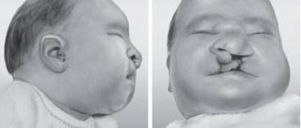
In children, symptoms and developmental mechanisms are the same as in adults. But when it comes to infants, things are different. Why does atelectasis develop in young children? Most often, this condition accompanies premature babies (children born prematurely) or children with developmental defects (improperly formed organs, such as the lungs).
The occurrence of atelectasis is associated with the entry of meconium (the so-called feces of a newborn), amniotic fluid, and mucus into the respiratory tract. In premature babies, the surfactant layer may not be sufficiently developed (a special substance that covers the structures of the lung and is involved in the act of breathing, helps to hold the frame of the alveoli, preventing them from “collapsing”). Possible head trauma during childbirth and damage to the respiratory center in the brain.
Pathogenesis
With atelectasis, there is a diffusion reduction in the surface of the lungs, a collapse of part of the alveoli and a reduction in their number that are able to function normally.
It is known that in the structure of the lungs there are more than 300 million alveoli, in which blood is perfused in parallel and sequentially during ventilation. This ensures gas exchange between the air in the alveoli and in the pulmonary capillaries. In the case of atelectasis, there is no perfusion in non-ventilated areas and gas exchange does not occur, which leads to respiratory failure. Aggravation of the processes causes extravasation and the formation of local edema. Subsequently, pneumosclerosis - the functional parenchyma is replaced by connective tissues.