Fluorography of the lungs is the study of the chest organs using X-rays that penetrate the lung tissue and transfer the pattern of the lungs onto film through fluorescent microscopic particles.
A similar study is carried out on persons over 18 years of age. The frequency of this is no more than once a year. This rule applies only to fluorography of healthy lungs, when further examination is not required.
It is believed that fluorography of the lungs is not a sufficiently informative examination, but the data obtained with its help make it possible to identify changes in the structure of the lung tissue and become a reason for further more detailed examination.
The organs of the chest absorb radiation differently, so the image appears heterogeneous. The heart, bronchi and bronchioles look like light spots; if the lungs are healthy, fluorography will show the lung tissue homogeneous and uniform. But if there is inflammation in the lungs, on fluorography, depending on the nature of the changes in the inflamed tissue, either darkening will be visible - the density of the lung tissue is increased, or lightened areas will be noticed - the airiness of the tissue is quite high.
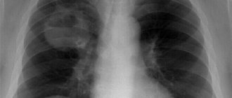
Healthy lungs
An image of the pulmonary system in a diseased or healthy state displays identical structures. On any x-ray you can see:
- Lung fields located on either side of the spine.
- In the central part there is a shadow from the main organ - the heart.
- At the top, the collarbone will be visible.
- In the lower zone is the top of the diaphragm.
- The shadow of the ribs should be visible in the image.
Taking a picture of a healthy patient’s lungs, the doctor immediately determines the condition of the bones, tissues, other important elements, and the diaphragm. An x-ray showing the healthy state of the lungs is shown in the photo.
Lung image is normal
What diseases can be diagnosed?
Most people think that FLG is only used to check the lungs for tuberculosis and do not know what diseases can be recognized with just one image. Indeed, the fluorographic method is capable of providing comprehensive information about such pathology, but this is not all of its capabilities. Then why else is this procedure needed?
Its results help diagnose other diseases of the lungs and mammary glands. Thus, based on the results of diagnostics, the following can be identified:
Darkening in the lungs on fluorography
- neoplasms of both benign and malignant nature;
- areas of the inflammatory process (when it spreads to a large volume of tissue);
- pathologically formed cavities - cysts, abscesses, cavities, and it is also determined what they are filled with - gases or liquid;
- sclerotic changes (replacement of normal connective tissue);
- fibrosis (scarring and thickening of connective tissues).
If a person has had an incessant cough, shortness of breath, general weakness, or lethargy for a long time, then he or she should definitely undergo fluorography to rule out tuberculosis or pneumonia. According to fluorography, it is possible to determine the anatomical features of the cardiovascular system and respiratory organs, some diseases that can sometimes have a non-standard clinical picture.
For example, foreign objects in the respiratory tract are not always accompanied by typical symptoms, so doctors find it difficult to make a diagnosis. But timely FLG quickly helps to determine the causes of certain pathological manifestations. The procedure is indispensable for the diagnosis of malignant neoplasms and, in particular, central lung cancer.
It is typical for such a disease to develop latently for a long time and without affecting the patient’s condition in any way, since the lung tissues themselves are not subject to pain. And only when a person begins to feel certain symptoms, the disease may already be at a stage at which surgical intervention is not performed. Latently developing pathologies include sarcoidosis of the lungs and lymph nodes of the thoracic region.
Fluorography does not take much time
Medical nuances
Experienced specialists argue that if an x-ray does not reveal significant pathological changes in the structure of the organ, this does not mean that there are no hidden diseases.
Often, doctors miss such data in the image, considering them insignificant:
- Small inflammatory foci, less than 2 mm in diameter.
- Small, barely noticeable lightened lines.
- Wall thickening up to 1 mm.
- Small dark areas.
- Miniature focal thickenings of tissue in the lungs.
- The presence of liquid in a volume of less than 200 ml.
Unfortunately, these symptoms may indicate the beginning of the development of some life-threatening illness for the patient. Given this fact, the doctor should know what an x-ray of the lungs looks like for pneumonia, tuberculosis, inflammation in a smoker and a person who does not abuse addictions.
Indications and contraindications for X-ray examination
X-rays of the lungs are often used in pulmonology.
The appointment of the appropriate technique is carried out according to the following indications:
- prolonged cough that cannot be treated with traditional methods;
- shortness of breath due to swelling of the lower extremities;
- cough, accompanied by expectoration of purulent sputum and a parallel increase in body temperature;
- suspected lung cancer;
- rib fractures.
Fact! X-ray of the lungs is a routine and safe procedure for the patient, allowing the doctor to monitor the progress of the disease in a particular person. However, there are situations when the use of the appropriate technique is not justified.
Contraindications:
- pregnancy;
- the presence of open wounds of the chest, internal bleeding;
- open pneumothorax.
In each specific case, the doctor independently decides whether to conduct an appropriate examination.
How to read a picture of a healthy organ
Knowing what healthy lungs look like on an X-ray, the doctor should know how to interpret and describe the image.
The traditional protocol for describing a healthy organ on an x-ray should include the following points:
- Clarity and definition of lung fields.
- Structurality and non-expansion of the root system.
- The presence of clear contours of the diaphragm.
- The shadow of the heart must be clearly visible in the central part of the image.
- Soft tissues should not be distinguished by the presence of pathological processes.
Pathology in the picture
If a smoker comes for examination, the specialist should know what lung cancer looks like on an x-ray. After all, patients who smoke for many years in a row are often exposed to the development of cancer. An equally rare pathology diagnosed during an X-ray examination is pneumonia. Unfortunately, both adult patients and children are affected by this disease. Pneumonia in the image appears as small focal shadows, which a specialist must recognize.
What does pulmonary tuberculosis look like on an x-ray? Any physician working in an X-ray room should be able to identify this dangerous disease. Tuberculosis is characterized in the image by foci of inflammation in the upper part of the pulmonary lobes. In an advanced form, multiple darkened areas are noted on the surface of the organ, which indicate the progression of the disease.
Why do you need routine fluorography?
Pneumonia and tuberculosis are the most dangerous diseases that can cause irreparable harm to the human body. According to statistics, in Russia alone, 1 person dies from this pathology every hour. This means that timely diagnosis can prevent possible complications and even save a person’s life.
For prevention, it is recommended that every person undergo fluorography annually. The radiation exposure a person receives during this procedure is negligible compared to the damage the disease can cause.
You should not try to independently decipher the data obtained after undergoing fluorography. Only an experienced doctor can do this correctly. After making a diagnosis, the specialist will issue a prescription and select the most effective medications. This way you can quickly cope with pneumonia and protect yourself from the serious and dangerous consequences of the disease.
What does a photo of a healthy person's lungs look like?
A photo of the lungs of a healthy person looks typical:
- Lung fields located on both sides of the chest.
- The cardiac shadow and sternum are in the center of the radiograph.
- The collarbone is at the top.
- The domes of the diaphragm are located below the pulmonary fields.
- Linear shadows of the ribs over the projection of the lungs.
A photograph of the chest organs of a healthy person with a description of the structures
In the photo, a radiologist evaluates the following structures:
- Soft fabrics.
- Osteoarticular system.
- Diaphragm.
- Mediastinum.
- Costophrenic sinuses.
These structures look different on x-rays in negative and positive versions.
It should be understood that the absence of light pathological shadows in the photographs does not mean that the person is healthy. For which pathologies are no changes observed on lung images:
- inflammatory foci up to 2 mm in diameter are not visualized on an x-ray, since the method has a resolution limit;
- small clearings;
- small darkening with infiltrative changes in the bronchi;
- fluid accumulation in the costophrenic sinus up to 250 ml;
- thickening of the bronchial walls less than 1 mm thick;
- slight focal thickening of tissue.
What is the radiation dose
Since fluorographic examination is considered one of the safest and most accessible diagnostic methods, the radiation exposure and radiation dose during fluorography remains negligible. From one procedure, 0.5 mSv (millisieverts) of radiation enters the body, and when using film technology, even less - 0.3 millisieverts.
Advanced equipment provides only 0.05 mSv.
The permissible limit for gamma radiation is 100 mSv. If a patient receives more than one Sievert (1000 mSv), the probability of developing oncological pathologies reaches 5%. A dose of 5 units is fatal.
Possible negative consequences
Some people take the position that radiation during fluorography is very harmful to the human body. But in practice this is far from reality, since only non-compliance with the permissible dosage and number of sessions can cause a number of serious complications. From one procedure per year, the patient is exposed to no more radiation than from household appliances.
How can you neutralize negative influences?
To reduce the negative impact of fluorographic equipment, it is recommended to drink a large amount of fermented milk products after the examination and eat fresh fruit.
What should not appear on an x-ray of healthy lungs?
Fluorography is a screening method of x-ray diagnostics. According to the order of the Ministry of Health, its implementation is mandatory for each person once a year. Using fluorography, doctors detect diseases such as tuberculosis and cancer in the early stages. Of course, an X-ray of the lungs is more informative, but it is characterized by a significant radiation examination of a person, and therefore cannot be used for mass examination of the population.
An ordinary person does not understand what clean lungs look like, so he will not be able to differentiate between normal and pathological shadows. Many diseases create shadows in the image, which is why “smears” and “white shadows” appear on them.
X-rays passing through the chest are reflected from some tissues and absorbed by others. As a result, dark, white and gray shadows are formed on radiographs. Their combination with each other causes a normal x-ray picture.
Some difference in the display of shadows arises due to the technical features of the radiography and fluorography method. During a fluorographic examination, the image is obtained from a fluorescent screen.
X-rays, penetrating through tissue, expose the X-ray film. This is how an x-ray is formed.
What should not be normal on a fluorogram:
- blackouts;
- enlightenment;
- additional shadows.
The figure shows a normal x-ray of the lungs. Remember her appearance. If you find additional spots in the picture that differ from the photo above, most likely, such a picture will reflect a pathological process.
What is pneumonia
Pneumonia is an inflammatory disease that involves the lung tissue. The provocateur of the pathological process is bacteria, fungi or viruses.
If the pathology is not treated, inflammatory processes can lead to serious consequences, including death. The disease poses a greater danger to older people and young children. Treatment for pneumonia begins after diagnosis. One of the effective diagnostic methods is fluorographic examination.
How does a screening grid affect an X-ray image?
The screening grid of the X-ray machine affects the quality of the images. When radiographs are taken on the same equipment, they have similar colors of anatomical structures, making it easy for the radiologist to identify pathological opacities.
However, images of the chest organs from the same X-ray machine may differ depending on the patient's position. In the photo above, the radiographs were taken of the same person in a standing (a) and lying (b) position.
Pay attention to the cardiac shadow and the intensity of the reflection of the pulmonary fields (they differ in color). The diaphragm domes are also located at different levels.
The X-ray picture of normal lungs is formed by the ability of X-rays to pass differently through the same tissues with varying degrees of physical thickening.
The quality of X-ray images is also affected by diagnostic equipment. In Soviet times, X-ray diagnostics were carried out at the RUM complex. In such equipment, radiography modes were set manually using regulators. The voltage, current, and exposure time of the x-rays were adjusted by x-ray technicians. In photographs of the lungs taken on the same person, differences in the intensity of the shadows and their hardness could be observed.
The emergence of modern diagnostic systems has made it possible to get rid of this problem. Exposure settings are now adjusted automatically.
How long to wait for the result and is it necessary to collect it in person?
The results of fluorography are not considered to be the direct image that the radiologist receives a few seconds or minutes after the procedure, but its interpretation. It is done within several tens of minutes or even hours. The final period during which the image is deciphered depends on the experience of the specialist and the quality of the image, as well as on the results of fluorography. As a rule, when pathological or potentially pathological foci are detected on images, the radiologist is faced with the task of finding the correct answer to the question - what it could be and how to classify the deviation.
It is quite problematic to receive a fluorography report on the day of the examination. The problem is that deciphering a fluorogram takes a certain amount of time: the doctor needs to look at the image, check its indicators with the norms, determine the presence of deviations and describe them in detail in a special form. Therefore, results in most clinics are issued no earlier than 2-3 hours after fluorography.
The timing of when the results are ready depends on the equipment of the fluorography room. For example, clinics that have digital equipment installed can immediately receive results and issue documents with transcripts to the patient within an hour. This is exactly what happens in private medical centers that conduct appointments strictly by appointment: in them, the radiologist can deal with the description of the image in a measured manner. You have to wait a little longer for decryption at municipal clinics. Sometimes the time frame for receiving the result can be up to 5 working days.
Good to know! In order not to waste time on an unsuccessful request for a transcript of OGK fluorography, it is worth finding out the deadlines for preparing documents in advance. In many clinics it is possible to call the receptionist and find out if the transcripts are ready.
In many clinics, radiologists are advised to appear in person for the results of fluorography, since third parties are not allowed to view the patient’s medical information. However, in reality, at the reception desk of the radiology department, the results can be given to the husband or wife, and parents can take a transcript of the examination of their children. To do this, it is enough to have with you documents confirming your relationship: a passport, marriage or birth certificate.