Author Peter Deryabin
03/06/2016 14:00 (Updated: 03/19/2020 10:10)
Health » Healthcare
Why is modern man increasingly faced with colon diseases? How to avoid the development of malignant tumors? And what intestinal diseases are most common today in Russia and the world? The director of the Federal State Center for Coloproctology, Doctor of Medical Sciences Yuri Shelygin, spoke about this.
“Stomach problems have become a “disease of civilization”
Intestinal anatomy
How many meters is the intestine of an adult? The organ is structurally divided into two main parts - the small and large intestine. The length of the first section can reach four meters. The small intestine is shorter in women than in men. It consists of three main departments:
- duodenum;
- skinny;
- iliac.
This department is responsible for digesting food. It has a small diameter and thin walls. Moreover, this structure covers almost the entire lower space of the abdominal cavity and even partially the pelvis. The thin section is also responsible for moving feces further along the intestinal tract, hormonal secretion and strengthening the immune system. The overall work of the enzymes of the small intestine, gall bladder and pancreas ensures the breakdown of the food bolus into monocomponents.
Attention! The average length of the human intestine is four meters. The thin section is longer than the thick section.
The thick section can reach one and a half meters. Anatomically, it consists of the following parts:
- blind;
- ascending;
- descending;
- transverse;
- straight;
- sigmoid.
After death, the length of the human intestine can reach eight meters. This is due to muscle relaxation. There are no villi on the mucous membrane of the large intestine. There is no active absorption of nutrients here.
This section of the intestine is necessary for the proper formation of feces. Here the absorption of water and the formation of feces from chyme occurs. Along the intestinal wall there is an accumulation of lymphoid tissue. She takes an active part in the processes of the immune system.
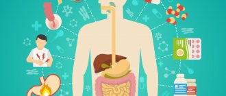
The photo shows the structural features of the gastrointestinal tract
Diagnosis of diseases of the small intestine
In the arsenal of small intestine research:
- examination and palpation of the abdomen by a doctor of any specialty;
- consultation with a gastroenterologist;
- laboratory tests (coprocytogram, blood and urine tests, ferment composition of blood and juices);
- Ultrasound of the abdominal organs for space-occupying formations;
- CT, MRI of the abdominal cavity;
- endoscopic methods (FEGDS, double-balloon enteroscopy with biopsy, duodenoscopy with special equipment);
- capsule endoscopy;
- X-ray examinations with contrast of the intestine;
- angiography of mesenteric vessels.
Read more: Methods for examining the small intestine
In continuation of the topic, be sure to read:
- Rectal cancer: symptoms, stages, treatment and prognosis for life
- Details about bowel cancer: stages, symptoms, treatment and prognosis
- Details about the coprogram: preparation, conduct and interpretation of the analysis
- Duodenum: location, structure and functions
- Jejunum: location, structure and functions
- Ileum: location, structure and functions
- Caecum: location, structure and functions
- Sigmoid colon: location, structure, functions and diseases of the organ
- Ileocecal valve (Bauhinian valve): concept, location, structure and functions
- Colon: sections of the intestine, structure and functions of the organ
Departments
Let's talk about the two main sections of the intestinal tract: the small and large intestine.
Thin
The small intestine is a multifunctional organ, the activity of which determines the coordinated functioning of the entire digestive system. It performs a secretory function, that is, it secretes juice necessary for the breakdown of food. Digestive secretions include mucus, which prevents the intestines from self-digesting.
In addition, the organ performs an absorptive function. Nutrient compounds are absorbed through the mucous membrane. The structure of the mucous layer of the intestinal wall ensures the absorption of exclusively beneficial elements. Endocrine function is ensured by the ability of cells to secrete peptide hormones. They affect the functioning of not only the intestinal tract, but also the entire body.
The muscle structures of the organ are responsible for motor function. The contraction of these muscles ensures digestion, separation of the food bolus and its further pushing. Diseases of the small intestine can have an inflammatory, functional or tumor nature. Some pathologies are congenital, while others are acquired.
Let's look at the most common diseases of the small intestine:
- enteritis. Inflammation of the mucous membrane can be caused by exposure to viruses, bacteria, parasites, and fungi. As the disease progresses, the mucous layer swells and turns red. The disease can be acute or chronic;
- allergy. The immune system reacts violently to certain foods that come into contact with the mucous membrane of the small intestine. The pathology provokes the appearance of skin rashes, swelling of the mucous membranes, as well as the appearance of dyspeptic disorders;
- tumors: lipomas, polyps, fibromas;
- peptic ulcer. Most often the disease is localized in the duodenum. The main etiological factor in the occurrence of the disease is Helicobacter pylori infection. An important role is played by genetic predisposition, the presence of bad habits, and emotional stress;
- ileitis. The disease causes inflammation of the final section of the small intestine. The cause of the disease is usually a viral or bacterial infection. Ileitis causes abdominal pain, nausea, as well as a disturbance in the general condition, up to an increase in temperature.
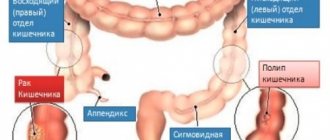
Thick
The thick section has larger dimensions and a wider diameter. Three muscle bands are responsible for peristalsis and the movement of feces. Muscle structures are unevenly distributed. Upon examination, it looks like a cluster of bulges and constrictions.
Attention! Most of the beneficial bacteria live in the large intestine.
The main function of this department is the formation of feces. After entering the thick sections, chyme loses liquid, so its structure changes, it becomes denser and takes on the appearance of feces. A common disease of the large intestine is ulcerative colitis. Chronic inflammation of the mucous membrane causes destructive changes and the formation of ulcers.
The reasons are still not fully understood, but scientists note a connection between UC and genetic factors. The influence of smoking and oral contraceptives on the development of chronic intestinal inflammation was also recorded. Patients develop diarrhea with the release of scarlet blood. There is pain in the abdominal area.
Another common pathology is Crohn's disease, which causes granulomatous inflammation. The disease can affect any part of the gastrointestinal tract and causes intestinal upset. The number of bowel movements per day can reach up to twenty times a day.
According to statistics, in developed countries, older people are at risk of developing diverticulosis. The pathology is characterized by the appearance of protrusions on the intestinal wall. The main role in the formation of the disease is played by poor nutrition, in particular, an addiction to flour and meat dishes, along with a lack of plant foods.
The intestinal tract consists of two main sections: the small intestine and the large intestine.
duodenum
The beginning of the structure of the small intestine opens - the duodenum, stretching behind the pylorus of the stomach, fitting the head and partly the body of the pancreas, thereby forming the shape of a “horseshoe” or semi-ring and flows into the jejunum.
Consists of four parts:
In the middle of the descending part, at the end of the longitudinal fold of the mucous layer, there is the nipple of Vater, which includes the sphincter of Oddi. The flow of bile and digestive juice into the duodenum is regulated by this sphincter, and it is also responsible for preventing the penetration of its contents into the bile and pancreatic ducts.
Physiology
Digestion of food begins in the mouth. Chewing thoroughly helps make this process easier. Next, the food bolus enters the esophagus, stomach and duodenum. In the initial part of the organ, food combines with bile secretions and pancreatic enzymes. Under the influence of these excreta, the food bolus is broken down.
The muscle layer ensures uniform distribution of nutrients along the inner wall. In addition to digestive function, the human intestine is responsible for endocrine and immune processes. Special microflora improves digestion processes and is responsible for the secretion of vitamins.
The intestines are responsible for the flow of hydrochloric acid into the stomach, due to which the primary processing of food occurs. Next, the eaten foods are broken down into individual components. From them the body takes for itself the necessary microelements and water. Then the formation of feces and their further evacuation occurs.
Important! The structure of the human intestine begins with the pylorus of the stomach and ends with the anus.
The job of the intestines is to absorb nutrients from food digested by the stomach. All these processes are supported by bacteria that form microflora. In addition, the intestine is an organ of the immune system. It serves as a barrier to pathogens trying to attack the human body.
Causes of diseases
Impaired epithelial functions occur due to a decrease in the number of cells and changes in their structure. For this reason, digestion time is reduced, and therefore absorption. When the muscle layer suffers, the movement of blood and lymph worsens. The delivery of incoming food elements is reduced.
As a result, the digestion process is disrupted and food is in transit. Pathogenic intestinal bacteria begin to use it, and their increased growth occurs. Therefore, it is important to prevent destruction and damage to the epithelial layer. Otherwise, a deficiency of elements occurs and inflammation develops.
Diseases of the small intestine are divided into functional, congenital, inflammatory and tumor. The following reasons can provoke them:
- infections;
- injuries or surgery;
- eating unhealthy food;
- alcoholism and smoking;
- stress;
- some medications.
Congenital pathology is detected during the first years of a child’s life. The development of the tumor process is typical for older people.
Microflora
The intestinal tract is inhabited by the following bacteria:
- lactobacilli;
- bifidobacteria;
- bacteroides;
- enterococci;
- coli;
- Proteus;
- staphylococci;
- fungi.
The first three names refer to the main group of microorganisms present in the intestines. In addition to beneficial bacteria, the microflora also consists of opportunistic microorganisms. Under the condition of strong immunity, these bacteria do not cause any disturbances in the body, but when the immune forces are weakened, these same microorganisms get out of control, begin to actively multiply and can cause serious abnormalities in the body.
Interesting! The human intestine is inhabited by microorganisms that are seventy times more numerous than the number of inhabitants of the globe.
Bacteria present in the intestines are divided into two main groups: anaerobes (do not require oxygen) and aerobes (live on oxygen). The overwhelming number of microorganisms in the intestinal tract are anaerobes: lactobacilli, bifidobacteria, bacteroides. And, for example, E. coli and enterococci are aerobes.
Skinny
Next in the order of the human intestinal structure diagram is the jejunum. It is separated from the duodenum by the duodenojejunal sphincter, located in the peritoneum at the top left and smoothly flows into the ileum.
The anatomical structure separating the jejunum and ileum is weakly manifested, but there is still a difference. The ileum, relatively lean, is larger in diameter and has thicker walls. It was called skinny due to the absence of contents in it during the autopsy. The length of the jejunum can reach 180 cm; in men it is longer than in women.
Violations
Poor functioning of the digestive organ can be associated with several factors. The more factors that affect the intestines at the same time, the more severe the pathology and the more difficult it is to treat. The following reasons play a role in the development of intestinal tract diseases:
- genetic predisposition;
- weakened immunity;
- poor nutrition;
- bad habits;
- passive lifestyle;
- some medicines;
- intestinal infections.
The following symptoms unite intestinal diseases:
- Abdominal pain. The pain syndrome can be intense aching or even sharp paroxysmal. In some cases, it appears in episodes or is associated with food intake. In some diseases, patients can name a clear localization of pain, while in other disorders, the pain outbreak is diffuse. For example, when the small intestine is damaged, discomfort occurs in the umbilical region. Diffuse pain is more characteristic of intestinal bloating due to stretching of the walls by gases.
- Flatulence. This symptom occurs due to excess accumulation of gases. The cause of this condition may be fermentation processes, intestinal atony or decreased motor function.
- Decreased appetite. In fact, patients develop a fear of eating. This is explained by the fact that after a meal the intestines begin to actively contract and secrete digestive juices, which provokes painful attacks.
- Constipation or diarrhea.
Intestinal diseases usually develop against a background of weakened immunity
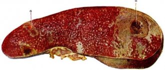
Heart attack
A heart attack is the death of the intestinal wall. Impaired blood flow can occur due to blockage or spasm. The insidiousness of this pathology lies in the difficulty of diagnosis. Without an angiographic study, it is almost impossible to make a diagnosis.
The pathology manifests itself in the form of sudden cramping pain in the abdomen, nausea, vomiting, and diarrhea. Given the fact that most often the disease is detected in late stages, treatment is mainly surgical. It is advisable to use conservative therapy until signs of peritonitis develop.
Dyskinesia
The pathology is based on deterioration of intestinal tone and motility. Organic damage is not detected during examination, but functional activity is significantly reduced. Dyskinesia causes indigestion. Pathology often develops against the background of neurological disorders. This is why dyskinesia is most often diagnosed in women.
Dyskinesia is divided into hypertonic and hypotonic types. In the first case, persistent spastic contractions of the intestine are observed. They can cause chronic constipation and painful colic. The pathology causes acute cramping pain in the lower abdomen and iliac regions.
The painful outbreak subsides for some time after defecation, and after eating it returns again. Chronic intoxication of the body leads to mental and physical decline in performance. With hypertensive dyskinesia, there may be no stool for several days, and then a large amount of feces is released.
With hypotension, on the contrary, peristalsis is weakened. Patients are bothered by dull painful cramps in the abdomen, a feeling of fullness, and bloating. Feces pass with great difficulty and in small quantities. This causes poisoning of the body.
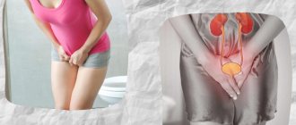
Endometriosis
A benign neoplasm occurs due to the entry of endometrial cells of the uterus into other organs. Hormonal changes, hereditary predisposition, and weakened immunity play a major role in the formation of the disease. When the external intestinal muscles are affected, nausea and abdominal pain occur during menstruation. If the sigmoid colon is involved in the process, the pain attack is localized in the left lower abdomen.
The following symptoms are typical for endometriosis:
- pain in the depths of the pelvis and in the anus during menstrual periods;
- constipation or diarrhea;
- painful bowel movements;
- the appearance of blood and mucus in the stool;
- increased bowel movements during menstruation.
In women, intestinal endometriosis can cause pain during intercourse, as well as prolonged and heavy menstruation. Drug treatment is aimed at normalizing hormonal levels, since intestinal endometriosis is only a secondary process.
Types and signs
The clinical picture of the diseases is very diverse. Manifestations depend on the cause of the pathology, the severity, duration of the course and the presence of complications.
Irritable bowel syndrome
The disease is caused by a psychoemotional disorder, a violation of intestinal motility. The pain is located in the lower abdomen and radiates to other parts. Colic occurs, a feeling of intestinal distension, rumbling, and bloating are observed.
After an urgent urge, loose stools begin. There is a feeling of incomplete bowel movement. Diarrhea may give way to constipation. The condition is accompanied by aggression, irritability, or vice versa, depression, fears. A person is haunted by thoughts about his own health, and there is a fear of going out into the street where there is no toilet.
Colostomy placement
In most cases, colostomy is only the first stage of surgical intervention, the second is restoring the integrity of the intestinal tube, that is, closing the stoma. The second stage is the most important for the patient; as a rule, for cancer it is planned with a delay of several months, for benign and inflammatory processes - for several weeks.
Planned surgical intervention with active preliminary preparation of the patient is always preferable, which improves the course of the postoperative period and the patient’s recovery. An emergency colostomy is performed in case of intestinal obstruction of any etiology or “acute abdomen”, that is, when the operation is performed for health reasons.
Surgery requires general anesthesia and serious anesthesia, because the operation is quite long.
During planned surgery with a high probability of a colostomy, before making a skin incision, the surgeon uses a marker to mark the location of the intestine on the abdominal wall. There should be no old scars in this area; the site chosen for the stoma should be sufficient for attaching the colostomy bag plate.
Depending on the location of the pathological process, the outlet can be located in the hypochondrium or just above the iliac region on the right or left. The hole is formed where the colostomy bag will not deform during body movements or interfere with the patient, that is, not at the waist line, not in the groin, not near the pelvic bones, not at the navel.
After dissection of the abdomen, a revision is carried out - a thorough examination of the abdominal cavity, then the intestines are dissected. After resection of the pathological area or hemicolectomy, or extirpation of the rectum, the surgeon begins to create a colostomy. Then a second revision is carried out and the final stage of the operation is closure of the wound. A special postoperative colostomy bag is placed on the patient’s operating table to monitor the condition of the stoma and bandage the wound.
The complexity of the postoperative period depends on the initial disease and the condition in which the patient entered the operating table.
We will call you back
Leave your phone number