Coxsackievirus is one of the representatives of enteroviruses that lives and multiplies in the digestive tract, but can infect other tissues and organs. The virus is resistant to freezing and the action of a number of disinfectants (ether, Lysol, 70% ethyl alcohol). It remains viable in feces for more than six months. The Coxsackie virus is diagnosed in adults much less frequently than in children; children under 5 years of age are most susceptible to it. The infection can be transmitted from person to person; after the illness, the patient remains with intense type-specific immunity.
Coxsackievirus is an enterovirus that multiplies in the digestive tract
The virus is an intracellular parasite. The entry points for infection are the mucous membranes of the pharynx and gastrointestinal tract. After entering the body, primary replication of viral particles occurs and their accumulation in the cells of the mucous membranes of the nasal cavity, pharynx, and small intestine. Then the infectious agent enters the blood and circulates for some time in the general bloodstream, as a result of which it can migrate to different organs and tissues, leading to the development of an inflammatory process in them. Different serotypes of the Coxsackie virus have different tropism for the tissues of the human body. The largest amount of the virus is usually localized in nerve cells, internal organs, striated muscles, and skin. Depending on the serological type of the virus, as well as on the individual characteristics of the body, the outcome of the disease can be complete recovery, transition of the disease to a chronic form (with prolonged persistence of the infectious agent in the affected tissues and organs) or virus carriage.
Infectious diseases caused by the Coxsackie virus usually occur in a mild or moderate form. The prognosis in the vast majority of cases is favorable, complete recovery occurs within 2-3 weeks.
Inactivation of the Coxsackie virus occurs by drying, exposure to ultraviolet radiation, heating to 50 ° C, disinfection with a 0.3% formaldehyde solution or chlorine-containing preparations.
General information
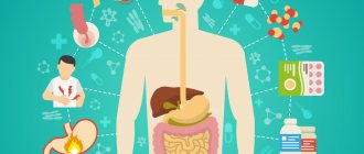
Coxsackie virus and ECHO virus belong to the group of enteroviruses and are the main causative agents of enterovirus infection (EVI).
In recent years, there has been a tendency towards activation of Coxsackie viruses and an increase in infectious diseases caused by enteroviruses. The incidence rates of EVI in the CIS countries vary between 11.5-16.2/100 thousand population. The majority of cases are children (90%). The specificity of EVI is the polymorphism of clinical manifestations, which is due to their ability to affect various tissues/organs (kidneys, heart, central nervous system, lungs, liver and others), long-term virus carriage, the presence and wide distribution of asymptomatic forms, the absence of a clear pronounced dependence of nosological forms on the serological type of the pathogen , as well as the lack of specific methods of prevention, which makes enterovirus infections practically uncontrollable. Diseases often occur in the form of sporadic morbidity, but every 3-4 years epidemic outbreaks caused by different serotypes of Coxsackie viruses are recorded around the world. Every year, the proportion of certain infecting virus serotypes changes significantly, which many authors explain by the accumulation of a “critical mass” of susceptible children, which is necessary to maintain the epidemic process. It is also important that one serotype of the Coxsackie virus in adults and children can cause clinically completely different diseases (even at the same time in the same family), while at the same time different serotypes of the virus can cause diseases whose symptoms are very similar, which complicates the diagnosis and treatment of patients .
Enterovirus infection Coxsackie in the everyday lexicon is called “Turkish flu” due to partial similarity with the symptoms of classic flu and the introduction of the Coxsackie virus into the CIS countries by people vacationing at resorts in Turkey.
Incubation period
From the moment pathogenic microflora enters the body until the development of a symptomatic picture, it can take from 4 to 7 days. During this period of time there are no signs of the disease. How long the incubation period will last depends on the type of disease and which mucous membranes the virus has affected:
- Coxsackie flu-like form. From the moment of infection to the appearance of the first symptoms, similar to a common cold or acute respiratory viral infection, it takes from 1 to 3 days.
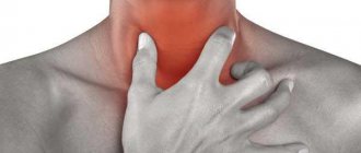
- A sore throat of the herpetic type, which is provoked by infection with a virus of the mucous membrane in the oral cavity, develops within 7-15 days.
- Conjunctivitis of hemorrhagic type. As a form of the disease, it has the shortest incubation period, lasting 1-2 days.
What causes such a long incubation period? Entering the human body through the intestines or oral cavity, the virus begins to accumulate in the mucous membrane of the oral cavity and nasal sinuses, on the intestinal walls.
When the concentration of pathogenic microflora reaches its highest level, it enters the bloodstream, with which it spreads throughout the body. Migrating along with the blood, pathogenic microflora settles on the mucous membranes of some internal organs, penetrates the cells and provokes the onset of inflammation.
Pathogenesis
For Coxsackievirus A and B, the entry gates are the mucous membranes of the oral cavity, upper respiratory tract and intestines. Enteroviruses freely overcome the “gastric barrier” and penetrate the cells of the small intestinal mucosa. Subsequently, the process of virus replication occurs in the cells of the intestinal epithelium, lymphoid tissue and mesenteric lymph nodes. The virus then enters the bloodstream and causes primary viremia. The Coxsackie virus exhibits the greatest tropism for cells of the central nervous system/muscle tissue, but a variety of organs are also involved in the pathological process.
Dissemination of the pathogen occurs in the heart, liver, lungs, kidneys, eye vessels, pancreas, where it multiplies and accumulates. Clinical symptoms, severity and outcome of the disease are determined by a number of factors - the biological properties of the virus serotypes, their tropism, the state of the humoral/cellular immunity of the human body. Swelling, inflammation and areas of necrosis develop in the affected organs. The stages of pathogenesis are shown schematically in the figure.
Persons who have recovered from the disease develop long-lasting (over several years) type-specific immunity .
Forms of the disease
Coxsackie viruses are divided into two groups:
- Group A – 24 serological types. Viruses of this group are localized in the skin, mucous membranes, and can cause acute hemorrhagic conjunctivitis, upper respiratory tract diseases, aseptic meningitis, enteroviral vesicular stomatitis, etc.
- Group B – 6 serotypes. These viruses affect the heart, pleura, liver, pancreas, and can cause hepatitis, inflammation of the heart muscle (myocarditis), inflammation of the visceral and parietal layers of the pericardium (pericarditis), pericardial effusion, etc.
The infection can be transmitted from person to person; after the illness, the patient remains with intense type-specific immunity.
Some of the serotypes of group A, as well as all types of group B, are capable of multiplying in embryonic cell culture. Viruses of both groups have a common complement-fixing antigen; some strains have hemagglutinating properties against group O red blood cells.
The infectious process caused by the Coxsackie virus can occur in the following forms:
- catarrhal;
- spinal (poliomyelitis-like);
- encephalic.
Classification
There is no unified classification of enteroviruses due to the wide polymorphism of clinical symptoms. In the Russian Federation, a classification according to the type of disease has been adopted.
- Typical forms of enterovirus infection: herpangina , enteroviral fever , acute respiratory diseases, exanthema , epidemic myalgia , aseptic serous meningitis , encephalomyocarditis , hepatitis , myocarditis , mesadenitis , meningoencephalitis , gastroenteric form , hemorrhagic conjunctivitis , vesi cular stomatitis .
- Atypical forms of enterovirus infection: erased, inapparent.
By current: light, medium-heavy. heavy.
Etiology
Coxsackie viruses were first isolated by GJ Dalldorf and G. M. Sickles (1948) in Coxsackie, New York (USA); they are close to the polio virus, but, unlike it, are non-pathogenic for monkeys and adult white mice. A characteristic feature of viruses is the ability to cause myositis in suckling mice. The diameter of Coxsackie viruses is 15-30 nm. They are among the simplest in chemistry. structure of animal viruses and consist of protein and RNA. Sensitive to chloramine and formaldehyde; inactivated at t° 60° for 20 minutes. Resistant to ether, 70% alcohol, 5% Lysol solution, chemotherapeutic agents and antibiotics. Frozen at t° 70°, they do not lose activity for several years. They do not tolerate drying well.
30 immunological serotypes of the virus have been identified. Based on the decision of the WHO Committee on Enteroviral Infections, Coxsackie viruses are usually divided into two subgroups: A and B. Subgroup A includes 24 immunol, serotypes, biol, the peculiarity of which is the ability to cause widespread hyaline necrosis with an inflammatory reaction in skeletal muscles in experimental animals, liver and spinal cord, as well as pulmonary atelectasis. Subgroup B includes 6 immunol. serotypes. Their distinctive biol feature is the ability to cause focal necrosis in experimental animals not only in skeletal muscles and liver, but also in the myocardium, pancreas, nervous tissue and other organs.
When studying the biological properties of Coxsackie viruses, it was also found that some of them cause degenerative phenomena in tissue culture cells. Cytopathogenic changes in the cells of monkey kidney cultures are caused by serotypes A 9, B 1, B 3, B 4, B 5; serotypes B 1, B 3, B 5 are active in cultures of human embryonic tissue fibroblasts; serotypes A 13, A 18 and B 1 - B 5 are active in Hela cells, etc.
Coxsackieviruses of subgroup A are considered causative agents of herpangina (serotypes 2, 3, 4, 5, 6, 8, 10, 16, 22); they are also associated with aseptic serous meningitis (serotypes 1, 2, 3, 4, 5, 6, 7, 9, 10, 14, 16, 23, etc.). Coxsackie viruses of subgroup B are the causative agents of Coxsackie myocarditis (serotypes 2, 3, 4), aseptic pericarditis (serotypes 4, 5), epidemic pleurodynia (serotypes 1, 2, 3, 4, 5) and Coxsackie meningitis (serotypes 1, 2, 3, 4, 5). Coxsackieviruses A and B are also causative agents of various acute respiratory, polio-like and enteroviral diseases.
Causes
Coxsackieviruses of group A (serotypes 1-22, 24) and group B (serotypes 1-6) are the etiological factor of a number of enteroviral infections.
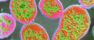
Morphology and biological properties
Coxsackie enterovirus is a small RNA virion (28 nm) with cube-shaped symmetry and the ability to form crystals inside affected cells. Virion capsid without envelope. The appearance of the Coxsackie virus is shown in the figure below (Wikipedia).
Based on their antigenic structure, Coxsackie viruses are divided into two large groups: A-26 and B-6 serological types. The virus is highly resistant in environments and on environmental objects. But they are quickly inactivated at temperatures exceeding 50 degrees, they can persist for 2 months at a temperature of 37°C, survive for a long time in river/tap water, but especially for a long time in waste water.
Stable in acidic environments (pH 3-5). The activity of enteroviruses remains for several years even when frozen, and when stored in a refrigerator at a temperature of +4-6°C - for several weeks. At the same time, repeated freezing and subsequent thawing does not lead to a decrease in their activity. They quickly die when dried, under ultraviolet irradiation and when exposed to formaldehyde , even in small concentrations. They are destroyed by boiling (at 100°C - instantly, 60°C in 6-8 minutes).
Epidemiology
Coxsackie viruses are ubiquitous: their reservoir in nature is water, soil, and food. In the human population, the human body. The main epidemiological feature is the widespread ability to form healthy virus carriers in the human body with a long period of virus release into the external environment (up to 3-6 weeks). Virus carriage in healthy individuals varies between 17-40%. Among children, the percentage of virus excretors reaches 20.0%, and at the age of under 1 year it is even higher - 32.6%. This feature contributes to the survival of the virus even in conditions of high levels of immunity in the population.
The level of natural immunity increases with age, reaching 90% already at the age of 5-10 years. Among the adult population, antibodies to the most common Coxsackie serotypes are found in 30-80% of the population. At the same time, the seropositivity of the human population is much higher in regions/countries/cities with a low social and hygienic standard of living. Some authors even consider these data as an indicator of the standard of living of the population and even the effectiveness of anti-epidemic protection.
The source of infection is a sick person, a recovering person or a virus carrier. Infection with Coxsackie viruses occurs throughout the year, but in the northern hemisphere the peak incidence of EVI occurs in the summer-autumn season. In warm regions and the tropics, the incidence does not have a pronounced seasonality. EVI affects people of all age groups, but incidence rates are inversely proportional to age. About 75% of EVI cases occur in children under 15 years of age, and children under 1 year of age get sick several times more often than older children and adults. Males get sick more often.
The leading mechanism of transmission is fecal-oral. Its implementation is carried out by water, food and household contact. Airborne and transplacental (from mother to fetus) routes are possible. The main condition for the implementation of the infection transmission mechanism is poor sanitary living conditions. The infection rate of children living in unfavorable sanitary conditions can reach 50%.
The most important way for Coxsackie viruses to spread is through contact with another person's contaminated hands and objects, followed by inoculation of the virus through the nose, mouth or eyes, and through contaminated food and water. Those infected are most contagious during the first week of illness.
Infection, detection and characteristics of the course of infection in pregnant women
The question “is the Coxsackie virus dangerous during pregnancy” does not have a clear answer. It all depends mainly on the immunity of the expectant mother.
To detect the presence of the virus in a woman, she may be prescribed the following tests:
- serological blood tests (for the presence and quantity of specific antibodies);
- PCR of the virus, which allows you to determine its genotype.
Pregnant women, like other groups of people, become ill with this infection through airborne or nutritional contact with the virus. The incubation period lasts from 4 to 6 days. In summer and autumn, as well as with high humidity, the activity of the virus increases, and it becomes even more dangerous for the expectant mother.
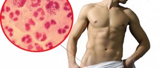
Pregnant women are especially susceptible to infection with Coxsackievirus type 16A. The first signs of infection are disturbances in the digestive tract, a rash in the form of small blisters on the feet, hands and mouth. Such blisters strongly resemble stomatitis.
There are several ways of infection of the fetus from the mother:
- Perinatal (through vaginal secretions, blood).
- Transplacental if the concentration of the virus in the blood is very high, or if there is an exacerbation of a chronic infection.
- Postnatal (via breast milk or other sources).
The virus poses a danger to the expectant mother because, due to the special state of her body, her immunity is already reduced, and Coxsackie suppresses it even more. In such cases, there is a high probability, firstly, of the disease progressing in severe forms, and secondly, of infection with other infections or exacerbation of existing ones.
Infection with the Coxsackie virus in the first trimester of pregnancy can lead to the death of the embryo or delayed fetal development, spontaneous miscarriage, and prematurity. In the second trimester of pregnancy, infection leads to respiratory failure and global cognitive defects of the fetus.
To prevent infection, pregnant women should be especially careful about their own hygiene: keep their hands and food clean, drink more fluids, and avoid contact with sick people.
If such contact is unavoidable, you must wear a medical mask and gloves and carry out thorough disinfection.
Symptoms of the Coxsackie virus
The incubation period of the Coxsackie virus varies for different nosological forms and can range from 2 to 35 days. The average incubation period is 7-10 days. The clinical symptoms of the Coxsackie virus in adults, as well as the symptoms of the Coxsackie virus in children, are extremely varied. Among the main clinical syndromes caused by the Coxsackie virus of various serotypes are:
- Coxsackie A viruses - herpangina , serous meningitis , acute pharyngitis , paralysis, exanthema , pneumonia of newborns , exanthema of the mouth and extremities, contagious rhinitis , hepatitis , diarrhea of newborns/young children, acute hemorrhagic conjunctivitis .
- Coxsackie B viruses - serous meningitis , pleurodynia , severe systemic infection of newborns, pneumonia and upper respiratory tract disease, myocarditis and meningoencephalitis , fever , hepatitis , rash.
Let us briefly consider the clinical manifestations of the most common nosological forms of enteroviral infections in adults caused by the Coxsackie virus.
- Enterovirus fever : the most common form of enterovirus infection in recent years. It is characterized by an acute onset with short-term fever for up to 2-4 days, less often up to 1-1.5 weeks. It occurs with mild general infectious symptoms - moderate headache, minor catarrhal symptoms, muscle pain, and less often occurs with an enlarged liver and spleen. Often not diagnosed or diagnosed in the presence of a local epidemic outbreak in a community.
- Epidemic exanthema : an increase in body temperature, during a decline/at a height of temperature, the appearance of a small maculopapular, less often hemorrhagic rash, which is localized mainly on the torso, face and arms. The rash persists for several hours or 1-2 days, after which it disappears without a trace. On the oral mucosa there is enanthema . A more common variant of enteroviral exanthema in children is damage to the skin of the hands and feet, as well as the mucous membrane of the oral cavity - the so-called “hand-foot-mouth syndrome” (see photo below), caused by serovars 5,10,16 of the Coxsackie A virus. Against the background moderate general intoxication on the skin of the hands, feet, cheeks and mucous membrane of the tongue, rashes appear in the form of vesicles with a diameter of 1-3 mm with hyperemia around them.
- Herpetic sore throat : sore throat, increased body temperature. On the palatine arches and mucous membrane of the soft palate there are papules, which quickly transform into vesicles and after 1-2 days ulcerate and become covered with a white coating. Hyperthermia persists for 2-3 days and gradually decreases. Changes in the pharynx persist for 6-7 days. With the addition of bacterial flora, general infectious symptoms increase. Often combined with other forms of EVI.
- Gastroenteric form: begins with an increase in body temperature to a subfebrile level, weakness, loss of appetite, hyperemia of the pharynx mucosa, and after 1-2 days watery diarrhea , abdominal pain, bloating, flatulence , an admixture of mucus appears in the stool, stool frequency up to 10 times , sometimes vomiting. The duration of the disease is 1-2 weeks.
- Serous meningitis : headache, fever up to 39-40 C, anxiety, vomiting, sometimes convulsions. At the height of the fever, meningeal signs appear - stiff neck and specific symptoms. Abdominal pain, abdominal reflexes are reduced. During a spinal puncture, there is an increase in cerebrospinal fluid pressure. Sometimes undulating fever. After meningitis, asthenic syndrome persists for a long time, and in some cases intracranial hypertension .
- Enteroviral encephalitis : headache, high body temperature, drowsiness /excitement, impaired consciousness, convulsions, vomiting. Subsequently, symptoms develop depending on the area of brain damage (hemispheric, brain stem, cerebellar). Sometimes the pathological process in the brain is combined with damage to the spinal cord, which is manifested by flaccid paresis/paralysis of the muscles of the limbs and torso, and autonomic disorders. With the development of meningoencephalitis, meningeal symptoms appear. The course of the disease is severe, with a high probability of death.
- Epidemic myalgia : pain in the muscles of the chest, diaphragm and abdomen. Often the pain is localized in the epigastric/iliac region or migrates, sometimes combined with pain in the extremities, fever to febrile levels, difficulty breathing, especially inhalation, headache. Muscle pain is paroxysmal, lasting 10-30 minutes. The duration of symptoms is 3-10 days.
- Mesadenitis : symptoms of general intoxication, abdominal muscle tension, frequent vomiting, pain and bloating.
- Paralytic form: weakness in the legs/arms, muscle tone and tendon reflexes are reduced on the affected side, increased body temperature, gait disturbance. Paresis and paralysis quickly disappear, atrophy does not develop.
- Mesadenitis : inflammation of the mesenteric lymph nodes. Increased body temperature, abdominal pain, vomiting, bloating, abdominal muscle tension.
- Enteroviral myocarditis : weakness, fatigue, discomfort/pain in the heart area, the boundaries of the heart are expanded, tachycardia , muffled heart sounds, ECG shows focal/diffuse changes in the myocardium.
- Enteroviral hemorrhagic conjunctivitis: redness, lacrimation, pain in the eyeballs, swelling of the eyelids, photophobia , hemorrhage in the conjunctiva of the eyes/sclera. Increased body temperature, over time, many patients develop epithelial small focal keratitis or uveitis. In severe cases - iris dystrophy, uveal cataract, corneal opacification, atrophy of the eyeball.
- Enteroviral hepatitis: clinical symptoms resemble the anicteric form of viral hepatitis A. Abdominal pain, increased body temperature, myalgia, enlarged liver. Deviations in liver function tests are insignificant.
Photo of Coxsackie virus in children (hand-foot-mouth syndrome)
Modern studies convincingly prove the role of EVI in the occurrence of isolated encephalitis, glomerulonephritis and hemorrhagic cystitis.
Forecast of the course of the disease and recovery
Timely consultation with a doctor and correct diagnosis of the virus increases the likelihood that the disease will pass quickly and will not cause significant harm to the body. In case of a mild infection, the person returns to the usual rhythm of life after 3 days. In more severe cases, recovery lasts up to two weeks, in rare cases, for example, with manifestations of encephalitis - up to 6-8 weeks.
With properly selected therapy, the child’s recovery occurs in approximately 10-14 days. After two weeks, the last lesions and blisters on the skin heal. Additionally, in the period after the end of the active stage of the disease, it is useful to give the baby vitamin complexes containing vitamins A, , , and B. For six months after recovery, it is not recommended to give the child any vaccinations.
There are several possible outcomes of the disease:
- the person completely gets rid of the virus and recovers;
- nerve cells and organs retain virus particles for some time;
- a person becomes a carrier of the virus.
Tests and diagnostics
The diagnosis of a specific nosological form of EVI is made on the basis of complaints, clinical manifestations, data from laboratory and instrumental examinations.
Laboratory tests include: CBC, OAM, analysis for Coxsackie virus (nasopharyngeal swab, vesicle contents, cerebrospinal fluid for enteroviruses using PCR), blood for CSC/PHA.
Additional diagnostic examinations: ECG, biochemical blood test (bilirubin, creatinine, AlAt, AsAt, urea), blood coagulogram , comprehensive ultrasound, MRI.
Returning to the Turkish issue...
No hotel will allow itself the luxury of being quarantined during the high tourist season. Yes, and complete disinfection is difficult. Given the several routes of transmission of the virus, it is quite difficult to interrupt the chain of infection once it has begun. Tourists can be advised to carefully read the reviews of vacationers who have just returned from the hotel to which you are about to go. And if there are sick people there, it is better to ask the travel agency to change the hotel.
While on vacation, drink bottled water, wash your hands often, always with soap, ignore swimming in the pool, visiting the children's playroom and public events, including evening animation, if you have a child under 10 years of age with you.
Have you already become acquainted with the Coxsackie virus?
Treatment of Coxsackie virus
Treatment of Coxsackie virus in adults is aimed at relieving symptoms of intoxication and clinical recovery, normalizing biochemical parameters of blood/cerebrospinal fluid and preventing complications. Hospitalization of patients is carried out according to clinical indications (symptoms of central nervous system damage, peripheral paralysis, the presence of meningeal syndrome, encephalomyocarditis, intense muscle pain that requires relief, severe symptoms of intoxication, severe background pathology, secondary infection, and others). For outpatient treatment, if necessary, bed rest. Since medications for etiotropic therapy have not yet been developed, treatment of patients is reduced to pathogenetic therapy and relief of symptoms of the disease in accordance with the clinical manifestations, form and severity of the disease.
For severe pain syndrome - NSAIDs, non-opioid analgesics ( Ketoprofen , Diclofenac , Ketorolac , Lornoxicam ), for hyperthermic syndrome (over 39.5 C) - antipyretic drugs ( Acetaminophen , Paracetamol ). In case of severe intoxication, detoxification therapy is carried out by infusion of solutions of Hemodez , Reopoliglyukin , Glucose (5-10%), Sodium chloride (0.9%). If necessary (meningitis and meningoencephalitis), dehydration therapy ( Mannitol , Furosemide , Diacarb ). For desensitizing therapy - Chloropyramine . For the paralytic form of EVI, Proserin . For myalgia: Ibuprofen , Prednisolone . For encephalitis and convulsions - Phenobarbital , Diazepam . In severe cases, cerebral edema is treated with oxygen therapy .
As for antibiotics, their prescription is justified only in cases of the addition of bacterial flora and the development of bacterial complications. For this purpose, Azithromycin and beta-lactam antibiotics ( Amoxicillin , Ampicillin , Oxacillin , Ticarcillin /clavulanate and others) are prescribed.
How to treat EVI in children?
There are no fundamental differences, but children have a number of characteristics. Treatment of Coxsackie virus in children includes drugs that have antiviral and immunomodulatory effects - human recombinant interferon α-2b. Immunoglobulins ( Pentaglobin , Sandoglobulin ) and recombinant interferons ( Viferon , Roferon ) are also used. In newborn children of patients and children with myocarditis, immunoglobulin preparations can be prescribed to prevent complications. With the most common forms of EVI in children ( herpangina , hand-foot-mouth syndrome), according to a number of authors (Komarovsky), specific treatment is not required and bed rest, symptomatic treatment and plenty of fluids are quite sufficient.
Indicators of the effectiveness of treatment are the absence of symptoms of intoxication and fever, the disappearance of the rash, the absence of motor disorders, pain, normalization of blood and CSF parameters.
Is there any treatment for coxsackieviruses?
There is no specific treatment for the Coxsackie virus. Symptoms go away “on their own” after about 2–10 days, and this without specific antiviral therapy. At the same time, symptomatic treatment is still necessary. Medicines to reduce fever, relieve itching - antihistamines, sore throat - sprays, lozenges, etc. At the same time, antiviral therapy is prescribed in our country to quickly relieve intoxication. Most often, these are interferons or homeopathic remedies designed to fight a viral infection.
In some cases, the Coxsackie virus is confused by its clinical manifestations with herpes or bacterial tonsillitis and the antiherpetic acyclovir and antibiotics are prescribed accordingly. You need to know that acyclovir does not help at all with Coxsackie enterovirus, but it can harm the body, because is hepatotoxic. Antibiotics do not affect viruses, so taking them is not only useless, but can aggravate the course of the disease.
It is recommended to prevent dehydration by drinking fluids, but juices, fruit drinks, and acidic foods can irritate mouth ulcers. Cool milk will come to the rescue, which will help soothe this pain and soften the throat. It is useful to drink cool chamomile tea, as well as brew chamomile in milk.
Relatively rare complications of coxsackievirus infections (for example, heart or head infections) require special individual treatment, which is prescribed by an infectious disease specialist and often in a hospital setting.
Medicines
- Azithromycin.
- Galantamine.
- Dexamethasone.
- Dextran.
- Diazepam.
- Ibuprofen.
- Mannitol.
- Paracetamol.
- Human immunoglobulin (IgG+IgA+IgM).
- Interferon alpha 2b.
- Sodium chloride.
- Prednisolone.
- Phenobarbital.
- Furosemide.
- Chloropyramine.
How it manifests itself
During the latent period of viral action, pathogens actively “travel” through the bloodstream. The result of their harmful action is specific rashes in the form of ulcers , inside of which there is a cloudy yellow liquid. Both the diagnosis and treatment methods depend on how the rash looks.
Important! The rash usually appears a day after the first manifestations of the disease. Skin rashes are called polymorphic exanthema.
Main locations:
- palms,
- feet,
- space between fingers
- area around the lips.
The rash bothers the sick child, causing discomfort with severe obsessive itching . Ulcers appear gradually.
| Period | Nature of the rash |
| 2nd day of illness | Pink small spots, without contour. They quickly become bright, turning into bubbles. The skin around the vesicles does not undergo changes. A similar rash is characteristic of allergic rashes, measles, rubella, and scarlet fever. Itchy sensations appear in the first hours after the appearance of papules. |
| Day 3 | The rashes turn into blisters with liquid inside. Reminds me of chickenpox rashes. The baby suffers from unbearable itching, itches, screams, and rushes around the bed. The attacks worsen at night. |
| 4 day | The ulcers begin to break out and partially turn pale. Skin with profuse rashes becomes dry, the skin bursts, peels and begins to peel off. The process is very active on the feet and palms. The itching subsides. |
| 5-7 day | The rash is less noticeable, turns pale, and gradually disappears. Small areas may remain for a short time; the skin continues to peel off. The skin renewal process can take up to 10 days. |
Photo of a child's throat:
Coxsackievirus in children
Despite the commonality of the manifestations of the Coxsackie virus in the body of adults and children, there are a number of features. First of all, in most children, Coxsackie infection is asymptomatic. According to Dr. Komarovsky, the most common symptoms of the Coxsackie virus in children are herpangina and hand-foot-mouth syndrome, which affect almost all children. In his opinion, no special treatment is required for such children; symptomatic therapy and plenty of fluid intake are sufficient. Diseases disappear quickly.
Sometimes parents are interested in whether the child is contagious and how contagious he is. A child is a source of the EVI pathogen with the possibility of transmitting it to others throughout the disease, and in many cases even after the disease for 1-2 weeks. Therefore, a sick child should be isolated as soon as possible.
Newborns and young children are a special risk group. In most cases, EVI manifests itself in the form of a relatively benign fever, in some with a rash on the body. At the same time, Coxsackie viruses are the most common cause of encephalomyocarditis . A child becomes infected mainly from relatives in the family, sometimes intrauterinely. Characterized by lethargy, vomiting, increased body temperature, symptoms of heart damage: shortness of breath , acrocyanosis , tachycardia , cardiac arrhythmia, expansion of the boundaries of the heart. With encephalitis - focal symptoms. The course is severe and death is possible.
Another extremely serious disease is sepsis-like disease, the etiological agent of which is Coxsackie viruses. Very often, the infectious disease is extremely severe, lightning fast with liver necrosis and damage to the heart, lungs, pancreas and brain and ends in death. The severity of the disease is determined by the level of maternal antibodies, the state of health of the child and the virulence of the infectious agent.
Possible complications after illness
How dangerous is Coxsackie for children and adults? Not only does it cause symptoms that are very similar to a dozen different diseases, but it can also cause the appearance of some diseases. Incorrect and untimely consultation with a doctor increases the chances of getting complications from the virus. Among them are encephalitis, viral meningitis, paralysis, myocarditis, insulin-dependent diabetes mellitus, and pleurodynia.
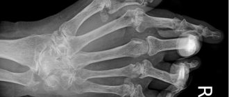
In children, in addition to the problems listed, a purulent sore throat, partial paralysis, as well as changes in the nails may occur (as a rule, 4-8 weeks after recovery, the nails may partially peel off, change color and shape, become brittle and brittle, and even fall off completely, followed by replacement of the renewed nail plate).
Prevention of Coxsackie virus
There is still no specific prevention, which is due to the large number of serotypes of the virus. Nonspecific prevention includes:
- compliance with personal hygiene rules (thorough hand washing with disinfectant solution after visiting the toilet/before eating);
- Thorough washing of fruits and vegetables before consumption and rinsing with boiling water;
- using bottled water for drinking;
- swim only in permitted places; do not swallow water while swimming;
- avoiding contact with persons with clinical manifestations of EVI.
When a focus of EVI is identified, in order to localize it, the following is carried out:
- Active identification of patients with EVI and persons in contact with patients and establishment of monitoring of them.
- Isolation and hospitalization of patients (if necessary).
- Disinfection measures - disinfection (final and current).
- Strengthening sanitary supervision over catering, the water supply system, maintenance of the territory, compliance with the regime of organized children's groups and health care facilities.
- In case of an unfavorable epidemiological situation, it is recommended to administer 0.3 ml/kg of gamma globulin to all persons in the foci of infection.
What is the prognosis for coxsackievirus infection?
The overall prognosis has been excellent until recently. Most children recovered completely and did not require hospitalization. In Alabama (USA), in 2011–2012, 12% of sick children required hospitalization for treatment of infection with the Coxsackie virus. Therefore, the forecast is difficult to predict because it changes.
In our country we are faced with a strange picture. Doctors treat enterovirus infection, all the symptoms of the Coxsackie virus are obvious, but patients are discharged with a simple diagnosis of ARVI. Although acute respiratory viral infections are sometimes caused by enteroviruses, Coxsackie infection is not indicated. This is due to paperwork when detecting such an infection, the high cost of tests, the tendency of patients to excessively panic, as well as the fear of doctors to involve inspection services. How normal this situation is is perhaps a rhetorical question.
Based on materials from https://www.medicinenet.com
Consequences and complications
EVI, which occurs in mild forms, ends in the vast majority of cases with complete recovery. However, with meningitis , encephalomyocarditis and meningoencephalitis in newborns, complications such as cerebral edema, hemi/monoparesis, epileptoid seizures, and decreased muscle tone are common. Severe EVI can contribute to acute respiratory failure and the development of pneumonia, and in case of eye damage - cataracts .
Routes of transmission and methods of infection
The virus is transmitted through unwashed fruits, vegetables, water, and airborne infection is also possible . The Coxsackie virus can survive for a long time in tap water and feces. The period for maintaining its activity is up to two and a half years (780 days)
. This causes the spread of infection and group epidemics.
More often the virus has seasonal manifestations. Its outbreaks occur during the warm season, when most unwashed fruits are eaten and raw tap water is used.
Interesting: in the tropical climate, Coxsackie is able to “rage” all year round.
How can you neutralize Coxsackie? The virus is afraid of ultraviolet radiation (sun) and disinfectant solutions (bleach, chloramine). In such conditions he dies almost instantly. Boiling kills the virus in 20 minutes.
List of sources
- Bondarenko, V. I. Enteroviruses in river water Text. / V. I. Bondarenko, V. I. Zadorozhnaya, K. V. Yashchenko // Hygienic aspects of biological pollution* of the environment: materials of the X All-Union Conference. -M., 1988. Part: 2. - P. 79.
- Kozhevnikova, N.V. Increasing role of enteroviruses in modern infectious pathology Text. / N.V. Kozhevnikova, T.N. Karavyanskaya, E.B. Golubeva // Far Eastern Journal of Infectious Pathology. 2007. -No. 10. - P. 52-53. — Bibliographer: p. 53.
- Kokoreva S.P. Modern complex therapy of viral neuroinfections in children / S.P. Kokoreva, N.P. Kuprina, O.A. Panina // Children's infections, 2007. – T. 6, No. 4. – P. 47-53.
- Resolution of the Chief State Sanitary Doctor of the Russian Federation dated July 27, 2011 N 106 SP 3.1.2950-11 “Prevention of enterovirus infection.”
- Sabitova A.M., Alexandrova T.A. Modern clinical and epidemiological features of enterovirus various clinical forms of infection // Collection of materials of the X Russian conference “Pediatrics and pediatric surgery in the Volga Federal District”, Kazan, November 26-28, 2013. - P. 60.
Examination methods
You can recognize the symptoms of Coxsackie virus in children after examining the patient, collecting anamnesis, and examination results. The main factors in making a diagnosis are the results of the following examinations:
- diagnostics using the PCR method - determines the causative agent of the virus in the blood;
- enzyme-linked immunosorbent assay (ELISA) – detects antibodies to the virus;
- blood and urine tests.
If necessary, the doctor may prescribe other research methods that will help obtain a complete picture of the disease. A sick child must be referred to other specialists: a cardiologist, an ophthalmologist, a neurologist, a gastroenterologist. Before the doctor makes a final diagnosis and prescribes treatment, the child must exclude diseases such as rubella, polio, mumps, scarlet fever, scabies or eczema.
Reviews from mothers and vacationers
Natalya: We weren’t in any Turkey, the child just went to first grade. A couple of days later he had a headache, his temperature rose to 38 degrees, then even higher, his throat became sore, and he even started vomiting. The pediatrician prescribed augmentin and isoprinosine. The temperature dropped, but rashes like stomatitis or herpes appeared. For the baby (she is 1.9 years old), the thermometer showed more than 38°. I have some discomfort in my stomach, my neck muscles are seized. This is the kind of Coxsackie we have...
Anastasia: We were in Turkey with my husband and three-year-old daughter. Before leaving, the baby’s temperature rose, her body was covered with rashes, severe itching appeared, and she couldn’t even sleep - I scratched her legs all night. And we flew home, and my husband and I fell ill. They also say that adults don’t get sick! Fever, body aches, skin pain, itching - you wouldn’t wish it on your enemy. And no medications help, only cold water soothes the itching. It hurts to walk, my legs are swollen, especially my husband’s. They say that my nails will peel off - I’m especially afraid of this, I’m a manicurist myself. Overall, terrible...
Irina: We picked up the infection not in the hotel or on the beach, but in the apartment, where it was not so crowded. But that didn't save us. What I would like to recommend is that before going to the resort, take care of your child’s immunity, let him get enough sleep, not get hypothermic, and eat well. My son slept poorly the day before his arrival, then he immediately dived into a pool of cold water, and this is the result - a day later he developed a fever and an itchy rash appeared. Take some local antiseptic with you, such as baneocin. Otherwise, the rashes will begin to fester; they must be treated in advance. Take anti-itch medications and antihistamines with you when traveling.
It is unlikely that you will be able to completely protect yourself and your children from contracting a hand-foot-mouth infection. But strengthening the immune system and basic hygiene skills, which a child should be taught from an early age, will help reduce the risk of infection.
And, if it was not possible to avoid the disease, it is important to contact a competent specialist and start treatment on time.
How to get rid of illness during pregnancy
Women during pregnancy are especially susceptible to enterovirus infections due to decreased immunity.
Pregnant women in the early stages if infected with the Coxsackie virus have a risk of miscarriage , and in the later stages there is a risk of premature birth.
Intrauterine infection of the fetus may occur, which may result in pathologies of the heart, liver, and damage to the central nervous system.
Mother infection in the 3rd trimester is less dangerous for the fetus. The infection can be transmitted through the placenta, but it does not threaten the health of the unborn baby.
Pregnant women are usually not prescribed antiviral therapy. Treatment is symptomatic:
- interferon-based drugs - to increase the body's resistance;
- antipyretics – if the temperature is above 38.5°C. The drug is prescribed taking into account the trimester to avoid complications;
- antiseptic sprays and gels - to alleviate the condition of a pregnant woman with herpangina;
- solutions of sea water - rinse the nose for congestion and runny nose;
- Regidron and other solutions based on glucose and salt - to prevent dehydration in the presence of diarrhea.
Also, the expectant mother is advised to have bed rest, drink plenty of fluids, and be monitored by the attending physician.