Hydrocephalus is a disease in which excess cerebrospinal fluid accumulates in the ventricles of the brain. In case of untimely diagnosis and treatment, the consequences of hydrocephalus in children can be severe. Hydrocephalus also develops in adult patients. Neurologists at the Yusupov Hospital examine patients using modern equipment from leading companies in Europe and the USA. Professors and doctors of the highest category working in the neurology clinic adhere to European recommendations for the treatment of hydrocephalus. Neurologists take into account the individual characteristics of the body, the causes and severity of the disease.
The cause of hydrocephalus in children under one year of age can be viral infections that the mother had during pregnancy - rubella, herpes, cytomegalovirus infection. Infection with chlamydia or genital herpes can occur during the passage of the fetus through the birth canal. Hydrocephalus develops in children after birth trauma, hypoxia, hemorrhage in the subarachnoid space or cerebellum.
Description of the disease
Today, almost every fifth newborn child can be diagnosed with increased intracranial pressure. Although in most situations it does not have any tragic consequences. But it is still worth checking the head for the presence of excess fluid in the head of a newborn. And if the diagnosis is confirmed, then you should definitely think about taking all the necessary measures for treatment.
Hydrocephalus of the newborn (or dropsy in other words) is the name of a complication due to which in the area of the brain in newborns there is an accumulation of cerebrospinal fluid, otherwise called cerebrospinal fluid. There are several variations of the disease, however, in children under two years of age, all its symptoms are very similar to each other.
The term "hydrocephalus" is derived from two Greek words that mean "water" and "head". In other words, this disease consists of excess fluid (water) in the head. This is where the second name of the pathology comes from, which sounds like dropsy of the brain. True, strictly speaking, this name is not entirely correct. The fact is that in the presence of hydrocephalus in the head of newborns, there is an excess not of water at all, but of cerebrospinal fluid, that is, cerebrospinal fluid. Liquor is a liquid that is vital for the functioning of nerve tissues. It can be found in the spinal cord. We will consider the fluid levels in the head of a baby below.
In addition, it is also present in the brain. In it, a substance such as cerebrospinal fluid is concentrated in four ventricles, which are located in the center of the skull. The upper two are located in both hemispheres, and the lower ones are located along the central cerebral axis. The ventricles usually communicate with each other through a system of pipes called the cerebral aqueduct. In addition, cerebrospinal fluid can enter the subarachnoid space, which separates the meninges with special cisterns located at the base of the skull.
Classification
Infantile hydrocele of the brain is classified according to many characteristics.
So, according to the time of formation of hydrocephalus of the brain in children, it happens:
- Intrauterine – diagnosed during pregnancy, mainly at 16-20 weeks. The disease can develop in the fetus after a pregnant woman has suffered infections or viruses, against the background of a genetic predisposition, as well as the expectant mother’s abuse of such addictions as alcohol and drugs.
- Congenital - occurs due to congenital abnormalities of the brain or central nervous system, premature birth, as well as during difficult labor and resulting intracranial injuries.
- Acquired - develops in children aged 1 year and older under the influence of certain factors: past infectious diseases affecting brain tissue; pathologies of the vascular system; intracranial tumors and injuries.
According to morphological characteristics, hydrocephalus is divided into:
- communicating (open) – occurs due to an imbalance in the production and absorption of cerebrospinal fluid;
- occlusive (closed) - appears due to blockage of the liquor-conducting pathways caused by a pathological process.
In turn, the described types of hydrocephalus are divided into the following subtypes:
- internal - cerebrospinal fluid accumulates in the ventricles and overwhelms them;
- external (external) - fluid fills the subarachnoid space, accumulating between the membranes of the brain;
- mixed - does not have a clear localization of fluid accumulation, which can fill the ventricles and subarachnoid space at the same time.
According to the criterion of stability of manifestations, the following types of hydrocephalus of the brain in children are distinguished:
- progressive – with increasing symptoms and rapid deterioration of well-being;
- regressing - a decrease in the intensity of symptoms followed by its disappearance;
- stabilized – the symptoms are stable, while the state of health does not change, either for the better or for the worse.
Hydrocephalus is also divided into two types depending on the stage of the disorder:
- compensated - despite the diagnosed hydrocele of the brain, there are no signs characteristic of this pathology, while the child develops normally and feels well;
- decompensated – accompanied by severe symptoms and a significant deterioration in well-being.
Timely identification of the type of hydrocephalus in a baby allows you to select the necessary treatment and thereby prevent the development of complications and serious consequences in the future.
Types of disease
There are only three main forms of this pathology, in which fluid is observed in the head of a newborn:
- open hydrocephalus;
- closed, or occlusal form;
- hypersecretory form of pathology.
The closed type of the disease occurs in cases where there is a physical obstacle that interferes with the outflow of cerebrospinal fluid from the cranial container intended for it into the systemic bloodstream. This type is mainly caused by cysts along with tumors or hemorrhages.
The open type of the disease is usually observed when the mechanism of absorption of cerebrospinal fluid into the systemic circulation is disrupted. With this variant of the development of pathology, the cause of the disease is often previous infections. For example, meningitis or the presence of blood in the subarachnoid space.
Hypersecretory hydrocephalus is a relatively rare type of the disease in question and is observed in approximately five percent of cases. It usually occurs as a result of excessive production of cerebrospinal fluid. A similar situation can happen, for example, due to pathology of the choroid plexus.
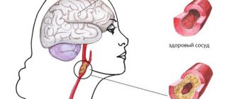
What leads to the development of the disease
Moderate hydrocephalus is based on impaired outflow of cerebrospinal fluid into the spinal cord canal, where it is normally absorbed into the venous network of the circulatory system.
Factors leading to the occurrence of the disease:
- Arterial hypertension;
- Stroke;
- Atherosclerosis;
- A cyst or tumor compressing the ventricle and blocking the circulation of cerebrospinal fluid;
- Osteochondrosis;
- Hernia of the cervical spine;
- Neuroinfections – meningitis, encephalitis;
- Alcoholism;
- Traumatic brain injuries caused by head contusions from a fall from a height, an impact, or a car accident.
Functions of fluid in the head
The volume of cerebrospinal fluid is, in fact, relatively small. Normally, in newborns it is, as a rule, 50 milliliters, and in adult patients - from 120 to 150 ml.
The functions of the fluid in the head of a newborn are very diverse:
- protection of nervous tissue from external mechanical influence;
- removal of harmful substances from the brain and delivery of nutrients to it;
- maintaining stable intracranial pressure values.
The fluid in the baby's head, like blood, can circulate inside the cranial cavity. Against this background, its composition is constantly updated. In adult patients, on average, this can happen three times a day, and in infants much more often - up to eight times a day. Every minute, adults produce 0.35 milliliters of cerebrospinal fluid, and approximately 500 milliliters per day. CSF pressure in adults can vary quite widely, namely from seventy to one hundred and eighty millimeters of mercury.
CSF is mainly formed in the ventricles of the brain. Two thirds of this fluid can be generated by their choroid plexus, and the rest - with the help of membrane elements and meninges. In special veins that are located inside the skull, in its occipital parietal part, namely within the venous sinuses, it is absorbed.
Consequently, if for some reason the circulation processes of the cerebrospinal fluid are disrupted, and it is formed in greater quantities than necessary, or is simply not absorbed quickly enough, then an excess of this fluid is observed in the newborn in the cranial cavity. It is this syndrome in children that is called hydrocephalus.
Excess cerebrospinal fluid can manifest itself differently in children and adults. For example, adults have hard skull bones, so excess fluid usually leads to increased intracranial pressure. The situation is completely different for young children under the age of three. They have rather soft skull bones, and therefore hydrocephalus very often manifests itself in the form of an abnormal expansion of the head circumference.
Anatomy
The brain and spinal cord are constantly bathed in a fluid called cerebrospinal fluid. Thanks to cerebrospinal fluid, the brain is protected from all kinds of damage:
- the liquid creates a kind of “airbag”,
- shock absorbing.
This is not the only function of cerebrospinal fluid:
- it also delivers nutrients to tissues,
- supports the microenvironment,
- promoting the normal functioning of the thinking organ, etc.
- CSF is produced from the blood.
In a healthy adult, the volume of cerebrospinal fluid is about 150 ml, in a newborn - 50 ml.
Normally, cerebrospinal fluid is in a state of constant circulation: cerebrospinal fluid is produced and absorbed in such a way that its total volume remains unchanged. In some cases, the fluid stagnates, accumulating in the ventricles of the brain or under its membranes. It is the disruption of circulation that causes the development of hydrocephalus. In this case, both an increase in the volume of produced cerebrospinal fluid and a disruption in the process of its absorption can cause pathology.
Causes of fluid in the head of a baby
Dropsy in newborns can develop from simple prematurity. And in addition, from the presence or previous infectious diseases. For example, factors such as smoking, drinking alcohol and other bad habits of the mother, not just during pregnancy, but also in everyday life, can contribute to the development of this pathology in the newborn.
During the first few years of life, any head injury is very dangerous, as it can lead to an increase in the production of cerebrospinal fluid. A tumor that occurs in the brain can significantly interfere with the healthy flow of fluid in the head of a newborn baby. Which, in turn, will create excess pressure.
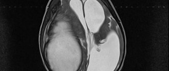
How does the disease develop and what are the consequences?
The activity of the endocrine glands, which produce cerebrospinal fluid, is disrupted. As a result, there is too much cerebrospinal fluid, and the small body is unable to ensure its normal outflow. On the left of the diagram is a normal brain, and on the right is one affected by hydrocephalus.
Hydrocephalic babies are easily recognized by their disproportionately large heads. Water cannot compress and increases the volume of the skull, and since in children under one year old rigid connections between the cranial bones have not yet formed, the increase in volume of the head occurs unhindered.
When hydrocephalus of the brain develops in an older child, excess fluid manifests itself exclusively as damage to the central nervous system.
The disease is quite dangerous: the cerebrospinal fluid does not leave the head and begins to put pressure on the nervous tissue, destroying neural connections. Without proper treatment, this usually leads to swelling of the brain and ultimately the death of the child.
How does this pathology manifest clinically?
The fluid in the head of a newborn must circulate in a correct and normal manner, and if this is disrupted, this will certainly lead to hydrocephalus. The most important symptom is a change in the shape of the head over a fairly quick period of time. In this regard, it will be necessary to visit a pediatrician every month, who must measure the head, checking the condition with the normal fluid levels in the baby’s head.
In addition, the fontanel in a newborn baby is characterized by an increased size, since the sutures of the skull are not yet fully formed. Over time, the symptoms may become more pronounced: a network of veins will appear on the face, and the shape of the forehead, in turn, will become more disproportionate. Cramps may occur from time to time. Newborns with hydrocephalus tend to be lethargic and cry frequently.
Such children are noticeably lagging behind in development, their psychomotor skills are impaired. This usually manifests itself in the fact that the child’s head is very poorly supported. In addition, such children begin to crawl, walk and sit up late. In addition, newborns with this disease often spit up and even vomit. Among other things, they experience constant drowsiness. All such symptoms may indicate that the baby has increased intracranial pressure.
However, you should not rush to make a diagnosis based on individual similar characteristic signs, since usually only the appearance of the entire complex of the listed symptoms can indicate the presence of hydrocephalus. Only the attending doctor, as part of the examination of the child, will be able to make the correct diagnosis and prescribe the required treatment for the accumulation of fluid in the head of the newborn.
Myths about increased intracranial pressure
The diagnosis of “increased intracranial pressure”, “intracranial hypertension (ICH)” or “hypertensive-hydrocephalic syndrome”, as already mentioned, is often made and in some cases - without reason. How does increased intracranial pressure (ICP) manifest? As already noted, in children under 2 years of age such manifestations are, first of all, accelerated growth of head circumference, bulging and enlarged fontanelle, possible eye movement disorders, and delayed psychomotor development. Most often, all these disorders manifest themselves in a complex. In children over 2 years of age, these are headaches with nausea and vomiting, more often in the morning, changes in the fundus of the eye (detected during examination by an ophthalmologist). Of course, the clinical picture may be different, but without the above symptoms, the diagnosis of “increased intracranial pressure” is doubtful.
This is how pediatrician Evgeniy Komarovsky describes the diagnosis of “increased intracranial pressure.” “Since the main symptom of hydrocephalus is a rapid increase in head size, measuring head circumference is included in the standards of any preventive examination, starting, of course, from the moment of birth. It is very important to emphasize here that it is not the specific size expressed in centimeters that matters, but rather the dynamics of this indicator. That is, stating the fact that the boy Petya at 3 months has a head circumference of as much as 45 cm is not at all a reason to become depressed and urgently save this boy. But the fact that the head circumference has grown by 7 cm over the past month is already alarming and dangerous, and requires both serious attitude and active control. Let me emphasize once again - not immediate treatment, but control. And if the trend continues, then measures should be taken.”
Symptoms such as sleep and behavior disorders, hyperactivity, attention deficit, bad habits, poor academic performance, hypertonicity in the legs, “marble” skin pattern, including on the head, nosebleeds, chin trembling, tiptoe walking, are not in themselves indicate increased intracranial pressure. And yet, some neurologists make a diagnosis of ICH based on these complaints. Neurosonography, having become a huge boon for pediatrics and neurology, has made a significant contribution to the excessive and false diagnosis of “hypertensive-hydrocephalic syndrome.” NSG makes it possible to quickly obtain an image of the brain substance and measure the size of the ventricles. However, to clarify the diagnosis, as we have already said, CT and MRI are mandatory.
Basics of diagnosis and therapy in newborns
After determining the primary diagnosis, children are prescribed neurosonography along with ultrasound examination of the brain, computed tomography or magnetic resonance imaging.
If the diagnosis is confirmed, ventriculoperitoneal shunting is often performed. The essence of this operation is that the cerebrospinal fluid from the cerebral ventricles of the newborn is sucked through silicone catheters into the abdominal cavity. Less commonly, fluid may be drained into the spinal canal or right atrium.
If the operation was performed on time, then the child has every chance of further normal life, which includes attending child care and school institutions. However, it is also necessary to take into account that the size of the head after surgery most likely will not decrease, since changes in bone tissue are always irreversible.
How to identify fluid in a baby's head?
Diagnosis of the disease
There are several ways to determine the development of hydrocephalus. It is worth noting that this disease is much easier to identify in children. But in adult patients, recognition of the described disease is sometimes difficult and problematic. Previously, many adults who suffered from hydrocephalus were diagnosed with various neurological and mental disorders. However, of course, their therapy was not very effective. Only after the advent of modern diagnostic techniques the situation changed radically. And for the better.
If there is a lot of fluid in the baby's head, this is mainly detected by the pediatrician during a thorough examination of the child. Doctors can turn their attention to obvious manifestations of hydrocephalus in the form of an enlarged head, bulging fontanel, divergence of the sutures of the skull, and in addition, changes in the appearance of the skin and characteristic neurological symptoms. To facilitate the diagnostic procedure, parents are recommended to record the baby’s head circumference values at a certain time interval. If a pathology is suspected, the pediatrician can write a referral to a neurologist, pediatric surgeon or neurosurgeon.
Prevention
In order to minimize the chance of developing hydrocephalus, follow these rules:
- Protect your child from head injuries in every possible way: wear helmets for rollerblading/scooter/bicycle skating, carry him in a car seat, and do not walk in places where there is a danger of injury.
- During pregnancy, a woman should be examined for the TORCH complex of infections, followed by consultation with an infectious disease specialist.
- If ARVI, rubella or any other disease occurs during pregnancy, additionally undergo an ultrasound scan of the fetus and consult with a medical geneticist and infectious disease specialist about the further management of the period of gestation.
- It is imperative to undergo routine examinations by a neurologist, ultrasound or MRI if the child was born premature.
- The previous rule applies to conditions after meningitis, meningoncephalitis, intracranial hemorrhages, and head injuries. Do not neglect visiting a doctor.
Author:
Krivega Maria Salavatovna resuscitator
Treatment
Recently, great progress has been made in medicine in the treatment of hydrocephalus and the presence of fluid in the head of an infant. If just a few decades ago more than half of patients with this disease died, today the mortality rate is no more than five percent.
The choice of treatment method for hydrocephalus directly depends on the etiology of the pathology, and in addition, on its form and degree of development. In some situations, etiotropic therapy is possible. However, in most cases, treatment is aimed at removing fluid from the cranial cavity. Treatment of the progressive course of hydrocephalus in children can be carried out only by surgical methods. Unfortunately, conservative therapy is ineffective.
How to remove fluid from a baby's head? The operations that are performed for closed and open forms of hydrocephalus may differ slightly. Previously, open dropsy of the brain was considered an almost incurable pathology. But in the middle of the last century, new technologies were developed that make it possible to save the majority of young patients.
To remove excess fluid from the cranial cavity, shunting is usually performed. It consists of laying a kind of pipeline through which liquor is pumped to the rest of the body cavities. Such tubes are located under the skin surface for most of their length. Typically, the fluid is drained to the peritoneum (in ninety-five percent of cases), the chest area, or the atrium. In some situations, it has to be diverted not from the brain, but from the spinal cord, from where it is sent to the abdominal cavity.
When such an operation is performed on a child, as the baby matures and grows, the catheters will require lengthening and replacement. It is worth noting that modern catheters are equipped with special valves that allow you to regulate the fluid pressure in the brain vessels. If there is no threat to life, bypass surgery is performed as planned. As a temporary measure to reduce the pressure of brain fluid, a puncture in the spinal region is used.
Closed hydrocephalus often requires rapid surgical intervention, since in this form of the disease compression of the respiratory centers can occur. Therefore, in such a situation, a temporary operation can be performed with the installation of a special container to drain the cerebrospinal fluid.
If the patient has closed hydrocephalus, all the surgeon’s efforts are aimed at eliminating obstacles that interfere with the normal circulation of cerebrospinal fluid. In some cases, such an obstacle (in the form of a vascular aneurysm, cyst, hematoma, tumor) can be eliminated. Often, an endoscopic system inserted into the ventricular cavity is used for this purpose. The surgical operation is carried out using special instruments, a laser or an electrode, which allows the functions of the brain pipelines to be restored.
However, sometimes, for example, with tumors, regardless of their benignity or malignancy, such operations are simply impossible. In this case, the surgeon lays a pipeline from the container within which the cerebrospinal fluid accumulates into an alternative container, where it becomes possible to absorb it directly into the blood.
In absolutely all cases, the main goal of the operation is to restore the balance of cerebrospinal fluid output and generation that has been disturbed for various reasons. Of course, when the disease is secondary, then the main efforts must be directed to the treatment of the underlying disease, which caused an excess amount of cerebrospinal fluid.
Complications of hydrocephalus
In the absence of treatment for the presence of fluid in the head of an infant, the disease in question can progress in most situations. This can lead to extremely negative consequences, which can also threaten the patient with death. The main complications of hydrocephalus are usually:
- the appearance of cerebral edema;
- the occurrence of epileptic seizures;
- displacement of the child's brain;
- development of coma, stroke and respiratory failure.
Not everyone knows why excess fluid in a baby’s head is dangerous. With the development of hydrocephalus in children during infancy, a slowdown and stop in the formation of new brain tissue are often observed. And this leads to a lag in the mental, mental and emotional development of the baby.
Pathology prognosis
The prognosis for the development of hydrocephalus in a newborn directly depends on how quickly and, moreover, how timely the baby is diagnosed and therapy is started. Children with hydrocephalus can live a normal life, although, unfortunately, they face a number of problems associated with the maintenance of surgical shunts.
But if treatment for this disease in an infant is not started in a timely manner, its further progression threatens the baby with serious developmental delays, as well as speech impairment and irreversible changes in the brain, which will subsequently lead to disability.
Signs of hydrocele in a child
Signs of congenital hydrocephalus
- Disproportionally large head. The bones of the child’s skull are soft, pliable, they are not yet tightly connected to each other, their growth continues. Therefore, due to high intracranial pressure, the skull increases in size. The diameter of the head can reach very large sizes, up to 80-100 cm.
- A large fontanelle bulges. On the front of the skull, where the frontal bone connects to the parietal bones, the child has a small diamond-shaped area that is not covered by bone. This is a large fontanel and can be easily felt under the skin. With hydrocephalus, due to high intracranial pressure, it bulges outward and is tense. Normally, the large fontanelle closes completely at 1 year of age, but with hydrocele this occurs later.
- The child is restless, irritable, or, on the contrary, constantly sleepy.
- The skin on the baby's head is thin and shiny. The veins are clearly visible through it.
- Decreased appetite. A sick child eats poorly and refuses feedings.
- Increased muscle tone in the arms and legs.
- The child is lagging behind in psychomotor development. He begins to sit, crawl, and stand later than normal for his age.
- Characteristic appearance of the eyes. The pupil is close to the lower eyelid; it covers the eye. This is called the "setting sun sign."
- Frequent regurgitation, vomiting.
- Low body weight. Children with hydrocephalus are delayed in gaining weight and often develop bedsores. This occurs due to disruption of tissue nutrition.
- Visual impairment. Up to complete blindness due to compression of the optic nerve as a result of increased intracranial pressure.
- The child's activity is reduced. It is difficult for him to hold his head, so he is less active compared to his peers, his skills are slower and difficult to form.
- With a significant increase in intracranial pressure, the child experiences convulsions.
Signs of hydrocephalus in older children (mostly all of them are associated with increased intracranial pressure)
- headache;
- lethargy, drowsiness;
- increased irritability;
- poor appetite;
- nausea, vomiting;
- visual impairment, double vision;
- urinary incontinence;
- convulsions and breathing problems (usually during an attack with open hydrocephalus).
What could be the consequences of hydrocephalus for a child in the future?
The child may develop the following problems
- decreased attention;
- autism;
- difficulties with learning at school, the child does not learn new information well;
- movement coordination disorders;
- memory problems;
- speech defects;
- visual impairment, up to complete blindness.
Advice from pediatricians
Hydrocephalus, or the accumulation of fluid in the head of a baby, is very often a congenital pathology. But is it possible to prevent such a condition long before its possible progression? In this regard, pediatricians advise, for the purpose of prevention, to examine both parents at once, and it is necessary to conduct research at the genetic level. Among other things, doctors recommend timely treatment of existing infectious pathologies and protect the mother’s body from their development throughout pregnancy.
In addition, in no case should you allow traumatic brain injury to a newborn during childbirth. It is necessary to carry out timely diagnosis and treatment of such pathological abnormalities as hydrocephalus. And it is best, according to pediatricians, for all parents to lead an exceptionally healthy lifestyle, and not just before planning a pregnancy.
The causes of fluid in the head of newborns are discussed. Thus, today every fifth newborn baby in the maternity hospital is diagnosed with increased intracranial pressure. But it is immediately necessary to reassure parents, since in ninety-nine percent of cases this diagnosis is made unreasonably and is put forward contrary to analyzes and research. But nevertheless, it is simply necessary to check suspicions of hydrocephalus. The accumulation of cerebrospinal fluid (CSF) in the brain cavity of a newborn must be treated.
The article described in detail the causes of fluid in the head of a baby. Get tested on time and stay healthy!
Signs and symptoms
Hydrocephalus in newborns has characteristic symptoms:
- The most basic symptom of the appearance of hydrocephalus in a child can be considered intensive growth of the skull. The brain grows inside the skull, pushing the bones apart in turn. In order to prevent the slightest changes in the size of the brain, the pediatrician should measure the head circumference of patients every two months during examinations.
- Another sign is increased enlargement of the forehead, which increases due to thinning of the cranial bones.
- A network of veins can be seen on the child’s forehead, and the position of the eyes may move downwards.
- Sometimes hydrocephalus is accompanied by seizures in children.
- Children are unable to hold their heads up on their own, they are lethargic and motionless, and cry constantly.
- Children may also complain of headaches.
- In older children, headaches are accompanied by nausea and vomiting or swelling of the eyes.
The most important thing is that the doctor makes the correct diagnosis in time, since the symptoms can be confused with ordinary gastritis, and the days count.
Frequent incontinence should also alert parents, and coupled with an enlarged thyroid gland, even more so. Due to hydrocephalus, children become absent-minded, may be in prostration and do not listen to parents and teachers.