Keratoses are a group of skin diseases of non-inflammatory origin. Pathology manifests itself in the appearance on the skin of benign neoplasms from single or multiple coarsened and keratinized tissues of the epidermis. The appearance of keratomas (size, color) can be different, but they all bring both physical discomfort (itching, itching) and aesthetic discomfort, since these dark growths look extremely unpleasant.
Seborrheic keratosis of the skin is also called senile keratosis, since it usually develops in people of retirement and pre-retirement age. Treatment consists of removing the formations. It is not recommended to treat the disease at home. Experts advise paying great attention to formations and monitoring their changes. If the keratoma begins to grow rapidly, it must be removed urgently.
What is seborrheic keratosis
One of the most common benign human skin tumors. It usually develops in old age due to the proliferation of the epidermis with pronounced keratinization. [1]
The epidermis is the top layer of skin that is constantly sloughed off and completely changes within about two weeks. Its thickness ranges from 0.07 to 1.4 mm. Thus, it becomes clear that the keratoma is a very superficial formation. Despite the fact that the foci of seborrheic keratosis can be very large in area, they do not penetrate deep into the skin.
Folk remedies
Before using folk remedies, be sure to consult your doctor.
Recipes for seborrheic keratosis:
- Aloe. For the procedure, we will need the leaves of a plant that is more than five years old. You need to wash them thoroughly and put them in the refrigerator for several days. Then take out one at a time and cut into thin plastic pieces. Leave everything overnight, and in the morning you need to wipe the skin in this area with a weak solution of salicylic alcohol. After half an hour, you can apply a new compress.
- Propolis. You need to roll out a piece of propolis into a thin leaf and apply it to problem areas, securing it with a bandage. Leave it like this for several days, maximum five. Then the bandage needs to be changed. The procedure must be repeated at least three times.
- Raw potatoes. Make a paste by grating a potato on a fine grater, place it on gauze folded in several layers and bandage it to the affected area of the skin. After an hour, the potato pulp should be replaced with fresh one. This should be repeated three times.
- Onion peel. Take 4 tbsp. l. clean dry onion skins and pour a glass of vinegar over it. You need to insist in a dark place for two weeks. Then strain the infusion and make lotions from it. For the first procedures, 30 minutes will be enough.
Who is more likely to develop keratomas?
It has been noted that most often these formations appear in people over 30 years of age [2]. I don’t really understand why in many articles (usually without an author) the statement about “seborrheic keratosis strictly after 30 years” is elevated to an absolute.
From my practice, I note that I have repeatedly seen these formations in younger people, and sometimes even children. According to this Australian study [3], 12% of patients aged 15 to 25 years had an average of 6 keratomas on the skin.
Preventive measures
After removing the tumor, it is important to protect the affected area from external influences and speed up the healing process. Dermatologists prefer to prescribe Levomekol or Solcoseryl.
When surgically removing a growth, the wound requires daily treatment with antiseptics, for example, chlorhexidine, followed by the application of antibacterial ointments and the application of a sterile bandage.
The patient is recommended to reconsider his lifestyle. You should give preference to natural, easily digestible foods with high vitamin A (liver, pumpkin, dried fruits). The diet should include fresh vegetables and fruits. It is advisable to give up bad habits or reduce your consumption of alcohol and nicotine.
It is important to avoid prolonged exposure to the street during periods of increased solar activity (in the summer from 10 a.m. to 4 p.m.). Rest in the open sun should be accompanied by the application of sunscreen and a wide-brimmed hat.
Problems of the body of an internal and external nature are less likely to affect people with strong immunity. Middle-aged people are advised to maintain a sleep schedule, not overwork, eat right and perform breathing and physical exercises. If skin changes are detected, you must go to the hospital and clarify the diagnosis of pathologies.
Risk factors for seborrheic keratosis
- Sunlight. At least one study shows a link between keratomas and ultraviolet exposure [3], although there are studies that question this claim. [2]
- Genetic predisposition. There are several reported cases of families with inherited large numbers of keratomas, sometimes at a very young age [4].
- Human papillomavirus. There are indications that human papillomavirus DNA can be detected on the surface of many keratomas using PCR [4]. At the same time, it should be noted that the same fragments of HPV DNA are also found on unaltered skin. The role of HPV in the development of keratomas is currently quite controversial.
- Immunity disorders. The emergence and progression of seborrheic keratosis lesions is possible in patients in a state of immunosuppression [15]. Long-term use of glucocorticosteroid hormones can lead to immunodeficiency.
Causes
Keratomas are benign skin formations that can be in the form of single or multiple elements and in rare cases degenerate into cancer. The causes of seborrheic keratosis have not been fully established.
Assumptions about viral etiology and the negative effects of solar radiation on the skin as a provoking factor have not found convincing evidence. Theories about the predisposition to the disease of people with oily seborrhea, about the occurrence of the disease in people whose diet contains insufficient amounts of vitamins, vegetable oils and excess animal fat are also unreliable.
Doctors suggest that the following factors may contribute to the appearance of seborrheic keratoses:
- age-related changes in skin structure (after 50 years);
- genetic predisposition (the chance of growths appearing in blood relatives is much higher);
- frequent microdamage to the skin surface (for example, chafing, peeling, calluses, tight clothing); regular and prolonged exposure to sunlight;
- exposure to chemicals (acids, alkalis, detergents, deodorants, fresheners, toilet water, work in a chemical laboratory, factories);
- chronic diseases of the endocrine system; immunodeficiency;
- poor monotonous diet, lack of vitamins and minerals;
- taking hormonal medications, including contraceptives;
- pregnancy period.
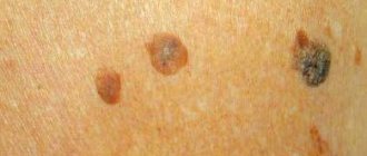
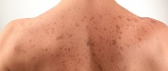
What does a keratoma (seborrheic keratosis) look like?
The appearance of a keratoma largely depends on how long it has existed.
In the initial stage, this formation protrudes very slightly above the skin level, differs slightly from it in density and has a color very close to flesh-colored. The shape of the lesions is round or oval.
Then, as the number of epidermal cells in the formation increases, the keratome increases in thickness and height. These formations are visible to the naked eye - milia-like cysts and comedon-like openings.
One common symptom for a keratoma, visible to the naked eye, is a surface that somewhat resembles cracked earth after rain. This pattern is formed by layers consisting of keratinized epidermal cells.
In addition to the clinical form - broad-based - there is a pedunculated form of seborrheic keratosis.
The human papillomavirus has a very distant relationship with this form of keratoma. In my experience, only in isolated cases did the histological examination of such formations indicate signs of viral damage.
Diagnostics + histology
If you discover any neoplasms in yourself, you should rush to see a qualified doctor; in no case should you draw your own conclusions by comparing your feelings with the symptoms from a medical reference book. It is not always possible to accurately determine the nature and danger of growths by external signs.
An experienced dermatologist-oncologist will be able to determine whether the neoplasm is a keratosis, the stage of development of the disease and the degree of its danger in terms of degeneration into an oncological disease. If factors predisposing to oncology are detected, the doctor prescribes removal of the growths using one of the available methods, followed by histological analysis of particles of excised tissue.