05/21/2018 Recently on Instagram and VK I published interesting prescriptions from a colleague regarding the diagnosis of seborrheic keratosis. The topic aroused keen interest, and as a result, I decided to write an article about this phenomenon. In it we will analyze in detail the reasons for the appearance of seborrheic keratosis (and, of course, myths), its varieties, possible health complications, methods of treatment and prevention.
To facilitate understanding: seborrheic keratosis and keratoma will be used as synonyms in the following.
Symptoms of actinic keratosis
The image in the photo allows you to see that at the age of 40 to 50 years, the initial stage of actinic keratoses looks like brown or black oval-shaped spots. Over time, the spots fade.
The sizes range from 3 to 6 mm. Location: neck, face, hands.
According to statistics, the risk of such a keratoma developing into skin cancer is 35%.
There are four stages of manifestation of actinic keratoses:
- Spot. At the first stage, spots appear on the human body. Especially typical for solarium lovers;
- Papular. The appearance of nodular formations on the skin, itching begins;
- The appearance of the keratoma itself or the so-called senile keratosis of the wart, which protrudes above the surface of the skin by several millimeters, with a rough surface;
- Cutaneous horn. The upper layer of the epidermis becomes keratinized.
Causes
The pathological process is polyetiological. The reason for the formation of neoplasms is, first of all, age-related degeneration of skin cells. The pathology is caused by two mutually opposite processes - aging and stabilization of cell activity. The lifespan of each cell depends on the balance of these processes. With age, skin cells partially lose their ability to withstand external negative factors, their RNA and DNA become vulnerable, the likelihood of tumor transformation increases, and changes in adaptation mechanisms are observed.
The main trigger for keratoma is ultraviolet radiation, the excess of which is neutralized by melanin. The pigment accumulates in the keratinocytes of the epidermis and is retained in the upper layers of the skin due to the cumulative effect. With age, melanin loses its ability to accumulate due to a slowdown in metabolic processes, while the intracellular secretion of melanin in keratinocytes increases with the simultaneous prevalence of hyperkeratosis processes. As a result, a keratoma is formed. With age, the immune system also loses some of its protective and control functions, which leads to intensive growth of epidermal cells and, as a consequence, the formation of keratinization areas.
Genetic failures provoke the formation of keratomas. In addition, the appearance of neoplasms is stimulated by neuroendocrine pathology, vitamin A deficiency, and impaired synthesis of sex hormones. Somatic diseases are important, affecting more than 70% of patients over 50 years of age. Keratoma also occurs against the background of pathogenic effects on the skin of chemicals, juices of poisonous plants, and long-term use of medications. In this case, the protective mechanisms of humoral and cellular immunity are activated, which, through macrophages and T-lymphocytes, pro-inflammatory cytokines and interleukins, activate inflammation with a predominance of proliferative processes and the development of hyperkeratosis.
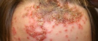
Symptoms of solar keratosis
The photo shows multiple examples of development as dots (depending on the development of various colors, from light pink to brown and even black) covered with peel.
Appears under the influence of ultraviolet radiation. Often the location is the hands, face, neck and forearms. People with light, sensitive skin are at risk.
Hair color in people prone to the disease ranges from light brown to red.
A tendency to the disease is also present in young people aged 25 to 30 years. This age is considered the most favorable for the development of cancer. Metastases appear 25% faster than at the age of 40 years. There is also a multiplicity of neoplasms.
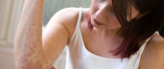
Symptoms of seborrheic keratosis
At the initial stage of development, these ulcers are predominantly yellow or brown in color.
After some time, nodules begin to form. The nodules have clear boundaries.
After some time, a seborrheic keratoma begins to form. It has a convex appearance and resembles a wart.
If you remove the scab, the wound will immediately begin to bleed and almost immediately the inflammatory process in the wound will begin.
The inflammatory process is accompanied by pain.
To diagnose this type of keratoma, doctors use a dermatoscope. With the help of which you can learn everything about the structure of the tumor. If there is a suspicion that the tumor is malignant, doctors should do a biopsy.
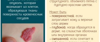
Classification
Benign hyperkeratotic skin neoplasms in dermatology are classified according to clinical manifestations and the degree of risk of malignancy.
There are senile, seborrheic, horny, follicular, solar keratoma and angiokeratoma.
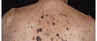
Seborrheic keratoma
Seborrheic keratoma appears approximately as often as senile keratoma. Its characteristics are unhurried growth with the formation of multilayer crusts.
The characteristics are as follows:
- by type - spots;
- by color - yellow;
- in size - no more than 3 cm in diameter;
- by location - most often on the chest, shoulders, back, scalp.
At the same time, there is a disruption in the functioning of the sebaceous glands - because of this, the spots in the lesion are covered with loose scales, which are easily separated.
With the development of seborrheic keratoma, the spots rarely remain separate from each other - they are grouped and can sometimes merge into a single whole. Also, such spots tend to grow along the edges. Crusts grow along with them - at the same time they begin to peel off and become covered with cracks. The thickness of such crusts in advanced cases can reach 1 cm. The keratoma becomes brown and sensitive to mechanical stress - even slight damage to it causes bleeding and pain.
Seborrheic keratoma is not prone to spontaneous disappearance or malignant degeneration.
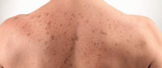
Senile (senile) keratoma
The first sign of the disease is the appearance of a light yellow or brown spot, which looks like a small hyperpigmented area of skin. As the spot develops, it acquires a darker color (burgundy, brown, gray), while increasing in size.
The growth of the neoplasm is combined with a change in its structure: it becomes loose, soft to the touch, while including keratinized cysts in the tissues of the base, which appear when the hair follicles are blocked. Infiltration, as well as accelerated growth of individual areas, leads to the formation of a lumpy surface with alternating protrusions and depressions, layers, veins, dark spots, etc. Later, the keratoma becomes rough; the layer of cells covering it flakes off, exfoliating in the form of small grayish scales.
Senile keratoma can range in size from 0.5 to 6 cm, most often 1-2 cm in diameter. Some keratomas may lighten over time, becoming pale brown or grayish. Often, age-related keratomas have a multiple distribution pattern, localized on the arms and lower extremities, face, neck, and occasionally on the body. If damaged, the tumor is prone to bleeding and inflammation, and can cause pain.
Senile keratomas, which can be treated in several ways, are often carried out in several stages, since the disease often recurs. Among the most effective methods of treating senile keratomas are:
- laser removal;
- electrocoagulation;
- radio wave method;
- cryodestruction;
- solcoderm;
- surgical excision.
If the keratoma is multiple in nature, the patient may be additionally prescribed a course of therapy with aromatic retinoids.
Other types of disease and their symptoms
In addition to age-related and seborrheic nodes, experts identify the following types of pathology:
- Follicular keratoma. This type of disease occurs near the hair follicles. The most common locations are the face, lips, and scalp. The appearance of small, colorless nodules on the body with a rough surface and a cone-shaped depression in the center is a sign of such a disease. As the papules grow, multiple clogged hair follicles (horny cysts) appear. Such a tumor is prone to recurrence even after removal.
- Solar keratoma (actinic keratosis). The main group that is susceptible to developing this form of the disease is men with fair skin. This precancerous disease is dangerous because the process of malignancy occurs hidden. At the initial stage, the patient may notice small multiple pink nodules on the body (most often on the face). As they grow, the sun seals become brown or brown in color and are covered with dense scales that come off easily.
- Angiokeratoma. This compaction does not have clear boundaries, often occurs in the scrotum area, and does not resolve on its own. Externally, it looks like a skin hemangioma. It may contain blood vessels, which is why the color of the neoplasm is blue or even black (depending on the number of capillaries). Nodes can be either single or multiple.
Large tumors covered with a thick crust can be damaged and torn off by clothing. These lumps often bleed and hurt.
Symptoms of Angiokeratoma
The image shows a tumor turning from red to black. The tumor node appears from the papillary dermis.
The size of the dermis does not exceed 10 mm, but there are no clear boundaries. There are cases when neoplasms appear in a child at birth under the guise of a hematoma bruise.
There is also an angiokeratoma of the scientist Fordyce - the scientist proved the existence of keratomas on the genitals.
As the disease manifests itself, it turns into a malignant tumor with painful bleeding wounds.
It is necessary to diagnose such a problem taking into account various types of cell cancer, papillomas and warts. To determine the nature of the origin, a biopsy is used in diagnosis and a histological examination is performed.
If the tumor is malignant, its removal is only possible through direct surgery, because any other effect can accelerate the development of cancer and its metastases.
Difference from melanoma
A benign skin growth differs from melanoma in that it does not contain atypical cells. The degree of malignancy of the tumor or its absence is revealed by histological examination. Melanoma has an uneven surface and contours, dark color and heterogeneous structure.
Keratoma is not contagious to surrounding people, since it does not occur against the background of viruses or infections. The disease can be diagnosed in a child as a congenital pathology.
Arsenic keratosis
Occurs only in men. The following image allows you to see cancerous changes in the skin.
What happens under the influence of arsenic. It looks like dark gray papules, ranging in size from a poppy seed to a pea.
Arsenic appears in the body during chronic poisoning with drugs containing it or when working with similar substances.
Affected areas are the feet, fingers, back of the bones.
The affected area also includes internal organs such as the bronchi and liver.
Malignant tumors of these organs appear, as well as cirrhosis of the liver and hepatitis. The histological characteristics are the same as for Bowen's disease.
This species is capable of degenerating into the solar stage of cancer. Inflaming the area around the tumor, with the appearance of pain, itching, and bleeding.
Etiology of the disease
The true reason that triggers the formation of the node has not been established. Doctors identify several factors that can influence changes in the epidermis:
- Old age of a person, especially a man.
- A skin nodule occurs in people whose parents or close relatives have experienced keratoses.
- The disease usually develops in exposed areas of the body. It has been established that ultraviolet radiation negatively affects the condition of the skin.
- The risk group includes people with freckles, nevi and a large number of moles.
- Skin condition worsens due to lack of vitamin A.
Types of keratomas
Diagnostics
Diagnosis is carried out based on:
- patient's age,
- absence of malignant tumors,
- biopsy of a piece of tissue infected with a wart.
Diagnosis is also carried out taking into account other diseases.
Especially if surgery with anesthesia is required, the presence of heart disease is determined. After all, if the patient has a weak heart, then the administration of anesthesia is prohibited.
In such cases, more loyal methods are used to combat the disease.
Diagnostic tests
The disease is treated by a dermato-oncologist. After clarifying the patient’s complaints, an external examination of the tumors is carried out. The doctor can suggest the type of pathology, but additional diagnostics will be needed to clarify the diagnosis:
- In older people, it is important to compare keratoma with other skin neoplasms, for example, warts and papillomas.
- The surface of the tumor is examined through a dermatoscope.
- Histology is performed after removal of the growth.
Types of keratomas
Treatment
There is no perfect cause, and therefore there is no cure as such. But after studying this problem, doctors came to the conclusion that with various complex simulations, the treatment gives results.
- Taking immune stimulants and antioxidants. That is, taking vitamin C in large quantities. This method does not get rid of keratosis, but it will reduce the size of the keratomas.
- Lubricating stains with hormonal ointments. This reduces the risk of contracting fungal infections due to mechanical damage.
- Fruit and vegetable diet. Proper nutrition will enrich the body with vitamins and restore the vitamin complex.
- Reducing stress. With normal health of the nervous system, the risk of keratoma developing into skin cancer is reduced.
- A way to avoid sun exposure. If you avoid ultraviolet radiation, this will not only get rid of new formations, but also improve the functioning of uninfected areas of the skin.
- Full sleep. Will provide a healthy look and rest for the whole body.
Dangers
It should be borne in mind that in the vast majority of cases, keratomas are not dangerous to human health. The extent of their damage to appearance as a cosmetic defect should be considered in each specific case. Of course, large or multiple keratomas significantly worsen the quality of life of a modern person, since they disfigure the appearance, especially if they are located on the facial segments.
Keratomas are dangerous when they are located in places subject to compression by clothing or friction, or if there is a risk of frequent injury.
An infection can easily get into a damaged keratoma, provoking the occurrence of serious diseases - eczema, pyoderma. The risk of infection with dangerous microorganisms, for example, herpes, papilloma virus, increases.
Eczema is a serious disease that requires serious treatment. Therefore, it is very important to recognize it at the initial stage and begin treatment.
The greatest danger is posed by certain types of keratomas, which, when unfavorable factors coincide, can degenerate (malignize) into malignant forms. This ability is inherent especially in the sunny and horny types of the disease.
Attention! When monitoring the development of suspicious hematomas, special attention should be paid to the areas around such keratomas, and if inflammation and pain around them, itching, and bleeding are suspected, the alarm should be sounded.
Indications for keratoma removal
- When a flaw appears;
- If there is a possibility of it developing into a cancer cell;
- If it interferes with life activities;
- If there is a lot of it on the body, especially on the face.
Method of surgical removal of warts
The photo above shows, as an example, the first and cheapest method of laser cleansing. With the help of which keratosis spots disappear immediately. This is the cheapest removal method.
Surgitron device (radio knife), for the radio wave method of removal.
This type of treatment is performed using local anesthesia. And it is much more effective than a laser, because thanks to the action of radio waves, the skin surface is less injured without scars.
This type of removal is used to remove keratomas on the face.
There is another method, removal using cauterization with liquid nitrogen. This method is not used often, because it is the most traumatic and the operation is performed by a surgeon. Healing after such an operation takes a long time.
There is also a method of surgical removal, namely electrocoagulation. This method removes only small formations. Using a device that generates current, the structure of the keratoma is destroyed, and a small wound remains in its place.
Preventive measures against the appearance of senile keratomas.
- Take vitamins. To increase the vitamin balance of the body. And reduce the aggressiveness of age-related and hormonal effects.
- DO NOT sunbathe. This is the basic principle of recovery, because the sun is the main factor influencing skin cells;
- Eat exclusively healthy foods. This improves blood circulation in the cells;
- More relaxation and less nerves.
When any keratomas appear on your skin. They need to be removed urgently, or if removal is prevented, consult a doctor regarding further actions and treatment.
Attention! Under no circumstances should you select treatment on your own, because due to your own ignorance you can aggravate the symptoms. And due to injuries, it can even turn into skin cancer.
If you decide to remove tumors, you should consult with an oncologist. It is not necessary to take tests before the operation itself, but it will not hurt.
It is important to know! Doctors recommend that immediately after the appearance of a keratoma, especially under the age of 40, it should be removed immediately. The fact is that in the future, after age-related hormonal imbalance, the risk of getting skin cancer exceeds 50%.
Preventive measures and forecasts
To prevent skin diseases in old age, it is recommended to avoid contact with direct sunlight. This is especially true for vacationers on the sea coast. Maximum solar activity is observed from 10 a.m. to 4 p.m. At this time, it is not advisable to leave the room. If you need to stay in the sun, it is important to use sunscreen with a high spf factor and move to shaded areas.
Preference should be given to loose, light-colored clothing made from natural fabric. A wide-brimmed hat or cap is suitable to protect your face. Glass sunglasses will keep your eye skin healthy.
Keratoma rarely affects the body of people with strong immunity. After 30 years, it is important to be physically active, perform breathing exercises, and maintain a sleep and work schedule.
Special attention is paid to the quality of food. It is recommended to include fresh vegetables or fruits in every meal. To replenish vitamin A deficiency, boiled or baked pumpkin, liver dishes, carrots, apricots and dandelion leaf salads are suitable.
If the keratoma has developed into cancer, survival rate is reduced due to the spread of atypical cells throughout the organs and systems of the human body. Fortunately, senile pathology rarely malignizes and a person lives to a ripe old age. If unknown changes appear on the body, you should consult a dermatologist to exclude malignant pathology and preserve a full life.
Treatment with traditional methods.
To cure the disease at home, folk methods from the list below will help you.
Aloe leaf recipe.
Young aloe coots need to be frozen. Freezing time is approximately three days.
After defrosting, make a compress from the leaves and apply to the new growths.
Recipe for using potatoes.
The grated potatoes should be placed on top of the wound, covered with a cotton cloth, and also wrapped with cling film on top.
Recipe for attracting castor oil.
Using castor oil, senile keratoma is removed. The method of use is very simple. It needs to be warmed up and used every day.
Preventing keratomas
After removal of the keratoma, it is necessary to take a number of measures to prevent relapse - the reappearance of the tumor. To do this, follow these simple recommendations:
- Avoid direct exposure of the skin to sunlight. Ultraviolet radiation provokes the formation of keratomas. Use creams with sunscreen filters, at least 40 SPF in summer.
- Avoid solariums, they contain direct rays of the same ultraviolet radiation.
- Take vitamin complexes, in particular vitamin C, it prevents the formation of keratomas. Or include foods rich in this vitamin in your diet: black currants, raspberries, citrus fruits.
- Lead a healthy lifestyle: exercise, balance your diet, eliminate fatty, fried foods, and sweets. Such measures will help improve metabolism and prevent the appearance of keratomas.
Surgeon and cosmetologist Vitaly Mikryukov will talk about keratomas and their removal:
Recipe using celandine.
You need to make lotions from the leaves and stems of dry celandine. Use for treatment and against keratoma restoration. The ratio of plant and water is 2 spoons per 25 ml of water.
Also, during treatment and preventive measures, you need to drink and eat foods high in vitamin P.
Products that contain this vitamin in large quantities:
- buckwheat;
- black chocolate;
- black currant;
- Bell pepper;
- lemon.
Also, tinctures that contain vitamin:
- burdock (drink as juice or make tincture),
- parsley (to make tincture),
- green teas,
- dill.
Recipe using sunflower and sea buckthorn oil.
The recipe is very easy to prepare and very effective. Mix the oil in a one to one ratio and lubricate the keratomas three times a day.
Dandelion recipe.
Dandelion juice and compresses from the leaves of the plant are suitable for use. It is also useful to drink juice from lemon and garlic, diluting them with water. Take three times a day, adding 7-8 drops to water.
Attention! Before using any product, be sure to consult your doctor.
Therapeutic baths
Since many doctors consider keratomas to be signs of skin aging, some of them themselves turn to traditional medicine, where there are many means to stop or significantly slow down such aging. To do this, you won’t need to take a bunch of vitamins or whole handfuls of antibiotics, it will be enough to just take baths - these methods are simple.
Baths with plant decoctions that contain vitamin P, that is, bioflavonoids, can have a remarkable effect on skin health. Use umbrella plants, legumes, rosaceae and buckwheat for water procedures - they are the most effective.