Tracheobronchoscopy (full name of the procedure) is a modern diagnostic and treatment method for visualizing the internal surfaces of the trachea and bronchi.
The examination is performed with a special optical device – a fiberoptic bronchoscope. Essentially, this is a multifunctional endoscope, which consists of a flexible cable with a light source and a video/photo camera at the end and a control handle with an additional manipulator.
About bronchoscopy - more details
Bronchoscopy is a procedure that is performed only in a hospital. Under local (treatment of mucous membranes with lidocaine) or general anesthesia, the doctor inserts a special device into the respiratory tract - a bronchoscope, which is either a flexible or rigid tube. At one end of the device there is an illuminator, the other ends with an optical system, where the doctor looks directly with his eyes.
There are holes on the side of the bronchoscope where you can connect:
- syringe: for rinsing the respiratory tract or for aspirating sputum for analysis;
- electric suction: it will “suck” sputum or blood - the contents of the trachea and bronchi;
- special forceps or brushes for taking biopsies;
- coagulator electrode – a device for cauterizing bleeding vessels.
For these instruments, there is a special channel in the body of the device through which they pass. In addition, the device can communicate with video equipment so that the doctor assesses the condition of the bronchi, looking not into the “tube” of the device itself, but by looking at the monitor.
Typically the bronchoscope is inserted through the mouth. Some doctors use a laryngoscope for this - a device that will simultaneously illuminate the path for the bronchoscope and squeeze the root of the tongue and the epiglottis - the cartilage into which the flexible bronchoscope can rest.
Since bronchoscopy is vital in many cases (for example, if there is an injury or malformation of the neck and breathing needs to be done using a breathing apparatus), the bronchoscope can be inserted through the nose.
Also, if the patient breathes through a tracheostomy (an opening in the trachea through which a special cannula connected to a breathing apparatus is inserted), the bronchoscope is inserted directly into the tracheostomy opening. In this case, separate anesthesia is not required.
What does bronchoscopy show:
- trachea;
- the main ones are the right and left bronchi;
- lobar bronchi: three on the right, two on the left.
The bronchoscope does not visualize smaller bronchi and bronchioles. If there is a suspicion that a tumor or inflammation is located there, a computed tomography or magnetic resonance imaging scan is performed.
We hope that it is clearly explained what it is - bronchoscopy of the lungs, although it is more correct to call this manipulation simply bronchoscopy (it means “visualization of the bronchi”).
Types of bronchoscopes
The newest bronchoscopes fall into two categories: flexible and rigid. The models differ in their advantages and scope of use. A flexible bronchoscope has a second name - fiber bronchoscope. The instrument is based on fiber optics. The device consists of:
- control handles;
- an elastic tube with a flat surface having an optical cable and a light guide;
- optical device - video camera;
- LED light source;
- controlled manipulator;
- a catheter for the delivery of medicines or removal of viscera;
- auxiliary ultrasound and surgical apparatus.
Positive aspects and advantages of a fiber-optic bronchoscope:
- the possibility of passage into distant parts of the bronchi, difficult for the introduction of a solid bronchoscope;
- reduced trauma to the bronchial membranes;
- the small diameter helps to use the device in pediatrics;
- General anesthesia is not used.
The instrumental device is used for:
- diagnostic measures of the trachea and bronchi, in particular the distant sections;
- viewing the mucous tissues of the air duct;
- elimination of small foreign objects.
The rigid model of a bronchoscope consists of:
- light source;
- manipulator for movement control;
- installation of solid hollow tubes;
- photographic and video equipment;
- devices for carrying out procedural actions - aspirators, a group of forceps and grippers;
- auxiliary laser device.
Model of a bronchoscope inside the lungs
The advantages and positive qualities of a rigid bronchoscope are reflected in:
- Widespread use of the device for therapeutic measures that are unattainable for a flexible bronchoscope model: increasing the bronchial opening, removing foreign objects that block the respiratory system.
- Possibility of introducing an elastic instrument through a solid bronchoscope for the analysis of thin bronchial membranes as an alternative to other devices.
- Removing and eliminating complicated processes identified during the examination.
- Use of the device for resuscitation of patients: in case of drowning, development of cystic fibrosis to eliminate fluid and mucous masses from the lungs.
- The use of general anesthesia, due to which there is no discomfort. This feature is important for people with increased anxiety and unreasonable fear.
The tool is used for:
- Normalization of bronchial patency and tracheal tracts caused by the formation of scars or growths, installation of stents to open and narrow the bronchi.
- Elimination of scarring, tumor growths and lumps of sputum.
- Detecting foci of inflammation of the air duct.
- Stop bleeding.
- Seizure of foreign objects.
- Washing the bronchi and introducing chemicals.
Bronchoscopy with removal of the capsule from the left main bronchus
Indications for bronchoscopy
You need to undergo a bronchoscopy if:
- there is shortness of breath in the absence of heart pathologies or bronchial asthma;
- I have a cough, but X-rays show nothing;
- there is hemoptysis;
- bronchitis and/or pneumonia often recur;
- foul-smelling sputum is produced;
- there is a feeling of incomplete inhalation or exhalation, while diseases of the heart and thoracic spine are excluded;
- there was rapid weight loss in the absence of any diets;
- have cystic fibrosis;
- An X-ray of the lungs reveals a disseminated process - many areas of darkening, which can be either metastases or pulmonary tuberculosis;
- according to computed tomography data, it is impossible to distinguish an area of suppuration from lung cancer with decay;
- a diagnosis of pulmonary tuberculosis has been established;
- it is necessary to establish the cause of severe pneumonia when the patient is on mechanical breathing;
- it is necessary to evaluate the dynamics of treatment after resection of the lung and bronchus;
- repeat bronchoscopy is needed after the tumor has been removed using this technique;
- if dilation or narrowing of the bronchi is visible on the x-ray.
This is a diagnostic bronchoscopy and is used to make a diagnosis.
There is also a treatment procedure that is used when:
- a foreign body has entered the respiratory tract;
- It is impossible to perform tracheal intubation in order to transfer the patient to artificial ventilation: for surgical intervention or in critical situations. This is a coma caused by various reasons; conditions when breathing is switched off (cervical spinal cord injuries, botulism, myopathies);
- you need to clear the airways of phlegm or blood. This is extremely important in the treatment of pneumonia, especially against the background of cystic fibrosis, when the sputum is very viscous;
- pulmonary bleeding must be stopped;
- one of the bronchi was blocked by a tumor, adhesions or sputum, resulting in atelectasis (exclusion of a section of the lung from breathing);
- it is necessary to remove pus from the lung abscess located near the bronchus;
- pneumonia is severe: it is better to inject an additional antibiotic directly into the desired bronchus.
Basically, bronchoscopy is performed using a flexible bronchoscope – fiberoptic bronchoscope. It is quite thin and can bend in different directions. But in some cases, it is necessary to introduce a rigid (metal) device that does not bend and cannot be inserted into the bronchi that extend at an angle.
Indications for bronchoscopy with a rigid bronchoscope are the removal of foreign bodies, expansion of bronchi narrowed by inflammation or adhesions. It is more convenient to put a stent (an expanding tube made of rigid corrugated plastic) on a rigid bronchoscope and install the latter into the narrowed bronchus. It is best used during thoracic operations - in the treatment of conditions associated with the entry of pus, air or liquid into the pleural cavity, as well as pulmonary hemorrhage. Then, using a bronchoscope, you can block the bronchus on the painful side, where surgeons work, and ventilate the second lung with the device.
Extended and Modified Methods
Sometimes advanced forms of visualization may be used as they can provide more comprehensive visualization. The following methods exist:
- Virtual bronchoscopy. During virtual bronchoscopy, a CT scan is used to see the airways in more detail. This procedure does not use a bronchoscope, meaning it is not endoscopic, but a type of CT scan.
- Endobronchial ultrasonography. Endobronchial ultrasonography uses an ultrasound probe that is attached to a bronchoscope to view the airway.
- Fluorescence bronchoscopy. During fluorescence bronchoscopy, a fluorescent light is additionally used, which is attached to the bronchoscope - this allows you to see the inside of the lungs.
New methods of bronchoscopy:
- Bronchial thermoplasty: This new technique is being developed to gently heat the airways of some patients with asthma. It reduces asthma exacerbations.
- Emphysema Volume Reduction: Small one-way valves are placed in the airway of the damaged lung, they reduce the volume of that part and leave room for the remainder of the normal lung to function.
- Repairing air leaks after lung resection: One-way valves are used to slow air leaks at lung suture lines. By slowing the airflow, these leaks can heal faster and prevent the need for further surgery.
- Sanitation bronchoscopy, which is performed for therapeutic purposes.
Virtual bronchoscopy
In addition to rigid and flexible bronchoscopy, another type of examination has been developed - virtual bronchoscopy. It is a computed tomography scan of the lungs and bronchi, which is processed by a special computer program that recreates a three-dimensional image of the bronchi.
The method is not as informative, but it is non-invasive. With it, you cannot take a sputum test, rinsing water or a biopsy of a suspicious area, you cannot remove a foreign body or rinse the bronchi from sputum.
No preparation is required for a virtual biopsy. According to the method of execution, it does not differ from computed tomography. The patient lies down on a couch that is placed inside the X-ray source.
Although X-ray radiation is low-dose, the method is not suitable for children and pregnant women.
Diagnostic and therapeutic capabilities of modern bronchoscopy
Today, more than a hundred years after the “father of bronchoscopy” Gustav Killian first inserted an endoscope into the trachea and removed an aspirated meat bone from a patient, bronchoscopy is one of the leading methods for diagnosing and treating respiratory diseases. Most lung diseases are in one way or another associated with bronchial pathology. The airways provide access to any areas of the lung, allow one or another instrument to be passed to them and obtain a variety of information about the state of the respiratory organs, and also represent an additional route for administering medications to pathologically altered areas of the lung.
History of development
The development of bronchoscopy can be divided into three stages. At the first, which began at the end of the 19th century. and continued until the end of the 50s of the twentieth century, bronchoscopy was performed under local anesthesia, usually using rigid bronchoesophagoscopes
, which had a dual purpose - examination of the tracheobronchial tree and esophagus. The progress of bronchoscopy during this period was greatly facilitated by the work of Ch. Jackson, J. Lemoine, A. Soulas, A. Olsen, N. Andersen, and in our country - A. Delens, V. Voyachek, V. Trutnev, A. Likhachev and Chalky. Bronchoscopy during this period was performed mainly for foreign bodies in the respiratory tract and was carried out mainly by otolaryngologists. The procedure was very traumatic, and the patients had a hard time bearing it.
With the advent and improvement of general anesthesia, pulmonary surgery began to actively develop and indications for bronchoscopy expanded significantly. This was facilitated by the creation in the late 50s - early 60s of respiratory bronchoscopes (N. Friedel; R. Hollinger; G.I. Lukomsky), which made it possible to perform bronchoscopy under anesthesia with myoplegia and injection ventilation of the lungs, which significantly alleviated the suffering of patients and made the study safer. The progress of bronchoscopy at this second stage of its development was facilitated by the appearance of lens telescopes with direct, lateral and retrograde optics, various instruments for biopsy, extractors, scissors, and electrocoagulators. At this stage, bronchoscopy passed into the hands of thoracic surgeons.
A true revolution in bronchology and the beginning of the third, modern stage in the development of bronchoscopy was the creation in 1968 of a flexible bronchofiberscope
[1], with the help of which it became possible to examine the lobar, segmental and subsegmental bronchi of all parts of the lung, perform a visually controlled biopsy, and administer medicinal solutions. The fiberoptic bronchoscope has significantly changed the technique of bronchoscopy. It was again performed under local anesthesia, causing almost no discomfort to the patients. Bronchofibroscopy began to be successfully performed on an outpatient basis, in pulmonology hospitals and offices, and in intensive care units. It seemed that the need for rigid bronchoscopes had disappeared forever. However, the creation of high-energy medical lasers determined a new direction in bronchology - surgical endoscopy, and rigid endoscopes were again required for it. Therefore, modern bronchoscopy is equipped with both flexible and rigid endoscopes and instruments that allow performing a wide range of diagnostic and therapeutic manipulations in the trachea and bronchi both under local anesthesia and under general anesthesia.
Indications for bronchoscopy
The main indications for bronchoscopy are shown in Table. 1. They should be divided into diagnostic and therapeutic.
Diagnostic bronchoscopy
Tumors of the bronchi and lungs
Tumors of the bronchi and lungs
Tumors of the bronchi and lungs are one of the main indications for bronchoscopic examination. Currently, verification of centrally located endobronchial cancer reaches almost 100% and is carried out by visually controlled biopsy with wire cutters. Diagnosis of the so-called “early cancer”, which also includes tumors in situ, is more difficult. Fluorescent chromobronchoscopy helps to identify such neoplasms, almost invisible to the naked eye.
with the introduction of special drugs - photosensitizers [2, 3].
Diagnosis of peripherally located tumors, especially small ones, can also be quite difficult, since getting to such tumors through the bronchi is very difficult.
Their biopsy is performed under the control of an X-ray television screen and, in addition to cutters, scarifying brushes and controlled curettes are used. However, even in experienced hands, verification of peripheral lung tumors rarely reaches 60-70% and in complex cases should be combined with percutaneous puncture biopsy under computed tomography (CT) control.
Diagnosis of peribronchial cancer also requires great skill, especially in the early stages. The presence of such a tumor can be suspected based on X-ray and CT data, and to verify it, a puncture biopsy
the bronchial wall in a suspicious place using a special needle. Thus, all darkening or cavitary formations in the lungs, hilar or located on the periphery, suspicious for the oncological process, are direct indications for bronchoscopy and various methods of bronchoscopic biopsy, the choice of which is determined by the doctor performing the study.
Mediastinal neoplasms and lymphadenopathy
Mediastinal neoplasms and lymphadenopathy may also be indications for bronchoscopy. With enlarged paratracheal and bifurcation lymph nodes and mediastinal tumors located in close proximity to the trachea, material for cytological examination can be obtained using transtracheal puncture biopsy
. However, the not very reliable results of such a study have now ceased to meet the requirements of practice and bronchoscopic methods have been replaced by more invasive, but much more informative methods: mediastinoscopy, pleuromediastinoscopy [4, 5] and video thoracoscopy [6]. They should be resorted to in cases where bronchoscopic biopsy methods are ineffective.
Diffuse lung diseases
The same trend can, to a certain extent, be attributed to the diagnosis of diseases accompanied by diffuse changes in the pulmonary pattern (the so-called diffuse lung diseases - DLD), which require morphological research for their verification. Since the early 70s, after the work of N. Andersen, transbronchial pulmonary biopsy
, performed using a bronchoscope, has become the leading method for diagnosing DLD [5]. Over time, however, it became clear that with transbronchial lung biopsy it is not always possible to obtain enough lung tissue to successfully carry out differential diagnosis in a number of DLD, especially those accompanied by fibrosing processes in the pulmonary parenchyma. And although the relatively less invasiveness still allows transbronchial lung biopsy to remain the method of primary endoscopic diagnosis of DLD, it is increasingly complemented by thoracoscopic biopsy, performed using wire cutters or endostapplers, hermetically stitching the lung parenchyma while simultaneously cutting off a section of lung tissue of the required size [6].
Important diagnostic information for many lung diseases, and primarily for DLD, can be obtained by studying material obtained using bronchoalveolar lavage (BAL)
.
The latter today is an almost obligatory study in the diagnosis and treatment of DLD such as cryptogenic fibrosing alveolitis and sarcoidosis. BAL performed repeatedly during the treatment of these diseases makes it possible to monitor the effectiveness of therapy and determine its prognosis. Bronchoscopy and BAL are also indicated for suspected fungal diseases of the bronchi
(bronchomycosis) and some
parasitic diseases of the lungs
(for example, with Pneumocystis pneumonia).
Inflammatory processes in the lungs
A bronchoscope allows you to look deeply into the airways. This makes it possible for patients with descending tracheobronchitis to determine the distal border of the lesion of the bronchial tree and the intensity of inflammation in it. Bronchoscopy is effective in searching for a draining bronchus in acute lung abscesses, as well as in the differential diagnosis of bacterial suppuration and disintegrating cancer in the presence of a cavity in the lung. The difficulties of absolute sterilization of bronchofibroscopes somewhat complicate the microbiological diagnosis of inflammation and require the use of special catheters that protect the material collected in the bronchi from contamination with the contents of the oral and nasal cavities. Our experience in the use of bronchoscopy in patients with acute and chronic inflammatory lung diseases allows us to give preference to the use of a rigid bronchoscope, amenable to thermal sterilization methods, if microbiological diagnosis of pulmonary suppuration is necessary.
Pulmonary hemorrhage and hemoptysis
If we strictly follow the terminological logic, hemoptysis is a manifestation, a symptom of pulmonary hemorrhage. However, in practice, pulmonary hemorrhage (or hemoptoea) refers to the release of pure blood or intensely bloody sputum when coughing, and hemoptysis (hemophthisis) refers to the coughing up of sputum tinted with blood or containing streaks of blood. Thus, there is a quantitative difference between hemoptoea and hemophthisis [4, 5]. Both pulmonary hemorrhage and hemoptysis are direct indications for diagnostic bronchoscopy, since this is the only way to determine the source of bleeding or at least its approximate location.
The causes of pulmonary hemorrhage and hemoptysis are extremely diverse. In addition to the pathology of the tracheobronchial tree and lung parenchyma, among them are diseases of the blood and circulatory system, hemorrhagic diathesis and capillary toxicosis, pulmonary embolism, pulmonary endometriosis, etc. The relative frequency of these causes has changed over time. Thus, in the 30s and 40s, destructive pulmonary tuberculosis was in first place among all causes of pulmonary hemorrhage. Currently, the most common cause of hemoptysis in the pulmonology clinic is chronic bronchitis accompanying bronchiectasis or focal pneumosclerosis, in which in foci of chronic inflammation against the background of decreased blood flow along the branches of the pulmonary artery, excessive vascularization develops due to dilation of the bronchial arteries and multiple anastomoses arise between the large and small circles of blood circulation.
Due to the shunting of blood from the bronchial arteries into the branches of the pulmonary artery, hypertension occurs in the microvasculature of the lungs, which the fragile walls of small vessels cannot withstand, and the blood enters the respiratory tract. Similar mechanisms are observed in the area of foci of destruction of lung tissue of specific and nonspecific etiology. During bronchoscopy in these cases, the source of bleeding, as a rule, cannot be seen, but it is quite possible to determine at least its approximate location, especially if the study is performed at the height of hemoptysis. This is very important for determining treatment tactics for each individual patient.
The causes of hemophthisis and hemoptoea diagnosed in patients in the thoracic surgical department are presented in Table. 2. Undoubtedly, the most serious cause of pulmonary hemorrhage and hemoptysis was and is bronchial tumors and, above all, cancer, which can only be verified using bronchoscopy. This allows us to conclude that in all cases of pulmonary hemorrhage and hemoptysis, bronchoscopy is a mandatory study, the main purpose of which is to identify or exclude a malignant neoplasm of the lungs
.
Chronic cough
Among the indications for diagnostic bronchoscopy, the so-called treatment-resistant cough should also be mentioned, i.e. cough that does not respond to intensive treatment for at least 1 month, the cause of which remains unclear. And although lung tumors, according to R. Irwin et al. [7], are rarely accompanied by an isolated cough syndrome (without any radiological manifestations), our experience in examining persistently coughing patients [4, 5] gives us reason to assert that bronchoscopy is one of the most important studies in the complex diagnosis of the causes of chronic cough.
Broncho-obstructive syndrome
Bronchoscopy plays an important role in the differential diagnosis of chronic obstructive pulmonary diseases and obstruction of the trachea and bronchi, accompanied by broncho-obstructive (asthmoid) syndrome. This primarily applies to tumors, foreign bodies (including those of endogenous origin - broncholitis) and cicatricial strictures of the trachea and large bronchi, in which radiological symptoms may be completely absent, and the clinical picture is very similar to an attack of bronchial asthma [8, 9].
Therefore, in cases where patients have signs of difficulty breathing that are not relieved by modern drug therapy, a bronchoscopic examination is indicated, which often reveals one or another organic pathology in the large respiratory tract.
Therapeutic bronchoscopy
Removal of aspirated foreign bodies
Removal of aspirated foreign bodies
The therapeutic capabilities of bronchoscopy have long been limited to the extraction of aspirated foreign bodies, and even now this is the only bloodless method of removing them from the bronchi.
The development of flexible extractors and the considerable experience accumulated to date suggests that most aspirated foreign bodies in adults can be removed using a bronchofiberscope under local anesthesia and even on an outpatient basis [5]. However, foreign bodies in the respiratory tract sometimes present the bronchologist with the most unpleasant surprises, forcing the use of general anesthesia and hard instruments and requiring maximum concentration of strength and skill, and sometimes inspiration.
Drainage of intrapulmonary purulent foci
The therapeutic effect of bronchoscopy as a method of draining intrapulmonary purulent foci, be it bronchiectasis or lung abscesses, is undeniable. Therapeutic catheterization of the bronchi during bronchoscopy makes it possible to unblock a significant part of the intrapulmonary abscess cavities [5], and long-term transnasal drainage [10] ensures the constant introduction of antibacterial drugs into the cavity and frees patients from repeated bronchoscopy and catheterization. A technique for immunoreplacement therapy has been developed in the form of intracavitary administration of a suspension of autologous macrophages [11], making bronchoscopic treatment even more effective.
Chronic obstructive bronchitis
The therapeutic role of bronchoscopy in chronic obstructive bronchitis (COB) has traditionally been limited to restoring airway patency with stimulation or imitation of impaired bronchial drainage function and local use of antibacterial and secretolytic agents. After the first publications by A. Soulas and P. Mounier-Kuhn, who described the method of treating patients with chronic nonspecific lung diseases using a bronchoscope, many different methods of bronchoscopic treatment of COB were proposed. Some of them were abandoned as having not been tested by practice, others took a strong place in the arsenal of therapeutic agents for patients with diseases of the bronchopulmonary system [5, 12].
Currently, sanitation bronchofibroscopies
, carried out under local anesthesia in a course method with a frequency of 1 time every 2-3 days.
The duration of the course depends on the severity of the pathological process and the effectiveness of treatment and ranges from 3 to 20 sessions. If the sputum is purulent and there is a significant amount of it, 10 ml of a 0.5-1% solution of potassium furagin, warmed to body temperature, is instilled through the channel of the bronchofibroscope into the bronchi with the addition of 1-2 ml of a mucolytic ( ambroxol, acetylcysteine
).
Before removing the bronchofibroscope, antibiotics are injected into the lumen of the bronchi in a daily dose (in accordance with the sensitivity of the bronchial microflora to them). In the presence of purulent sputum with an ichorous odor, instillation of a 1% dioxidine solution in an amount of 5-10 ml is used. At the end of the procedure, the patient is placed alternately on each side for 5-7 minutes, after which he is asked to actively cough.
The emergence of new technical devices is also reflected in the endobronchial therapy of inflammatory lung diseases. The publications of E. Klimanskaya, S. Ovcharenko, V. Sosyura and others describe the use of low-frequency ultrasound and radiation from ultraviolet and helium-neon lasers
during therapeutic bronchoscopy in patients with chronic bronchitis and pulmonary suppuration, including children. The authors have obtained good results from the use of these methods, which, in their opinion, contribute to better sputum production, increased concentration of antibiotics in the bronchi and improved local immune defense of the respiratory tract.
NOT. Chernekhovskaya and I.V. Yaremaya [13] obtained a positive effect from the intrabronchial use of the immunomodulator T-activin, which, according to the authors, contributes to the restoration of the immune reactivity of the bronchial mucosa. In patients with COB, the drug was injected during bronchoscopy using a needle into the mucous membrane of the spurs of the lobar and segmental bronchi in the places of the most visually pronounced inflammation. For severe inflammation in the bronchi, the authors recommended the use of intrabronchial immunotherapy in combination with endolymphatic administration of antibiotics into interbronchial spurs.
In conclusion, we consider it our duty to remind you that sanitary bronchoscopy is a rather crude and traumatic method of treatment and should be performed in patients with COB if there are appropriate indications, which primarily include purulent complications and a pronounced obstructive component of the disease. It is not necessary to expand the indications for therapeutic bronchoscopy in patients with serous forms of endobronchitis without severe bronchial obstruction, where it is quite possible to achieve good results using inhalation, injection or oral methods of administering therapeutic drugs. Bronchoscopy is a “cannon” method of treatment and is hardly worth using when “shooting at sparrows.”
Severe bronchial asthma
If there is a significant accumulation of thick, viscous sputum in the distal parts of the bronchi in cases of ineffective expectoration, which is often observed in severe bronchial asthma, therapeutic bronchial lavage
. For the first time, massive lavage of the bronchi through an endotracheal tube was described by H. Thompson and W. Pryor in patients with alveolar proteinosis and bronchial asthma. By modifying this method, we developed a technique for therapeutic bronchial lavage through a rigid bronchoscope under conditions of pulmonary injection ventilation [5, 12]. Therapeutic bronchial lavage in patients with severe respiratory failure requires highly qualified anesthesiological care and post-anesthesia monitoring in an intensive care unit or intensive care unit. When performed correctly, this procedure effectively helps remove sputum from medium- and small-caliber bronchi that are inaccessible to other methods of endobronchial aspiration. It is important to point out the dangers of using this technique in patients with purulent forms of endobronchitis, since the absorption of liquefied and not completely removed purulent sputum can lead to increased intoxication and worsening of the patients’ condition.
In several particularly severe patients with status asthmaticus and hypoxic coma, we performed bronchial lavage under conditions of extraorgan oxygenation. The experience of using such a resuscitation aid is relatively small, but it deserves attention and can be used in specialized intensive care units.
Early postoperative period
Bronchofibroscopy has proven itself to be an effective treatment procedure for impaired bronchial obstruction in patients in the early postoperative period and, especially, in patients requiring long-term artificial pulmonary ventilation (ALV). A flexible bronchofiberscope can be easily inserted into the patient’s respiratory tract through an endotracheal or tracheostomy tube, which makes it possible to perform sanitary bronchoscopy in patients on mechanical ventilation daily, and, if necessary, several times a day [5].
In addition to the fairly ordinary situations listed above that require the use of bronchoscopy, there are a number of more rarely occurring pathological conditions in which bronchoscopy can also have therapeutic value. These include isolated cases of destructive pneumonia complicated by pyopneumothorax
. In some patients with this disease, wide or multiple bronchopleural fistulas not only do not allow the lung to expand after drainage of the pleural cavity, but also do not allow successful sanitization of the pleural cavity due to the penetration of lavage fluid into the respiratory tract. In such a situation, it is possible to insert an obturator made of foam rubber or collagen sponge through a bronchoscope into the corresponding segmental or lobar bronchus and temporarily block it [5]. This seals the lung and stops the drainage of air. This creates conditions for effective lavage of the pleural cavity and reexpansion of the lung. Such a blockade of the bronchi is possible for a period of several days to 2 weeks. During this time, the pleural moorings manage to fix the lung in an expanded state, and small fistulas can close. Temporary bronchial occlusion is also successfully used for large solitary lung abscesses, helping to reduce and obliterate their cavity [14].
In patients with severe dystonia of the membranous wall of the trachea
, manifested by the clinical picture of expiratory stenosis,
transtracheal sclerotherapy
performed during bronchoscopy can help reduce its symptoms. According to the method proposed by A.T. Alimov and M.I. Perelman [15], using a flexible bronchoscopic needle-injector, a mixture of glucose and blood plasma is injected into the tissue between the walls of the esophagus and trachea through the membranous wall of the latter, which causes the development of retrotracheal sclerosis and fixes the excessively mobile tracheal membrane. In patients, the difficulties of exhalation and expectoration are reduced and the annoying and ineffective cough that torments them is alleviated.
Endotracheal and endobronchial surgical interventions
A description of the therapeutic capabilities of bronchoscopy will be incomplete without mentioning endotracheal and endobronchial surgical interventions. At first, they were performed using high-frequency current, but recently high-energy YAG lasers—neodymium and holmium—have been predominantly used. benign tumors of the trachea and large bronchi are successfully removed during bronchoscopy
, perform recanalization of the trachea with its
tumor, granulation and cicatricial stenoses
[16, 17]. The latter occur quite often, complicating prolonged tracheal intubation or tracheostomy in patients in intensive care units. To prevent re-stenosis of the trachea after its recanalization with a laser, for peribronchial tumors compressing the lumen of the trachea or main bronchi, as well as for collapse of the tracheal walls as a result of tracheomalacia, silicone stents of various designs are used - self-fixing with the help of protrusions, T-shaped or Y-shaped , bifurcation [17].
Such spacer stents can remain in the lumen of the trachea and main bronchi for a long time and provide free patency of large airways, in some cases making it possible to do without tracheostomy.
Contraindications to bronchoscopy
Contraindications to bronchoscopy are usually relative.
These include severe respiratory failure, cardiac arrhythmias, a tendency to bronchospasm, blood clotting disorders, and severe intoxication. In these cases, we are talking mainly about diagnostic studies. Where bronchoscopy is performed for therapeutic purposes, these contraindications often fade into the background and, according to vital indications, bronchoscopy can be justified in the most severe patients, being part of the resuscitation manual.
Complications of bronchoscopy
With the increase in the number and invasiveness of bronchoscopic techniques and the expansion of indications for them, the risk of the procedure has also increased, which, despite the increased level of anesthesia, is still occasionally accompanied by quite serious complications (Table 3).
Their prevention and treatment constitute a separate and very extensive problem that cannot be covered within the limited scope of this review. Our analysis of complications of bronchofibroscopy and the so-called rigid or rigid bronchoscopy in homogeneous groups of patients [5] showed that “flexible” bronchoscopy, performed for diagnostic purposes, is generally accompanied by a significantly smaller number of severe complications, in particular those caused by diagnostic manipulations, because it is associated with less trauma to the bronchi and biopsy objects. This allows us to speak about the comparatively greater safety of diagnostic bronchofibroscopy under local anesthesia, which is especially important in outpatient practice. It is impossible to compare the safety of therapeutic bronchoscopic procedures performed using rigid and flexible endoscopes, since the indications for their use, and therefore the severity of the patients’ condition, differ significantly. It should only be emphasized that bronchofibroscopy, as well as “rigid” bronchoscopy, cannot be considered an absolutely safe method of research and treatment. This procedure requires the endoscopist to not only perform it in different ways and understand endobronchial and pulmonary pathology, but also to be prepared for the development of various, sometimes severe complications, and requires certain knowledge and skills of a resuscitation, therapeutic and surgical nature. The room in which bronchoscopy is performed, whether it is a special room or an intensive care ward, must be appropriately equipped and equipped with all the devices for successful resuscitation or immediate treatment of any complication that is potentially possible during the introduction of a bronchoscope and endobronchial manipulation with its help. Literature
1. Ikeda Sh. Flexible Bronchofiberscope. Ann.Otol., 1970; 79 (5): 916–23.
2. Chissov V.I., Sokolov V.V., Filonenko E.V. and others. Modern possibilities and prospects of endoscopic surgery and photodynamic therapy of malignant tumors. Ross. oncological magazine 1998; 4: 4-12.
3. Lam S., MacAulay C., Palcic B. Detection and Localization of Early Lung. Cancer by Imaging Techniques. Chest, 1993; 103: 1 (Suppl.): 12S—14S.
4. Lukomsky G.I., Shulutko M.L., Winner M.G., Ovchinnikov A.A. Bronchopulmonology. M., Medicine. 1982; 399.
5. Lukomsky G.I., Ovchinnikov A.A. Endoscopy in pulmonology. In the book: Guide to clinical endoscopy. Ed. V.S. Savelyev, V.M. Buyanov and G.I. Lukomsky. M., Medicine. 1985; 348-468.
6. Porkhanov V.A. Thoracoscopic and video-controlled surgery of the lungs, pleura and mediastinum. Abstract of dissertation. Doctor of Medical Sciences M., 1996; 33.
7. Irwin R., Rosen M., Braman S. Cough: A comprehensive review. Arch.Intern.Med., 1977; 137 (9): 1186–91.
8. Danilyak I.G. Bronchoobstructive syndrome. M. Newdiamed. 1996; 34.
9. Perelman M.I., Koroleva N.S. Asthmatic syndrome in diseases of the trachea. Ter. archive 1978; 3:31-5.
10. Ovchinnikov A.A., Filippov M.V., Gerasimova V.D. and others. The use of long-term transnasal catheterization in the treatment of patients with lung abscesses. Gr.hir. 1986; 4:45-9.
11. Chuchalin A.G., Ovchinnikov A.A., Belevsky A.S. et al. The use of a suspension of autologous macrophages in the treatment of lung abscesses. Klin.med. 1985; 2: 85-8.
12. Ovchinnikov A.A. Endoscopic diagnosis and therapy of chronic obstructive bronchitis. In the book: Chronic obstructive pulmonary diseases. Ed. A.G. Chuchalina. Publishing house BINOM, 1998; 423-35.
13. Chernekhovskaya N.E., Yarema I.V. Chronic obstructive pulmonary diseases. M. RMAPO, 1998; 148.
14. Ivanova T.B. Prolonged temporary bronchial occlusion in the complex treatment of acute suppurative diseases of the lungs and pleura. Author's abstract. diss. ...candidate of medical sciences. M., 1987. 22.
15. Alimov A.T., Perelman M.I. Sclerosing endoscopic therapy for expiratory stenosis of the trachea and main bronchi. Gr.hir. 1989; 1:40-3.
16. Rusakov M.A. Endoscopic surgery of tumors and cicatricial stenoses of the trachea and bronchi. M., Russian Research Center of Chemistry, Russian Academy of Medical Sciences. 1999; 92.
17. Dumon J., Meric B. Handbook of endobronchial YAG laser surgery. Hopital Salvator, Marseille, France. 1983; 97.
| Applications to the article |
| All dark spots or cavitary formations in the lungs suspicious for an oncological process are direct indications for bronchoscopy with biopsy |
| The most common cause of hemoptysis in the pulmonology clinic is chronic bronchitis |
| Sanitation bronchoscopy is a rather traumatic method of treatment and in patients with COB should be performed according to indications |
How to prepare for manipulation
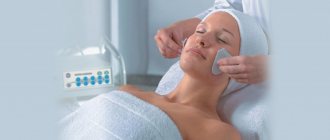
Preparation for bronchoscopy is very important, since the manipulation is very serious, classified as invasive and requires only special equipment and special skills from the doctor.
Therefore, you need to start with a detailed conversation with your therapist. He will tell you what specialist consultations are needed. So, if a person has suffered a myocardial infarction, he needs, in agreement with the cardiologist, to increase the dose of beta blockers 2 weeks before the study. If a person suffers from arrhythmia, he needs to reconsider antiarrhythmic therapy and possibly increase the dose of drugs or add some other antiarrhythmic drug. The same applies to diabetes mellitus and arterial hypertension.
Also, everyone needs to undergo the following studies and show their results:
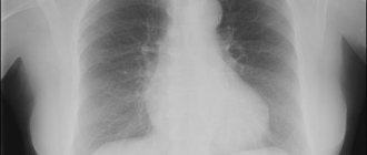
- X-ray or CT scan of the lungs.
- ECG.
- Blood tests: general, biochemical, coagulogram.
- Blood gas analysis. This requires venous and arterial blood.
The last meal is no later than 8 pm. Then you can take your last scheduled pills. The need to take them in the morning is discussed separately.
Be sure to empty your bowels in the evening using an enema, Microlax microenema (Norgalax), glycerin suppositories.
You are not allowed to smoke on the day of the test. Immediately before the procedure, you need to empty your bladder. You must take a towel or diaper with you so that you can dry yourself after the examination; for those suffering from arrhythmia - antiarrhythmic drugs; for those suffering from bronchial asthma - an inhaler. Removable dentures will need to be removed.
It is imperative to familiarize the doctor who will perform the procedure with previous diseases and allergies, as well as medications that you are constantly taking.
Course of the procedure
Learn more about how bronchoscopy is performed. First, let's talk about how this procedure is performed without anesthesia - under local anesthesia:
- The patient comes into the office, he is asked to undress to the waist and then either lie down on a couch in the middle of the room, or sit on a chair near the equipment.
- He is given an injection under the skin - in the shoulder area. Usually this is the drug “Atropine” - a drug that will suppress the secretion of saliva and bronchial contents. It makes your mouth dry and your heart rate increases.
- The drug can be administered intramuscularly. This is soothing to make the manipulation easier to bear.
- Also, the drugs “Salbutamol” or “Berodual” are sprayed into the mouth. They are needed to expand the bronchi.
- Next, the doctor administers local anesthesia. He sprays or lubricates an anesthetic (usually lidocaine 10%) at the root of the tongue and a little deeper. The outer part of the bronchoscope is also treated with the same solution.
- After this, they begin to carefully insert the bronchoscope into the mouth. Before insertion, a mouthpiece, a plastic device that holds the teeth, may be inserted into the mouth. This is necessary to ensure that the patient does not bite through the bronchoscope.
- If bronchoscopy is performed in a supine position, the doctor, going around the patient’s head, can insert a laryngoscope into his mouth and larynx. This is also accompanied by spray of local anesthetic into the respiratory tract. The laryngoscope will open the way for the bronchoscope, so the latter will be inserted faster and safer.
- Let's be honest: the introduction of a bronchoscope will be accompanied by a gag reflex, as well as a feeling of lack of air. The first is due to the fact that the root of the tongue is affected. But there is not enough air, since the bronchoscope will take up 3/4 of the diameter of the trachea. To eliminate both of these effects, you need to breathe frequently and shallowly (“like a dog”).
- The study is carried out quite quickly so as not to cause severe hypoxia. Oxygen levels should be monitored using a pulse oximeter. Its sensor – a “clothespin” – is put on your finger.
During bronchoscopy, do not bend over so as not to damage the airways with the bronchoscope (especially if a rigid device is used).
If bronchoscopy with biopsy is performed, it is painless. There is only discomfort behind the sternum. The bronchial mucosa has virtually no pain receptors. The introduction of lidocaine before manipulation is due to the need to turn off vagal (from the word “nervus vagus” - “vagus nerve”) reflexes from the root of the tongue and vocal cords, which can lead to cardiac arrest.
If bronchoscopy is performed under anesthesia, it is performed with the patient lying down. Then the injections are performed intravenously, and the person falls asleep as a result. A rigid polypropylene tube is inserted into his trachea, which is connected to a breathing apparatus. For some time, air is pumped into the lungs with a breathing apparatus (exhalation occurs spontaneously), then a bronchoscope is inserted through the tube, and bronchoscopy is performed. A person does not feel how a bronchoscopy is done.
The procedure under anesthesia is performed in children, people who are very afraid of the procedure, and people with unstable mental health. It is performed on patients who have already been on mechanical breathing, as well as when surgical intervention is necessary.
WHAT DOES A PATIENT FEEL AFTER BRONCHOSCOPY
Examination of the respiratory organs using a bronchoscope is, although a safe method, but rather unpleasant. At the end of the study, the patient typically develops the following sensations:
- numbness of the throat;
- nasal congestion;
- difficulty swallowing;
- sensation of a foreign body inside the body;
- cough with discharge of blood veins.
All these negative symptoms disappear on the first day after completion of bronchoscopy. To avoid the development of serious complications, the patient after gastroscopy is prohibited from eating, smoking, drinking and taking medications for some time. All this will only worsen the situation, so check with your doctor when you can return to a normal lifestyle. If after the end of the study the discomfort continues for more than two days, then you should immediately notify the doctor who performed the bronchoscopy.
Conducting a survey
Regardless of the reasons why you were prescribed the procedure, it starts the same. Therefore, anyone who has already encountered such a study can tell you how bronchoscopy of the lungs is done. Three specialists take part in the process: an endoscopist, an assistant and an anesthesiologist. Initially, the cavity of the pharynx and mouth is anesthetized. This is necessary not only to reduce discomfort, but also to suppress the cough reflex. The anesthetic is sprayed locally using a special device. The patient’s condition is constantly monitored: medical staff monitors pulse, blood pressure and oxygen levels in the body.
Bronchoscopy for lung cancer can be performed sitting or lying down, depending on the doctor’s opinion. The endoscope is inserted through the nose or mouth. The procedure itself lasts from several minutes to an hour. The exact time of its implementation depends on the goals that the doctor has. He may perform certain medical procedures, take a biopsy, or simply examine the surface of the respiratory tract.
If the patient is sitting during the procedure, he should slightly tilt his torso forward and lower his hands between his legs. At the same time, you need to tilt your head back. If a fiberoptic bronchoscope is used, a nasal examination is most often performed. But when using a rigid bronchoscope, the procedure is done only through the mouth.
After the procedure
After bronchoscopy you feel:
- heaviness or pressure behind the sternum - during the day;
- numbness of the oral cavity and larynx - for 2-3 hours;
- hoarseness or nasal sound – for several hours;
- You may cough up sputum streaked with blood.
The following rules must be followed:
- stay in the hospital for 3 hours under the supervision of staff;
- Do not eat, drink or smoke for 3 hours. Food and food can enter the trachea, while smoking impairs the healing of the mucosa after manipulation;
- do not drive for 8 hours, as drugs have been administered that significantly reduce the reaction rate;
- Avoid physical activity for the next 2-3 days.
It is also necessary to monitor your condition. Must not be:
- discharge from the respiratory tract of blood in the form of clots or liquid blood;
- shortness of breath;
- chest pain when breathing;
- temperature rise;
- nausea or vomiting;
- wheezing.
Why do they do it?
There is a fairly large list of cases in which bronchoscopy is prescribed. Pulmonologists recommend it to their patients to clarify the diagnosis or to carry out therapeutic procedures.
Therapeutic goals of bronchoscopy:
· elimination of bleeding;
· expansion of the lumen of the airways;
· removal of large foreign objects from the respiratory tract;
· cleansing the lungs of mucus or fluid;
· rinsing the branches of the windpipe;
· administration of drugs into the respiratory tract;
· removal of formations or scars.
When taking a biopsy or performing therapeutic manipulations, the bronchoscope is equipped with the necessary surgical instrument.
Who should not undergo bronchoscopy?
There are such contraindications to bronchoscopy (namely diagnostic):
- arterial hypertension with diastolic (“lower”) pressure more than 110 mm Hg;
- mental illness;
- immobility (ankylosis) of the lower jaw;
- recent myocardial infarction or stroke (less than 6 months ago);
- aortic aneurysm;
- significant rhythm disturbances;
- coagulation disorders;
- significant narrowing (stenosis) of the larynx;
- chronic respiratory failure stage III.
In these cases, virtual bronchoscopy can be performed.
The procedure should be postponed during an acute infectious disease, exacerbation of bronchial asthma, for women - during menstruation and from the 20th week of pregnancy.
When bronchoscopy is intended to assist intubation, or is needed to remove foreign bodies, bronchial stenting or other therapeutic purposes, there are no contraindications. This procedure is carried out jointly by an endoscopist and an anesthesiologist, under anesthesia, after proper intensive preparation.
Presence of contraindications
The algorithm for preparing for the study is that the patient should prepare himself morally and psychologically. But if the patient has some contraindications, then the procedure is prohibited. Such contraindications are:
- carrying a child during the first and third trimester;
- experienced a heart attack or stroke;
- acute respiratory failure;
- psychological disorders;
- tendency to intolerance to drugs and anesthetics;
- impaired blood clotting process;
- aortic aneurysm.
If you suffer from one of the above ailments, you need to warn your doctor about it. Otherwise, you are putting your life at risk.
Complications of the procedure
With bronchoscopy, the consequences may be as follows:
- bronchospasm - compression of the walls of the bronchi, which causes oxygen to stop flowing into the lungs;
- laryngospasm - the same as the previous complication, only the glottis (larynx) spasms and closes;
- pneumothorax – entry of air into the pleural cavity;
- bleeding from the bronchial wall (may occur during biopsy);
- pneumonia – due to infection of the small bronchi;
- allergic reactions;
- mediastinal emphysema - entry of air from the bronchus into the tissue surrounding the heart, large vessels extending from it, the esophagus and trachea;
- in those suffering from arrhythmia, it increases.
Bronchoscopy in children
Bronchoscopy can be performed in children from the neonatal period, provided that the hospital has a device of such a small diameter. The procedure is carried out only under anesthesia, and after it antibiotics are prescribed.
Bronchoscopy is performed in children when:
- severe difficulty breathing, most likely caused by a foreign body;
- accurately determining the presence of a foreign body in the respiratory tract;
- severe pneumonia, especially against the background of cystic fibrosis;
- bronchial tuberculosis - to make a diagnosis or stop bleeding;
- if, in the presence of shortness of breath, an area of atelectasis is visible on the x-ray;
- lung abscess.
Children are more likely to develop laryngo- or bronchospasm due to the rich blood supply to the respiratory tract. Therefore, general anesthesia is often supplemented with local anesthesia.
In addition, complications may include collapse (a sharp decrease in blood pressure) and anaphylactic shock. Tracheal perforations are extremely rare, since bronchoscopy is performed with flexible bronchoscopes.
Patient reviews
Those who are just about to undergo the procedure are certainly interested in reviews of those who have already undergone it.
Natalya, 34 years old: “I, like many, am a big coward. But I want to say right away that it doesn’t hurt at all. In this case, the main thing is to breathe correctly and not strain, otherwise everything will freeze. I think that those who undergo this procedure only for research should generally feel comfortable. And they did a medical procedure on me. When the lungs were washed with a special solution, everything was bubbling inside, and there was a feeling that I would suffocate. This is, of course, scary, but I repeat - it doesn’t hurt! And when the device was pulled out, I didn’t feel anything at all.”
Kristina, 23 years old: “The procedure, of course, is unpleasant, but what can you do if the doctor insists and assures that only it will definitely show what’s inside and allow you to make an accurate diagnosis. In the process, 2 moments turned out to be the most unpleasant for me. The first was freezing of the trachea when Lidocaine was infused through a bronchoscope. And the second is taking a biopsy. I really wanted to cough, but I couldn’t. The procedure took 20 minutes in my case. Personally, I remember this manipulation not as painful, but as very unpleasant.”
Of course, patients seen by a pulmonologist should definitely understand - pulmonary bronchoscopy, what is it? This will help him respond adequately to the doctor’s orders, be mentally attuned to the procedure and know what to be prepared for later. No matter how scary this manipulation may seem, it is important to remember that it is essential for making an accurate diagnosis or carrying out important therapeutic measures.
Bronchoscopy for tuberculosis
Bronchoscopy for tuberculosis is an important diagnostic and treatment procedure. It allows:
- using aspiration of bronchial contents and its bacteriological examination, isolate Mycobacterium tuberculosis (especially if the culture was negative) and determine sensitivity to anti-tuberculosis drugs;
- drain cavities (tuberculous cavities) from necrosis;
- administer anti-tuberculosis drugs locally;
- dissect fibrous (scar) tissue in the bronchi;
- stop the bleeding;
- assess the dynamics of treatment (this requires repeated bronchoscopy);
- inspect the stitches after surgery to remove a lung;
- cleanse the bronchi from necrotic masses and pus when they have broken through there from the cavity or intrathoracic lymph nodes;
- assess the condition of the bronchi before surgery;
- remove fistulas - connections between the focus of pulmonary tuberculosis and the bronchus.
Author:
Krivega Maria Salavatovna resuscitator
Tissue sampling
During the procedure for examining these organs, a specialist can take a tissue sample in order to conduct a detailed analysis in the future. Using a bronchoscope, you can examine the respiratory organs and perform medical treatment by administering pharmacological drugs. A bronchoscope allows you to carry out the procedure of washing the bronchi from the mucus that has accumulated in them.
As soon as the examination is completed, and its duration is no more than 30 minutes, the doctor will remove the bronchoscope from the oral cavity. After completing the diagnostic study, the patient should be under the supervision of medical staff for approximately two to three hours, so the patient is placed in a special ward. During this time, it is possible to determine the absence of diseases.