Achondroplasia (diaphyseal aplasia, congenital chondrodystrophy) is a genetic disease that results in impaired bone growth. This is one of the most common types of dwarfism. It is possible to judge whether a child has this pathology from the moment of birth.
The exact causes of the disease are unknown, but in approximately 80-90% of cases it is the result of new gene mutations and occurs in approximately one in every 25,000 newborns, regardless of race or gender.
The fact that this pathology was reflected in ancient Egyptian art makes it one of the most ancient birth defects recorded in human history.
brief information
Achondroplasia (also called: diaphyseal aplasia , Parrot-Marie disease , congenital chondrodystrophy ) is a cartilage disease that is one of the main causes of disproportionate dwarfism: affected patients have shorter-than-normal upper and lower limbs and a normal torso.
The causes of achondroplasia are genetic: a mutation in the FGFR3 gene, located on chromosome 4, causes its occurrence.
In addition to the characteristic features of short stature and lack of proportionality between the limbs and trunk, achondroplasia causes other clinical signs including : short toes, valgus of the knees or feet, large head and prominent forehead.
There are currently no specific treatments for achondroplasia .
Therefore, achondroplasia is an incurable disease.
Prevention
Prevention of achondroplasia consists of medical genetic consultation and prenatal diagnosis, which makes it possible to detect pathologies even at the stage of intrauterine development. Consultation with a geneticist is especially required for those who already have a history of dwarfism in their family. A special examination will allow you to assess the risk of having a sick child.
If parents already have achondroplasia, the disease cannot be prevented, as it is inherited.
No one can be protected from the birth of a sick child. This is a big blow not only for parents, but also for medical staff. This is especially true for pathologies that cannot be prevented or prevented. Achondroplasia is one of these diseases.
Achondroplasia is a genetic disease, the main characteristic of which is short limbs, while the body length remains normal. The average height of the patient is 130 centimeters, and in some cases even less. The spine of such a patient has a curved shape, the head is large, and the frontal tubercles protrude significantly.
The incidence of pathology among all newborns is 1:10,000, and this affects girls more often than boys.
This pathology cannot be cured, and methods for completely restoring growth are currently unknown. All methods of therapy are aimed at reducing all negative manifestations of the disease to a minimum.
What is achondroplasia?
Achondroplasia is a rare genetic (inherited) bone disorder that affects one in 15,000 to 35,000 live births.
Achondroplasia is the most common type of dwarfism, in which the child's arms and legs are short in proportion to their body length. In addition, the head is often large, but the body is of normal size.
The average height of adult men with achondroplasia is about 133 cm. The average height of adult women with achondroplasia is about 125 cm.
Forecast
The prognosis for achondroplasia is complex. Such patients adapt to disorders of their skeleton, socialize, and even find employment. But the quality of life may suffer.
Kovtonyuk Oksana Vladimirovna, medical observer, surgeon, consultant doctor
11 total today
( 65 votes, average: 4.11 out of 5)
Bursitis - symptoms, causes of development and treatment methods
Synostosis of the knee joint: causes, symptoms, treatment tactics
Related Posts
Signs and symptoms
This rare genetic disease is characterized by distinctive features:
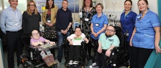
- short stature (usually under 5 feet [see photo]);
- an unusually large head (macrocephaly) with a prominent forehead (frontal prominence) and a flat (depressed) nasal bridge;
- short arms and legs;
- bulging belly and buttocks (due to the internal curve of the spine);
- short fingers that assume a three-pointed position during extension.
Signs in childhood.
Children born with achondroplasia usually have a “domed” (vaulted) skull and a very wide forehead. In a small proportion, there is excessive accumulation of fluid around the brain (hydrocephalus). Achondroplasia in infancy is also characterized by low muscle tone (hypotonia).
Increase in height (video)
– a congenital disease in which the growth of the bones of the skeleton and the base of the skull is disrupted. The disease was first described under this name in 1878, and in 1900 an expanded clinical description was given by Pierre Marie. Therefore, achondroplasia is sometimes called Parrot-Marie disease.
.
Achondroplasia is the main cause of dwarfism. The incidence of the disease is one case per twenty thousand newborns. The disease does not depend on gender or race. Every fifth patient suffering from achondroplasia inherits the disease; in the remaining patients it is associated with primary mutations of the FGFR3 gene. If the achondroplasia gene is present in both parents, then such children most often die in the womb.
According to geneticists, achondroplasia is most often diagnosed in children born to fathers over forty years of age. However, it is impossible to foresee the possibility of the birth of such a child due to the fact that genes can mutate even in healthy parents. Genetic counseling can only be useful for parents who have signs of achondroplasia in order to plan a family in the future.
Today, doctors continue research into the FGFR3 gene, the “culprit” of achondroplasia. As it turned out, this gene belongs to the group responsible for the formation of fibroblast growth receptor proteins. Normally, this protein ensures the passage of signals from chemical compounds and thereby stimulates cell growth and maturation. In people suffering from achondroplasia, signal transmission mechanisms are disrupted, which is why problems arise with the normal formation of bone tissue. Currently, scientists are studying the mechanism of achondroplasia, which can make a significant step forward in the treatment of the disease.
Today medicine knows several forms of the disease:
- Hyperplastic achondroplasia (characterized by accelerated but disordered growth of the epiphyses);
- Gelatinous softening of the cartilage of the epiphysis (this form of the disease is the main cause of intrauterine mortality);
- Hypoplastic achondroplasia (this form of the disease is characterized by underdevelopment of cartilage tissue).
Causes
The main cause of achondroplasia is a mutation of the FGFR3 gene, leading to degeneration of the epiphyseal cartilage. The cells of the growth zone are located chaotically, as a result of which the physiological process of ossification is disrupted. Because of this, bones begin to lag behind in development. At the age when cartilage ossifies in children, patients with achondroplasia have still unformed bone tissue with a predominance of cartilaginous tissue. Mostly bones that grow according to the enchondral type are affected, i.e. ossification occurs from within the cartilaginous bone. These are the bones of the base of the skull, tubular bones.
Symptoms
Symptoms of the disease appear already at the birth of a child - an increase in head circumference greater than normal and shortened limbs can be noted. The forehead is convex, the back of the head and crown protrude. Doctors often diagnose hydrocephalus in children suffering from achondroplasia. Due to improper formation of the skull bones, the facial skeleton also suffers - the child’s eyes are widely spaced, deeply set in the sockets, and the brow arches droop. The nose has an irregular shape - it is flattened, the upper part is wide, the wings of the nose are significantly removed on the sides. Due to the fact that the nasal passages are narrowed, patients often suffer from sinusitis and otitis media.
The proximal segments of the limbs - the hips and shoulders - are shortened, and curvatures are noted. The newborn's hands barely reach the navel. The feet are wide and short. The fingers of such babies are almost the same length, however, they are also short. In most cases, this symptom retains its severity - in adults suffering from achondroplasia, the middle finger in a straightened position of the hand barely reaches the inguinal fold. The torso in children suffering from achondroplasia is little changed, the chest is developed normally. Due to the curvature of the spine, the stomach protrudes forward; on the contrary, the buttocks protrude back more than in healthy babies.
Patients with achondroplasia from an early age have breathing problems, which in turn is associated with features of the facial structure, enlarged tonsils, and a high palate. Doctors often pay attention to the increase in the rate of sudden mortality in children with this pathology. It is assumed that compression of the medulla oblongata due to the irregular size and shape of the foramen magnum plays a key role here.
Often children with achondroplasia are developmentally delayed; later they begin to hold their heads, sit up and walk, but their intellectual development does not suffer. In some cases, children may have problems adapting to the world around them, since most services are not adapted to people with special needs.
As the child grows, his bones take on an increasingly irregular shape - they thicken and bend, and lumps form. Excessive elasticity in the epiphyseal and metaphyseal regions provokes bone deformations, which are aggravated when a load appears on them and an increase in body weight.
In general, patients with achondroplasia are characterized by the following disorders:
- curvature of the femur;
- “looseness” of the knee joints;
- formation of a flatvalgus foot;
- decreased muscle tone;
- incorrect relationship between the fibula and tibia;
- curvature of the upper limbs in the forearm area;
- growth deficit;
- change in the shape of the skull, malocclusion;
- a number of diseases accompanying achondroplasia;
- excess body weight.
Diagnostics
Usually it is not difficult for a doctor to diagnose a disease. The patient's appearance and body proportions primarily indicate achondroplasia. In childhood, the study is done more carefully to see the degree of deviation of the parameters from the norm. For children with achondroplasia, there is a special table of height and weight, with which the data obtained from children with suspected disease are compared. Thus, doctors can assess the child’s tendency to achondroplasia and observe the dynamics of his indicators. In case of significant deviations, consultations with other specialists are prescribed. The neurosurgeon examines the patient for hydrocephalus, and magnetic resonance and computed tomography of the brain is prescribed. An otolaryngologist examines the structure of the patient's throat and nasal passages to confirm or refute the diagnosis. In some cases, a consultation with a pulmonologist may be necessary.
To diagnose achondroplasia, patients undergo an X-ray examination of the skull, chest and pelvis. Based on its results, the proportions of the brain and facial parts of the skull, the size of the foramen magnum, the thickness of the ribs, their deformation, the presence of anatomical bends, the thickness and shape of the ilium are assessed.
Particular attention is paid to tubular bones, which with achondroplasia have a shortened thin diaphysis and widening of the metaphyses. The joints of patients with achondroplasia are deformed, with especially pronounced deformation in the knee and wrist joints.
Treatment
Treatment of the disease is currently not possible. Doctors can only minimize the consequences to improve the quality of life of patients. In childhood, conservative therapy is carried out for such children - massage, physical therapy. This helps strengthen the muscle corset and prevent severe deformation of the lower extremities. Patients are advised to wear special shoes that reduce pressure on the bones. It is also important to follow a diet to avoid burdening the skeleton with excess weight. Defects in the jaw area are corrected by wearing special plates.
There are cases when hormone therapy is prescribed in childhood, which helps to slightly compensate for the lack of growth. Hormone therapy is not used for adults.
If the parents decide to surgically solve the problem, then it is necessary to contact a specialized clinic that has sufficient experience in performing such operations.
Surgery is performed if the disease causes obvious discomfort to the patient. An unambiguous decision in favor of surgery is made when there is a threat of spinal cord entrapment, kyphosis of the middle part of the back, or an “o”-shaped leg.
It is also possible to lengthen the bones, for which several surgical interventions are performed in stages. At four to six years of age, children undergo surgery to lengthen their legs (up to six cm), hips (up to about seven to eight cm) and shoulders (possibly up to five centimeters). The duration of such stages is about five months with a break of two to three months. The next series of interventions is carried out at the age of fourteen to fifteen years. Here the patient also goes through three stages, the expected result is the same as the first time.
However, such actions do not completely eliminate the disease and its symptoms, since with short stature, elongation of bones even up to ten centimeters does not make patients look like ordinary people. In addition, not all patients are ready to undergo a series of operations and painful rehabilitation periods.
Contents of the article
Achondroplasia
(Parrot-Marie disease, chondrodysplasia, chondrodystrophy) The name does not accurately characterize the essence of the disease, since cartilage is formed, but it is sharply hypoplastic. The basis of the disease is a violation of the processes of enchondral growth, while periosteal ossification is practically unchanged.
Causes and risk factors
Achondroplasia results from specific changes (mutations) in a gene known as fibroblast growth factor receptor 3 (FGFR3).
For most patients there is no obvious family history of this condition. Increased paternal age (advanced paternal age) may be a contributing factor in the occurrence of sporadic achondroplasia.
Less commonly, familial cases of achondroplasia follow an autosomal dominant pattern of inheritance. Dominant genetic disorders occur when only one copy of an abnormal gene is needed to cause a specific disorder.
The abnormal gene may be inherited from either parent or may be the result of a mutation (change) in a gene in the affected person. The risk of passing the abnormal gene from an affected parent to offspring is 50% for each pregnancy. The risk is the same for men and women.
Achondroplasia-like disorders
Symptoms of the following disorders may be similar to those of achondroplasia. Comparisons can be useful for differential diagnosis:
Hypochondroplasia is a genetic disorder characterized by short stature and disproportionately short arms, legs, hands, and feet (i.e. short limb dwarfism). In those who suffer from this disorder, short stature is often not recognized until early or middle age or, in some cases, adulthood. Affected individuals may also develop bowing of the legs in early childhood, which often improves spontaneously with age. In some cases, additional abnormalities may be present, such as an unusually large head (macrocephaly), a relatively prominent forehead, limited elbow extension and rotation, and/or other physical abnormalities.
Complications
Complications that can accompany achondroplasia are most often:
- Otitis media – inflammatory lesion of the middle ear;
- conductive hearing loss – hearing impairment due to disturbances in sound conduction;
- respiratory failure - deterioration of respiratory function.
The first two complications develop due to the narrowing of the nasal passages, which occurs with achondroplasia, the third - due to obstruction (blockage) of the upper respiratory tract.
Diagnostics
The clinical and radiological features of achondroplasia are well characterized. Those with typical results generally do not need molecular genetic testing to confirm the diagnosis.
When clinical features are suspicious in a newborn, radiographic findings can help confirm the diagnosis. However, if there is uncertainty, identification of a genetic variant of the FGFR3 gene using molecular genetic testing can be used to make a diagnosis.
Standard Treatments
There is currently no way to prevent or treat achondroplasia, as most cases involve new, unexpected mutations. Treatment with growth hormone does not significantly affect the height of a person with achondroplasia. In some very specialized cases, leg lengthening surgery may be considered.
Detecting bone abnormalities, especially in the back, is important to prevent difficulty breathing and leg pain or loss of function. Kyphosis (or hunchback) may require surgical correction if it does not go away when the child begins to walk. Surgery may also help leg flexion. Ear infections caused by illness must be treated immediately to avoid hearing loss. Dental problems may need to be addressed by an orthodontist (a dentist with special training in straightening teeth).
First aid measures and comprehensive treatment
If your thigh muscles hurt, then the first thing to do is provide first aid measures. If there has been an injury, you should apply ice to the area of suspected injury, immobilize the limb, preventing its mobility, and go to the emergency room as soon as possible. If pain of inflammatory origin appears, then it is necessary to provide rest to the affected limb and apply a thin layer of anti-inflammatory non-steroidal ointment (Ortofen, Diclofenac, Nice, Ketorolac, etc.).
Complex treatment of pain in the thigh muscles is always closely related to those diseases that manifest themselves with a similar symptom. if it is deforming osteoarthritis of the knee or hip joint, then measures must be taken to restore cartilage tissue. In case of gout, it is important to normalize the balance of uric acid and ensure its excretion in the urine.
https://www.youtube.com/watch?v=s_vFCH40xOo
Treatment of thigh muscle pain should always be carried out under constant supervision by an experienced physician. Manual therapy, physical therapy and reflexology provide excellent results.
Tags: achondroplasia, doctor, give, what, prognosis, child, such
About the author: admin4ik
« Previous entry

