Medical terminology contains many different diseases about which the average person can say little concretely. One of them is angioma, which is a red mole. This disease refers to benign tumors, the structure of which is represented by lymphatic or blood vessels.
The main factor that provokes their development is the pathology of the functioning of the circulatory and lymphatic systems. Their formation occurs throughout a person’s life, but only up to the age of seven there is a possibility of these neoplasms disappearing naturally.
Types of skin angioma
The following types of skin angioma are recognized:
- Simple (capillary) angioma – formations of capillaries and vessels. May occur on the surface of the skin or mucous membranes. When you press them, they can change color to a lighter color.
- Cavernous angioma is a formation that protrudes above the surface of the skin and does not have clear edges. Inside the angioma there are cavities filled with blood clots.
- Branched angioma - consists of different types of vessels.
- Granuloma is a formation on the mucous membranes.
Regarding vascular damage, angiomas are divided into the following types:
- Hemangiomas are lesions of blood vessels: arteries, veins, capillaries.
- Lymphangiomas are lesions of the lymphatic vessels.
Also recognizes:
- Monomorphic - formation occurs from structures of one type.
- Polymorphic - originate from different structures.
Cavernous angiomas on MRI
Cavernous angiomas represent about 1% of all intracranial vascular lesions and 15% of cerebrovascular malformations. With the development and introduction of MRI, cavernous angiomas have become the most commonly detected vascular malformations of the brain. In early studies on autopsy material, the frequency of their occurrence was 0.02-0.53%. Using MRI, the incidence of formations similar to cavernous hemangiomas was 0.39-0.9%, and the detection of previously unidentified asymptomatic formations using MRI increased their incidence to 0.45-0.9%.
Get an MRI of the brain in St. Petersburg
On MRI, parenchymal cavernous angiomas are represented by a characteristic “popcorn”-type formation, clearly defined, with a smooth border. The internal part is represented by multiple foci of signal of varying intensity, which correspond to hemorrhages at different stages of resolution.
MRI signs of cavernous angioma. Large cavernous angiomas of the right frontal lobe and left occipital lobe on T1-weighted axial section. These two heterogeneous space-occupying lesions have a central mesh structure with alternating areas of high and low signal intensity, surrounded by a hypointense rim of hemosiderin.
A fresh hematoma containing deoxyhemoglobin is isointense on T1-weighted images and significantly hypointense on T2-weighted images. A subacute hematoma containing extracellular methemoglobin is hyperintense on both T1- and T2-weighted images due to the paramagnetic effect exerted by methemoglobin.
Intermediate fibrous elements are characterized by a weakly hypointense signal on T1- and T2-weighted images, since they contain calcifications and hemosiderin. The heterogeneous interior of the mass is surrounded by a hemosiderin rim, which has low intensity on T1-weighted images. The hypointensity of this rim becomes more pronounced, resembling a halo, on T2-weighted and gradient-echo images due to the higher sensitivity of these sequences to changes in the magnetic field.
Axial gradient-echo MR images provide better visualization of large cavernous angiomas in the right frontal and left occipital lobes. The hemosiderin rim appears as a halo due to the increased magnetic susceptibility of hemosiderin.
Smaller cavernomas appear as low-intensity nodules on T1- and T2-weighted images.
Small lesions are better visualized on gradient echo images due to the increased sensitivity to changes in the magnetic field that is inherent in such pulse sequences. It has also been shown that in sequential gradient echo images, small punctate formations are better visualized with longer echo times; these data suggest that such formations contain paramagnetic substances.
Gradient-echo MR imaging shows multiple bilateral small, punctate and round, low-intensity lesions in the periventricular and subcortical white matter. The largest lesion is visualized in the periventricular white matter of the frontal lobe anterior to the anterior (frontal) horn of the left lateral ventricle near the genu of the corpus callosum. Multiple smaller lesions are visible anterior and posterior to it.
On time-of-flight angiography images, methemoglobin in the center of a cavernous malformation may resemble moving blood. However, on a subsequent phase-contrast MR angiogram obtained with a low blood flow speed setting during encoding (10-20 cm/s), blood flow or pathological vascularization is not visualized, which makes it possible to exclude vascular lesions.
Typically, cavernous angiomas do not have a bulking effect on adjacent tissue or cause edema, and they do not have a feeding artery or draining vein unless they are associated with other similar vascular malformations. Cavernous angiomas are often associated with venous malformations, which are characterized by the presence of a draining vein. In such mixed cases, standard angiography may be useful.
T2-weighted image of a cavernoma of the pons.
Cavernous malformations detected on MRI include other occult vascular malformations (AVM/aneurysm thrombosis, capillary telangiectasia), hemorrhage in a primary or secondary tumor (metastasis of melanoma, choriocarcinoma, thyroid or kidney cancer), amyloid angiopathy, treated or primary infection (toxoplasmosis or cysticercosis), multiple hemorrhages associated with damage to the blood system (disseminated intravascular coagulation, leukemia), as well as the consequences of diffuse axonal damage.
Stages and degrees of skin angioma
The following stages of development of angiomas are recognized:
- period of intensive growth (period of external manifestations) - occurs due to external or internal factors (trauma, temperature effects, hormonal imbalances during pregnancy or menopause, metabolic disorders) that accompany the development of the tumor;
- growth arrest stage;
- stage of reverse development - lasts from several months to several years. Pathological vessels at this time are replaced by normal skin cells or scar tissue. The reversibility of the process does not occur in all cases.
Red moles on the body: what are they?
If you turn to specialists with this question, then, according to them, this is an intermediate stage of a pathological change in the skin, which can subsequently change into a tumor. The medical literature offers little information regarding this neoplasm. The fact is that a person should not be afraid of red moles on his body, since this does not have any negative effect on health. It is believed that their development is associated with a genetic factor.
Symptoms of skin angioma
Skin angioma, the symptoms and treatment of which depend on its type, has its own characteristics.
You can determine the type of angioma by its shape, color and contours:
- Flat angiomas or port-wine stains - in most cases they are located on the head and can have any shade of red: from purple to dark cherry. With age, they often enlarge and change shade. When you press on their surface, they turn pale. They are a dense interweaving of capillaries covered with endothelial cells into one ball. Found in 80-90% of all angiomas.
Skin angioma. Photos, symptoms and treatment depend on the type - Swollen - venous hemangiomas. They have a bluish tint.
- Protruding above the skin are cavernous hemangiomas. The skin in the pathological area is warmer to the touch and turns pale when pressed. It has a characteristic appearance: in the form of various shapes of uneven protruding nodes of a bluish color. When the body bends, the angioma tends to increase, as there is an influx of blood into its vessels.
- Colorless angiomas are lymphangiomas that are found on the skin near the mucous membranes. They protrude slightly above the surface of the skin, are identical in color, and are painless.
- Combined - combine undeveloped capillaries and cavities filled with blood. The color of the angioma is bright red, without clear contours. It protrudes slightly above the skin with an uneven, bumpy surface.
Cavernous angioma or tumor?
Brain scanning using CT and MRI in most cases allows one to clearly distinguish a cavernoma from other brain formations, including tumors of varying degrees of malignancy. However, in some cases, differentiation of these formations represents a diagnostic problem, the solution of which requires extensive experience. In this regard, the ability to attract a highly qualified diagnostician is critical. In addition, high-quality interpretation of CT and MRI images provides a solution to other diagnostic problems: exclusion of surrounding cerebral edema, identification of the severity of hemorrhage, description of details affecting the operability of the cavernoma. If you are in doubt about the diagnosis, you should consult a radiologist from a leading center specializing in brain pathology. A second opinion from such a diagnostician can be very valuable in the differential diagnosis of angiomas and other pathological conditions.
Causes of skin angioma
In order to explain the cause of the appearance of skin angiomas, it is necessary to understand the mechanism of its origin.
- Flat angiomas develop due to disruption of the innervation of the capillary vascular system. New vessels are generated that are woven into nearby tissues.
- Venous and cavernous angiomas appear due to a defect in collagen fibers. Collagen fibers are found around subcutaneous blood vessels. If their quantity is insufficient, the capillary walls lose the necessary support, expand and lose tone.
Disruption of the innervation of the vascular wall or a defect in collagen fibers occurs for the following reasons:
- maternal smoking during pregnancy;
- drinking alcohol during pregnancy;
- stress during pregnancy;
- taking certain medications;
- past inflammatory diseases during pregnancy.
The highest risk of angioma in a baby occurs if these factors were active in the first trimester of pregnancy.
Angiomas in adults are caused by the following factors:
- smoking;
- alcohol abuse;
- taking insufficient fluids;
- deficiency of B vitamins;
- chronic liver or kidney diseases;
- oncological diseases;
- cachexia.
There are also “senile” angiomas that develop by the same mechanism, but the cause is a physiological decrease in collagen in the body.
Locations
Angiomas are pathological neoplasms on the human skin, having different shapes and sizes. According to research, such neoplasms are localized not only on the surface of the skin, but also on bone tissue, internal organs, muscles and even in the brain . Red dots on the body like moles can appear both in childhood and in adulthood. In certain situations, the formation of angiomas is provoked by hormonal imbalances during pregnancy.
Growths on the body can be either single or multiple. Single formations do not pose a danger to human health. In contrast, multiple growths indicate the presence of serious pathological processes occurring in the body.
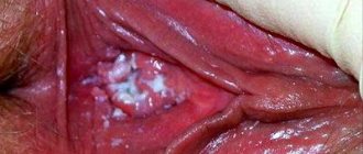
Simple angiomas are usually located on the surface of the scalp and face. These neoplasms are characterized by dimensions of several millimeters, round shape and blue color. In rare cases, growths are located in the cheeks, nose and external genitalia. More complex types of pathology form under the skin. The development of this form of angiomas leads to damage to muscles and bones. This pathology can spread throughout the internal organs. More than eighty percent of the growths are usually located in the upper torso, including the limbs, head and neck.
The head is considered to be one of the most unfavorable places for localization of growths. This feature is explained by the fact that the growth is easily injured when cutting or combing hair. Many women experience discomfort when pathology develops in the breast area. It should be noted once again that single growths do not pose a threat to health. But pathological growth, the appearance of pain, itching and color changes are symptoms that require increased attention. In order to prevent possible complications, it is imperative to visit a dermatologist.
Diagnosis of angiomas is based on examination, X-ray examination (angiography, lymphangiography), ultrasound
Diagnosis of skin angioma
Skin angioma, the symptoms and treatment of which must be confirmed diagnostically, has an examination algorithm.
It includes standard procedures, which consist of the following main points:
| Name of diagnostic method | Description | Which doctor performs it and the cost of the procedure |
| 1. Visual inspection and palpation | In most cases, the diagnosis can be established based on the external signs of formation. | The examination is carried out by a general practitioner or family doctor, if necessary - by an oncologist. If an angioma is detected in a newborn, the child is examined by a pediatrician. |
| 2. Ultrasound examination | It is necessary to carry out for cavernous or combined angiomas in order to determine the internal composition and volume of the formation, its connection with adjacent tissues. The examination does not require additional preparation and is carried out in every clinic or medical center. | The cost of the procedure is in the range of 300-700 rubles. A doctor performs ultrasound diagnostics. |
| 3. Dermatoscopy | Examination with a special device - a dermatoscope. Allows you to see the tumor at maximum magnification. | Dermatoscopy is performed by a dermatologist. Its cost is about 500 rubles. |
| 4. Histological analysis of the biopsy | It is carried out for the purpose of differential diagnosis with melanoma and basal carcinoma. | A biopsy is performed by an oncologist or dermatologist, and the analysis is sent to the laboratory for histological examination. In public clinics the procedure and analysis are free, in a private clinic the price is in the range of 3000-5000 rubles. |
| 5. Angiography | Examination of the vascular wall of the tumor. The procedure is simple, but not very pleasant. | Carried out by a qualified doctor. The cost of the examination is about 1000-2000 rubles. |
| 6. Computed tomography. | It is carried out in a specialized clinic. | The price of tomography depends on the area of education and its area. |
The most informative are histological analysis of the biopsy specimen and computed tomography. Although in most cases you can limit yourself to a visual examination and dermatoscopy.
Also for differential diagnosis it is necessary to conduct laboratory tests:
- general blood analysis;
- general urine analysis;
- blood chemistry;
- coagulogram.
Methods for diagnosing angiomatosis
Diagnosis is carried out depending on the degree of progression of the growth and most often consists of the following procedures:
- examination by a doctor;
- blood analysis;
- Ultrasound;
- X-ray;
- CT scan.
Let's celebrate! In cases where the lump causes unpleasant symptoms, additional examination may be required.
Which doctor should I contact?
When angiomas form, you must first contact a therapist, who will examine the growth and refer you to a specialist, depending on the location of the defect.
The following medical specialists treat this type of discomfort:
- dermatologist;
- otolaryngologist;
- surgeon.
If the presence of cancer cells is suspected, the treatment is carried out by an oncologist.
When to see a doctor
A skin angioma, the symptoms and treatment of which should be considered by a doctor, may not bother the patient at all.
The following are the main reasons for visiting a doctor and choosing further treatment:
- rapid growth of formation - may indicate malignancy;
- bleeding from angioma;
- tumor ulcer;
- high risk of injury;
- dysfunction of an organ or tissue at the site of formation.
If the angioma is only a cosmetic defect that the patient wants to remove, even without warning symptoms, this should be done only after a full examination.
Treatment is also necessary if atypical cells are detected during dermatoscopy or the malignancy of the process is confirmed during histological analysis of a biopsy specimen. Angioma is treated by a dermatologist or oncologist, depending on the stage and origin of the formation.
Classification of the disease
Depending on the size and structure, experts divide angiomas into the following classes:
Types of angioma according to construction features:
- monomorphic formation - this type of defect manifests itself as a result of damage to the walls of one of the buildings;
- polymorphic formations - occur as a result of damage to several vessels.
Depending on the shape of the growth:
- simple - small growths of red color;
- cavernous - seals that have a purple tint; most often, upon palpation, you can feel pulsation or a temperature higher than in other parts of the body;
- branched - this type of angioma occurs as a result of the accumulation of several blood vessels. The formation pulsates and can be large.
Let's celebrate! Damage to angioma on the skin can cause bleeding, so it is not recommended to comb such growths and remove them yourself.
Prevention of skin angioma
There is no specific prevention of skin angioma.
But some of its types can be prevented by adhering to the following rules:
- During pregnancy, do not drink alcoholic beverages or smoke.
- Limit exposure to direct sunlight.
- Avoid going to the solarium.
- Eliminate stressful situations.
- Undergo preventive examinations on time.
- Contact your doctor if you have warning symptoms.
- Do not delay treatment if there are direct indications for it.
Leading clinics in Israel
Assuta
Israel, Tel Aviv
Ikhilov
Israel, Tel Aviv
Hadassah
Israel, Jerusalem
What do angiomas look like in newborns? The neoplasm on the baby's face looks like a red or cherry mole. If it does not increase in size or itch, then there is nothing to worry about. If the tumor has formed near the eye or nose, it is recommended to remove it. A tumor on the child’s lip, palate and tongue looks like a colorless neoplasm formed from lymphatic vessels.
The phenomena in which multiple angiomas appear are called angiomatosis and angiokeratoma.
When vascular tumors appear in a child, you should constantly monitor whether they are increasing in size. If the growths do not increase, they do not require treatment and may disappear on their own over time. If you notice changes, you should seek help from a doctor.
Treatment methods for skin angioma
Treatment of skin angioma is divided into: medication and surgery. It involves removing the formation.
For this purpose, the following methods are used:
- Electrocoagulation – the use of a high-frequency electric stream to cauterize a vascular tumor. During the procedure, blood clots form and the vessel walls melt. Later, a dry scab forms, which will fall off on its own in 7-10 days.
- Laser excision is a non-contact method that is based on layer-by-layer excision of angioma tissue.
- Cryodestruction is the use of low temperature to remove a tumor. Local method, freezing duration 10-30 seconds. Used to treat punctate angiomas. Several formations can be removed in one procedure.
- Sclerosing treatment is based on the injection of alcohol into the subepithelial angioma. As a result, there is a disruption of the blood supply to this area, aseptic inflammation of the tissues with further scarring.
- Fulguration is a non-contact method of treatment using plasma.
- Surgical tumor removal – used to remove large and deep tumors. Its essence is ligation of large blood vessels and complete removal of the tumor, sometimes with a package of lymph nodes.
Medications
Drug (conservative) treatment is used less frequently, since it involves local administration of drugs, which causes fear and protest in most patients, especially children. The choice of treatment method for skin angioma should be individual, taking into account all the symptoms of the disease, their characteristics, the wishes of the patients and the safety of the procedure.
Drug treatment includes the following drugs:
- Systemic glucocorticosteroids (hydrocortisone, prednisolone) - the introduction of hormones accelerates regression and stops the growth of the tumor. Administered locally. The cost of the course is in the range of 1000-2000 rubles.
- Interferons (α-2a and α-2b) – reduces the proliferation of angioma, administered intramuscularly. 10 interferon injections cost an average of 200-300 rubles.
- Vascular endothelial growth factor blockers —inhibit angiogenesis and are used for pathological neovascularization. The cost of treatment is 500-1000 rubles.
The course of treatment is selected individually, in some cases 3-5 injections are enough, in others more than 10 will be needed.
Traditional methods
Traditional methods in the treatment of angiomas have been used for a long time, but doctors do not recommend experimenting on your own, since skin burns, damage or allergic reactions are possible if the products are used incorrectly.
The most common are the following recipes for the treatment of skin angiomas:
1. Kombucha treatment:
- separate a small piece of kombucha;
- remove the film;
- apply to the angioma and bandage it;
- leave overnight, then remove and rinse skin with warm water.
The procedure should be repeated every day for 2-3 weeks.
2. Treatment with Kalanchoe infusion, for the preparation of which you need:
- finely chop the Kalanchoe leaves;
- pour boiling water so that the water completely covers the leaves;
- leave for a week in a cool, dark place;
- strain.
Wipe the formation with infusion 2 times a day for 1 month.
3. Treatment with celandine juice:
- Grind the grass and flowers of celandine;
- fill with warm water;
- leave in a cool, dark place for 24 hours;
- strain.
Lubricate angiomas with infusion once every 2 days for 1 week. Take a break for 3-4 days, then repeat the procedure. Apply cream to the skin around the angioma before using celandine.
Other methods
Treatment of angioma using a yellow and green laser. Based on the concept of selective photothermolysis. The wavelength is 578 nm.
The effect of the laser occurs only on dilated defective skin vessels. As a result, the vessels are heated to such a state that they coagulate. Adjacent tissues and skin remain unharmed.
Visible results are observed after 3-5 such procedures. In the case of a large angioma, treatment is carried out in courses with a break of 1-2 months.
Preventive measures
Congenital disease can be prevented by leading a healthy lifestyle. Alcohol in the life of a pregnant woman has a huge impact on the brain of future offspring. When planning a pregnancy, it is important to identify all diseases of the future parents and undergo treatment before conception. Healthy children are mostly born to parents without harmful habits who live in a favorable environmental environment.
If the diagnosis has already been established, it is important for the patient to minimize the risks of complications:
- The daily schedule includes blood pressure measurement. If the patient has a history of hypertension, the use of corrective medications is mandatory.
- Alcohol-containing drinks and nicotine products are excluded from life.
- Aspirin use is minimized or stopped. The acetylsalicylic acid contained in the composition thins the blood and promotes cerebral hemorrhages.
- Psychological disorders and stressful situations affect adrenaline surges. When it is released, a jump in blood pressure can rupture the wall of the angioma.
- Oral contraceptives should be taken with extreme caution as they affect the consistency of the blood in the body.
- To prevent problems with the brain, it is important to maintain a sleep schedule, not overwork mentally and physically, and plan a daily routine with rest breaks.
- A patient with a cerebral angioma must undergo regular medical examinations and studies of brain structures. It is important to monitor the size of the tumor and, if an increase in volume is detected, promptly treat.
People all over the planet suffer from the disease, but only a minority find out about the presence of the pathology. Compliance with preventive measures preserves the health and life of patients.
Possible complications
Skin angioma, the symptoms and treatment of which have already been defined, has a number of complications. Some of them arise in connection with the treatment, others, on the contrary, due to its absence.
The most common among them are the following factors:
- Infection of angioma - the formation of superficial ulcers and the addition of a secondary infection. Symptoms depend on the location of the angioma:
- in the area of the nose, ear, eyes - dysfunction of leading organs with subsequent complete loss, in case of untimely or inadequate treatment;
- in the jaw area – difficulty chewing, pain when opening the mouth;
- in the ridge area – dysfunction of the musculoskeletal system and pelvic organs;
- in the joint area – the development of arthropathy.
- Bleeding is a complication characteristic of capillary angiomas; the risk of development is quite high.
- Heart failure - occurs due to the development of coagulopathy and threatens with cardiogenic shock. Occurs rarely, mainly with large angiomas.
- Kasabach-Merritt syndrome - large capillary angiomas tend to deposit platelets, which leads to thrombocytopenia.
- An allergic reaction is possible when treated with traditional methods or hormonal drugs.
- Degeneration into a malignant form.
Skin angioma is a pathology that requires attention and caution. Patients need to undergo mandatory diagnostic methods at least once every 2-3 years, and if alarming symptoms appear, they should immediately contact a specialist.
Treatment of skin angioma directly depends on its symptoms and type, individual characteristics and wishes of the patient. You can learn to live with an angioma, but if it causes inconvenience to your normal life or becomes a threat, you should consult with your doctor about the advisability of removing it.
Article design: Mila Friedan
What is the danger of the disease?
The appearance of angiomas on the skin can contribute to the manifestation of cancer cells, so the appearance of any change in the structure and color of the growth requires diagnosis. Also, depending on the location of the growths, bleeding may occur, which provokes the development of complex diseases.
Especially if such disorders occur in the spinal cord or in the head area. Therefore, if unpleasant symptoms occur, you should contact a medical facility.
The formation of angiomas can disrupt the normal course of a person’s life, especially if such lumps appear on open areas of the skin.
Let's celebrate! A small number of formations tend to disappear on their own, but compactions may occur that require urgent treatment. A medical specialist will help determine the correct treatment and duration of therapy after diagnosis.