Cutaneous basal cell carcinoma or basal cell carcinoma is a neoplasm of the skin epithelium characterized by a pink, scaly patch that occurs mainly on the face.
The tumor is a reddish single nodule that rises above the surface of the skin. The risk group includes older people with fair skin, as well as people who regularly expose themselves to sunlight. Among children and adolescents, the likelihood of basal cell carcinoma is virtually eliminated.
Basalioma is the most favorable skin tumor in terms of cure and subsequent survival. A distinctive feature of this malignant neoplasm is that the tumor does not metastasize, and therefore is relatively curable.
What it is?
Basal cell carcinoma (basal cell carcinoma) is a malignant skin tumor that develops from epidermal cells.
It gets its name due to the similarity of tumor cells to cells in the basal layer of the skin. Basalioma has the main signs of a malignant neoplasm: it grows into neighboring tissues and destroys them, and recurs even after proper treatment.
Unlike other malignant tumors, basal cell carcinoma practically does not metastasize. For basalioma, surgical treatment, cryodestruction, laser removal and radiation therapy are possible. Therapeutic tactics are selected individually depending on the characteristics of the disease.
Curettage and electrodissection
This removal method is usually used for superficial and nodular basal cell carcinomas. The operation is performed with local anesthesia. After the anesthesia begins to take effect, the surgeon uses a special curette in the form of a miniature spoon with a hole to scoop out the tumor tissue. Due to its loose and soft structure, tumor tissue is easily amenable to the process of mechanical removal in this way. After the entire area of basal cell carcinoma visible to the eye is removed, the remaining tissue and bleeding vessels are cauterized with an electric knife. Due to cauterization, the skin around the tumor also becomes loose and soft - this way, the required indentation in depth and area is achieved to eliminate the possibility of the presence of cancer cells in the surrounding structures.
The edges of the wound are not sutured; it heals under a crust that forms from electrical cauterization. If the surface of the basal cell carcinoma is very small, only electrodissection can be used.
The disadvantage of this method is the high probability of relapse if the doctor did not perform the curettage with sufficient qualifications. The tumor recurrence rate ranges from 7 to 40%.
Reasons for development
Despite many years of studying basal cell carcinoma, the causes of its occurrence have not been precisely determined. The appearance of these tumors is most often associated with skin diseases, most of which are more common in older people (after 50 years). They are very rare in childhood and adolescence, and when basal cell carcinoma is diagnosed in children, it is usually associated with congenital anomalies, for example, Gorlin-Goltz syndrome.
Factors that may contribute to the development of basal cell carcinomas include:
- ultraviolet irradiation;
- ionizing radiation;
- prolonged exposure to sunlight;
- exposure to carcinogenic and toxic substances;
- skin injuries (burns, cuts, etc.);
- impaired functioning of the body's immune system;
- damage by viral infections;
- genetic predisposition;
- heredity.
It has been proven that frequent and prolonged exposure to sunlight often causes most skin diseases, and the risk of developing basal cell carcinoma also increases. The tumor should not be ignored, even if it does not cause any discomfort to the patient; basal cell carcinoma is dangerous because during the development of the tumor, it grows into the deep layers, thereby destroying soft, cartilage and bone tissue.
Classification
This type of basal cell skin cancer can affect the skin tissue in different forms, each with its own stage of development.
- Nodular form. A cancerous tumor appears on the skin in the form of a nodule, the size of which reaches 3-4 cm. It can be pearl-colored and forms an erosion with a crust on the surface of the skin, which can bleed when removed.
- Pigmented. The tumor appears in the form of an ulcer with raised edges. Typically, its peripheral growth can reach 0.7 cm.
- Ulcerative. A dark gray ulcer forms in the center of the tumor, slowly enlarging and deepening. It destroys nearby healthy skin tissue.
- Scar. This solid malignant neoplasm has a dark pink tint; unlike other tumors, cicatricial basal cell carcinoma does not appear on the surface of the skin. During development, this type of skin cancer is characterized by the appearance of erosions that scar and very quickly destroy tissue, causing unbearable pain to the patient.
- Scleroderma-like. In appearance, it resembles a white atrophic scar. Malignant tumors are most often localized in various parts of the face (nose, cheeks and forehead).
- Superficial. It has different shades and grows on the surface of the skin to more than 10 cm in diameter, becoming covered with a thin erosive crust. This type of skin cancer is quite difficult to diagnose as it is often mistaken for eczema or psoriasis.
- Metatypical. This tumor appears in the form of a solitary node that spreads rapidly. This is the only form of basal cell carcinoma that has the ability to metastasize to internal organs and lymph nodes.
What types of basal cell cancer are there?
According to the cellular composition, basal cell carcinoma can be (WHO, 2006):
- superficial – changes affect only the superficial layers of the skin;
- nodular (nodular, solid) – a single node is formed in the skin;
- micronodular – carcinoma consists of several small nodules;
- infiltrative – growing into the underlying tissues, vessels and nerves;
- fibroepithelial - the tumor combines epithelial cells and connective tissue elements;
- with adnexal differentiation – cells similar in structure to ovarian cells mature in carcinoma;
- basal squamous cell carcinoma with keratinization - some of the cells in the tumor are completely filled with the dense protein keratin and die.
In their work, oncologists use the TNM classification, which allows them to assess the prognosis of basal cell cancer based on a number of characteristics. The letter T indicates the prevalence of the process:
- Tis – carcinoma “in situ”, that is, not extending beyond the boundaries of the basal layer of the epidermis. This is a microscopic formation consisting of several altered cells, which can be detected by chance during a histological examination of the skin;
- T1 – basal cell carcinoma spreads throughout the entire thickness of the epidermis and can involve the dermis, but its size does not exceed 2 cm. At the same time, no more than 2 high-risk factors are identified in the patient;
- T2 – basal cell carcinoma is more than 2 cm in size or the patient has more than 2 high-risk factors, regardless of the size of the tumor;
- T3 – tumor grows into the skull bones;
- T4 – the tumor grows into the skeleton bones or cranial nerves.
High risk factors for aggressive course and metastasis of basal cell skin cancer are:
- The thickness of the neoplasm is more than 2 mm;
- Germination into nerve trunks;
- Localization of carcinoma on the red border of the lips or in the ear;
- Low differentiation (degree of maturity) of tumor cells.
The letters N and M characterize metastases of basal cell carcinoma:
- N0 – no metastases in regional lymph nodes;
- N1 – metastasis in a single lymph node on the affected side, the size of the lymph node is less than 3 cm;
- N2 – metastasis in a single lymph node on the affected side (2a)/opposite (2b)/both sides (2c), lymph node size 3-6 cm;
- N3 – metastases in lymph nodes, the size of which exceeds 6 cm;
- M0 – no distant metastases (beyond regional lymph nodes);
- M1 – there are distant metastases.
Thus, the initial stage of basal cell carcinoma is designated as Tis N0 M0 - a microscopic tumor, only a few cells in size.
Symptoms
Symptoms of skin basal cell carcinoma (see photo) in the initial stage appear immediately after the tumor begins to grow.
Common places for basal cell carcinoma to appear: face and neck. Small, light pink or flesh-colored nodules look like pimples, are painless and grow slowly. Over time, a light gray crust forms in the middle of such an inconspicuous sore. Basalioma is surrounded by a dense formation in the form of a roller with a granular structure.
If the disease is not diagnosed at the initial stage, the process worsens in the future. The appearance of new nodules and subsequent fusion leads to pathological expansion of blood vessels and the appearance of “spider veins” on the surface of the skin. Often, scars form at the site of ulcers that form in the central part of the tumor. As basal cell carcinoma grows, it invades nearby tissues, including bone and cartilage tissue, which results in pain.
- The nodular variant is considered the most common type of basal cell carcinoma, manifested by the appearance of a small painless pinkish nodule on the surface of the skin. As the nodule grows, it tends to ulcerate, so a depression covered with a crust appears on the surface. The tumor slowly increases in size, and the appearance of new similar structures is also possible, which reflects the multicentric superficial type of tumor growth. Over time, the nodules merge with each other, forming a dense infiltrate that penetrates deeper into the underlying tissue, involving not only the subcutaneous layer, but also cartilage, ligaments, and bones. The nodular form most often develops on the skin of the face, eyelid, and in the area of the nasolabial triangle.
- The nodular form is also manifested by the growth of neoplasia in the form of a single node, but, unlike the previous version, the tumor does not tend to grow into the underlying tissue, and the node is oriented outward.
- The superficial growth pattern is characteristic of dense plaque-like forms of the tumor, when the lesion spreads 1-3 cm in width, has a red-brown color, and is equipped with many small dilated vessels. The surface of the plaque is covered with crusts and can be eroded, but the course of this form of basal cell carcinoma is favorable.
- Warty (papillary) basal cell carcinoma is characterized by superficial growth, does not cause destruction of underlying tissues and is similar in appearance to cauliflower.
- The pigmented version of basal cell carcinoma contains melanin, which gives it a dark color and similarity to another very malignant tumor - melanoma.
- Cicatricial atrophic basalioma (scleroderma-like) resembles an externally dense scar located below the skin level. This type of cancer occurs with alternating scarring and erosion, so the patient can observe both already formed tumor scars and fresh erosions covered with crusts. As the central part ulcerates, the tumor expands, affecting new areas of skin along the periphery, while scars form in the center.
- The ulcerative form of basal cell carcinoma is quite dangerous because it tends to quickly destroy the underlying and surrounding tissues. The center of the ulcer is sunken, covered with a gray-black crust, the edges are raised, pinkish-pearly, with an abundance of dilated vessels.
The main symptoms of basal cell carcinoma boil down to the presence of the structures described above on the skin, which do not bother you for a long time, but still an increase in their size, even over several years, the involvement of surrounding soft tissues, vessels, nerves, bones and cartilage in the pathological process is very dangerous. In the late stage of the tumor, patients experience pain, dysfunction of the affected part of the body, possible bleeding, suppuration at the site of tumor growth, and the formation of fistulas in neighboring organs. Tumors that destroy the tissues of the eye and ear, penetrate into the cranial cavity and grow into the membranes of the brain pose a great danger. The prognosis in these cases is unfavorable.
Signs of disease formation
When a malignant tumor forms on a skin area, it constantly increases in size. Sometimes basal cell carcinomas larger than 100 mm are recorded. Symptoms of the pathology in the early stages of formation are not pronounced - a small bubble of a pinkish-gray hue appears on the skin. On palpation, it feels like a dense formation covered with a crust on top.
Sometimes, with basal cell carcinoma, an erosive area may be observed that extends deep into the skin layer. Signs of this pathology include the presence of central ulcerations. If the crust separates from the nodule, blood discharge from the areas of ulceration is noticeable. There is a border of transparent bubbles around the affected area. The lesion constantly moves into the epidermal layer, and the surface layer begins to peel off.
The disease occurs in two forms - it can grow above the dermis or move inside. Plaques of different sizes gradually form above the skin. Pathologies developing inside can destroy bone structures.
Photo
What basal cell carcinoma looks like in the initial and advanced stages can be seen in the photo:
Complications
A long-term tumor process causes it to grow into the very depths of the body, damaging and destroying soft tissue, the structure of bones and cartilage. Basalioma is characterized by its cellular growth along the natural course of nerve branches, among the tissue layers and the surface of the periosteum.
Formations that are not removed in time are subsequently not limited to tissue destruction. Basal cell carcinoma can deform and disfigure the ears and nose, destroying their bone structure and cartilage tissue, and any associated infection can aggravate the situation with a purulent process.
The tumor may:
- affect the mucous membrane in the nasal cavity;
- go into the oral cavity;
- hit and destroy the bones of the skull;
- located in the orbit of the eyes;
- lead to blindness and hearing loss.
Particular danger is caused by intracranial (intracranial) tumor invasion by moving through natural openings and cavities.
In this case, brain damage and death are inevitable. Despite the fact that basalioma is classified as a non-metastasizing tumor, more than two hundred cases of basalioma with metastases are known and described.
Diagnostics
As stated earlier, basal cell carcinoma has several forms, each of which may be similar to other diseases. Correct and timely recognition of this neoplasm is the key to successful treatment.
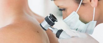
Usually, based on the above clinical signs of the nodular form, it is enough to simply suspect basal cell carcinoma. However, in the initial stages of growth, when the size of the tumor does not exceed 3–5 mm, it can easily be confused with a regular mole (especially if the tumor is pigmented), molluscum contagiosum or senile seborrheic hyperplasia. Hair can grow from a mole, which does not happen with basal cell carcinoma.
A distinctive feature of molluscum contagiosum and senile seborrheic hyperplasia is a small island of keratin in the central part. If there is a crust on the tumor, it can be confused with a wart, keratoacanthoma, squamous cell skin cancer and molluscum contagiosum. In this case, the crusts must be carefully peeled off. With basal cell carcinoma this is easiest. After the bottom of the wound is exposed, for greater confidence and scientific confirmation, it is necessary to make a smear-imprint from the bottom of the ulcer and determine its cellular composition.
Heavily pigmented basal cell carcinomas are easily confused with malignant melanomas. To prevent this from happening, you need to know that the raised edges of basal cell carcinoma almost never contain melanin. In addition, the color of basal cell carcinoma is often brown, while melanoma has a dark gray tint. The flat form of basal cell carcinoma can be confused with eczema, psoriatic plaques and Bowen's disease, but scraping the scales from the edge of the tumor reveals the true picture of the disease.
These clinical signs are intended to guide the doctor towards the correct diagnosis, and its confirmation should be carried out only after a biopsy, cytology or morphological examination of the tumor.
During an appointment with these specialists, the patient may be asked the following questions:
- How long ago did education appear?
- How did it manifest itself, was there any pain or itching?
- Are there similar formations anywhere else on the body? If yes, where?
- Is this the first time the patient has encountered it or have there been similar formations before?
- What is the type of activity and environment in which the patient works?
- How much time does the patient spend outdoors on average?
- Does it take the necessary protective measures against solar radiation?
- Has the patient ever been exposed to excessive radiation? If so, where and approximately what was the total dose?
- Does the patient have relatives with cancer?
If scales are present, they are carefully peeled off onto a glass slide, soaked in a special solution and examined under a microscope. When the ulcerative surface is exposed, a glass slide is applied to it, covered with a coverslip and also examined under a microscope. If the skin over the tumor is intact, then the only way to establish an accurate diagnosis is to perform a biopsy with the collection of tumor material for analysis.
Recovery after surgery
The wound after surgery can either be closed with stitches or left to heal openly. Treatment tactics depend on the type of surgery performed.
If the skin defect was covered with flaps, then moderate pressure is applied to this area using elastic bandages or special tampons during the first 2 days after surgery. In order to reduce swelling of the postoperative area, cold compresses are used. Both closed and open wounds are treated daily with antiseptic solutions.
In order to form a crust on the surface of the open postoperative defect, which is necessary for complete wound healing, baneocin powder, lincomycin, and erythromycin ointment are used 2 times a day. If signs of wound suppuration appear, then antiseptic solutions are used, for example, Miramistin. To speed up healing, multivitamins are also prescribed.
How to treat skin basal cell carcinoma?
The main method of treating basal cell carcinoma in the initial stage on the nose and other parts of the body has been and remains surgical removal of the tumor, after which the removed tissue is sent for further examination. The specialist not only removes the basal cell carcinoma, but also the surrounding intact, healthy tissue. After surgery, the patient needs observation by a dermatologist for timely detection and removal of relapse.
Older people (who have basal cell carcinoma in the ear or nose area) can undergo local chemotherapy (using fluorouracil-based ointment). During therapy, severe redness may occur. The ointment should be used until the treated area reaches the regeneration stage. An immunomodulating ointment can also be used, due to which immune cells become more active, thereby better protecting the skin from tumors.
If you refuse surgical intervention, or if the growth of the tumor is very active, specialists may recommend radiation therapy.
At the initial stage of the disease, treatment with liquid nitrogen (cryotherapy, cryodestruction of basal cell carcinoma) shows great effectiveness. First, the diseased tissue is frozen, and then the fallen part is sent for histological examination.
Recently, more modern approaches—Mohs treatment—have been growing in popularity. It is usually used during localized development on the face. During therapy, the basal cell carcinoma is removed layer by layer under a microscope. In this case, undamaged tissues are not affected, as a result, the chance of getting various cosmetic postoperative defects is minimized.
After removal of basal cell carcinoma (relapse)
Basalioma is a tumor prone to recurrence. This means that after removal of the tumor, the risk of basal cell carcinoma appearing in the same area of skin after some period of time is quite high. There is also a high risk that it will form on another area of the skin.
According to the results of modern studies and observations of people who have had various forms of basalioma removed, the probability of relapse within five years is at least 50%. This means that within 5 years after removal, the tumor will form again in half of the people.
Relapses are most likely if the removed basal cell carcinoma was localized on the eyelids, nose, lips or ear. In addition, the likelihood of relapse is higher when the tumor is large.
Treatment with folk remedies
Since the disease is characterized by a low level of malignancy, patients can use traditional medicine as an auxiliary therapy technique. All kinds of herbal preparations dry out ulcers, stimulate their death and destruction. We will share the most effective recipes.
Medicines from celandine
Celandine is the most famous plant for combating any tumors on the body. The disease in its early stages can be successfully treated with the juice of a fresh plant - just treat the sore spots with it several times a day. Celandine juice is preserved for the winter.
To do this, the plant is crushed in a meat grinder or juicer, and the juice is drained. The remaining cake is poured with a small amount of boiling water, after which the infusion is drained and mixed with pure juice.
Now all that remains is to add alcohol or glycerin (in a 50:50 ratio) and pour the finished canned juice into a glass container with a lid. For a better effect, you can not just soak the skin lesions with celandine juice, but also make compresses for the whole night.
To do this, apply cotton wool soaked in this medicine to the tumor and secure it with a band-aid. By boiling fresh celandine herb in pork fat (in a 1:1 ratio), you will get an ointment that can also be used to successfully fight our illness.
Tobacco tincture
Remove tobacco from 20 cigarettes, pour into a jar, add 100 ml of alcohol and 50 ml of honey. Place the mixture on the windowsill, close the lid and leave for 2 weeks (shaking occasionally). Strain the resulting liquid and use it to treat malignant spots.
Chatter based on Kalanchoe
A mash prepared according to this recipe works well:
- 50 ml Kalanchoe juice;
- 50 ml kerosene;
- 50 ml honey;
- 20 ml glycerin;
- 5 drops rosemary essential oil
Mix all ingredients and store in a glass bottle. Gently apply stains several times a day until they begin to dry out and break down.
Spurge
Patients who were treated with milkweed at an early stage completely got rid of their problem (after the tumor there was not even a scar left on the body).
Before the procedure, you need to steam the skin well - this can be done using a salt bath or lotions with warm saline solution. Then you need to apply a cotton swab or gauze soaked in fresh milkweed juice to the erosion and secure the compress for 2 hours (using a patch or bandage).
Such compresses are repeated daily until the disease disappears completely.
Forecast
The prognosis for life and health with basal cell carcinoma in the initial stage is favorable, since the tumor does not metastasize. Overall, 90% of people survive 10 years after tumor removal. And among those whose tumor was not removed in an advanced state, the ten-year survival rate approaches almost 100%.
A tumor that is more than 20 mm in diameter or has grown into the subcutaneous fat is considered to be advanced. That is, if the basal cell carcinoma at the time of removal was less than 2 cm and did not grow into the subcutaneous fatty tissue, then the 10-year survival rate is almost 98%. This means that this form of cancer is completely curable.
Skin cancer Sarcoma Esophageal cancer Rectal cancer Psoriasis Early stage melanoma
Disease prognosis
The prognosis for the pathology is favorable. The survival rate with adequate treatment exceeds 90%. People with cured forms of basal cell carcinoma at an early stage live on average up to 10 years or more.
In medical practice, there are cases of a new nodule appearing in the same place, which requires repeated therapy. To prevent relapse, it is recommended to be regularly examined by a doctor after treatment. Sometimes the patient is prescribed a special diet. A balanced diet helps support the body and boost the immune system.

