Diseases of the rectum are often detected at later stages. This situation is explained by late access to a doctor; the symptoms force the patient to go to the hospital. In the case of cancer, this leads to death. The year 2012 was marked by a record number of deaths from malignant tumors - about 8 million people, according to WHO statistics. 450 thousand patients died from damage to the rectum. 70-80% of deaths could be prevented if diagnosed at an early stage.
For it to be carried out, not only doctors, but also patients must have “oncological alertness”. If you notice the first symptoms of rectal cancer and the presence of predisposing factors, you should contact a medical institution for advice and diagnostic help.
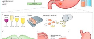
Special risk group: causes of colorectal cancer
In most economically developed countries, with the exception of Japan, colorectal cancer is one of the most common types of cancer, affecting both men and women. There is a statistically significant relationship between the incidence of colorectal cancer and a large amount of meat and animal fats consumed, a deficiency in the diet of coarse fiber and dietary fiber, as well as a sedentary lifestyle. Rectal cancer occupies a stable 3rd place in the structure of the incidence of malignant neoplasms of the gastrointestinal tract, accounting for 45-55% of intestinal neoplasms.
Only some of the factors contributing to the development of malignant tumors of the rectum have been reliably studied. Thus, a number of substances formed during the digestion of animal food, primarily meat (indole, skatole) are carcinogens, and with prolonged contact with the intestinal mucosa they contribute to epithelial metaplasia. This contact increases when food is depleted of dietary fiber, which disrupts the natural passage of food and contributes to chronic constipation due to the long stay of feces in the ampullary section of the rectum.
Precancerous diseases of the rectum include chronic inflammatory diseases of the large intestine: chronic proctitis, chronic nonspecific ulcerative proctosigmoiditis, Crohn's disease.
Get a treatment program
Forecast
An optimistic prognosis for the survival of patients with rectal cancer is observed in countries with a highly developed level of medicine, where 60% of patients live more than five years from the moment the tumor is detected. In countries with a lower level of medicine, this figure does not exceed 40%. Thanks to high-quality early diagnosis and the use of innovative methods for treating rectal cancer, the five-year survival rate of patients at the Yusupov Hospital exceeds 72%.
If you develop intestinal discomfort, undergo a comprehensive examination at the Yusupov Hospital. Call by phone and they will make an appointment with an oncologist. Contact center specialists will offer a convenient time for the patient to consult with an oncologist-proctologist specializing in the treatment of malignant tumors of the rectum.
Make an appointment
Intestinal polyps
The diseases with the highest oncogenicity include intestinal polyposis due to the high incidence of malignancy (malignancy). Transformation into cancer occurs both with single polyps in the rectum and with multiple foci. This is especially true for cases of hereditary polyposis in the family.
In accordance with the classification of the World Health Organization, intestinal adenomas are divided into three types: tubular, villous-tubular and villous. An important role is played by the primary histological diagnosis of polyp biopsy obtained during colonoscopy: for example, villous adenomas become malignant in 35-40% of cases, and in the case of tubular adenomas the risk of malignancy is lower - up to 2-6%. The risk of malignancy increases depending on the size of the adenoma, especially if its diameter is more than 1 cm.
According to biopsy data, from 0.2 to 11% of all intestinal adenomas removed during endoscopy contain cancer cells. Initially, “carcinoma in situ”, intramucosal carcinoma, high-grade dysplasia, or intraepithelial neoplasia develops. These terms refer to malignant tumors that are located in the most superficial layers of the mucous membrane. They are referred to as Tis or stage 0 cancer. These malignant tumors do not metastasize.
When a tumor grows into the submucosa, such cancer is already considered invasive, it can spread to the lymph nodes and give distant metastases.
Relapse of the disease
There is always a risk of cancer recurrence. This is possible with inadequate treatment, with incomplete excision of the affected tissue, with insufficient chemotherapy.
However, more often the cause of relapse is the inability to detect and eliminate cancer cells in full.
More than 60% of recurrent tumors form in the first two years after surgery.
The five-year survival rate for relapse of the disease is about 35%.
Stages of rectal cancer
Rectal tumors are classified according to the generally accepted TNM system, which takes into account the characteristics of the primary tumor (T), the presence of lesions in regional lymph nodes (N) and distant metastases (M).
The letter T can have the indices is, 1, 2, 3 and 4. Tis is a tumor that is located within the superficial layer of the mucous membrane, does not spread to the lymph nodes and does not metastasize. T4 – cancer that has grown through the entire thickness of the wall of the rectum and has spread to neighboring organs.
The letter N can have indices 0, 1 and 2. N0 – there are no tumor foci in the regional lymph nodes. N1 – lesions in 1–3 regional lymph nodes or mesenteric lesions. N2 – lesions in more than three regional lymph nodes.
The letter M can have indices 0 or 1. M0 – no distant metastases. M1a – distant metastases in one organ. M1b – distant metastases in two or more organs, or tumor lesions of the peritoneum.
Depending on these characteristics, five stages are distinguished:
- 0 - The tumor is located in the mucous membrane (sometimes inside the polyp) and does not spread deeper (Tis). Such neoplasms are called “cancer in situ”.
- I - The tumor has spread beyond the mucosa - into the submucosa (T1) or muscle layer (T2). In this case, there are no cancer cells in the lymph nodes (N0), and there are no distant metastases (M0).
- II - The tumor grows through the wall of the rectum and can spread to adjacent tissues. In stage IIa, the cancer has spread deep into the bowel wall but has not penetrated through it (T3). In stage IIb, the tumor invades the bowel wall but does not invade surrounding tissue (T4a). At all these substages, the lymph nodes are not affected (N0), there are no metastases (M0). At stage IIc, the cancer grows into neighboring organs (T4b), or does not grow through the intestinal wall (T1-2), but 1-3 regional lymph nodes are affected (N1), or adipose tissue in the area of the lymph nodes (N1c).
- III - the tumor spreads to neighboring organs and lymph nodes that are located next to the rectum. There are no distant metastases (M0)
- IV - there are distant metastases. At stage IVA there is only one metastasis (M1a), at stage IVB there is more than one (M1b), at stage IVC cancer cells spread to distant areas of the peritoneum (M1c).
Features of squamous cell carcinoma
Externally, this cancer looks like a non-keratinizing ulcer with undermined edges. These types of tumors are prone to early metastases, are characterized by rapid growth and have a disappointing prognosis.
These neoplasms are large in extent, tend to grow into the prostate, vagina, ureters, quickly penetrate the lymph nodes, and have a tendency to recur.
The survival rate for this type of cancer depends on the extent of spread of the cancer process, the number of metastases, the age of the patient and other factors. Patients who began treatment for the disease no later than six months after the onset of the disease have a greater chance. The 5-year survival rate for this type of cancer is almost 33%. The vast majority of patients with this diagnosis die within the first 3 years.
How can rectal cancer manifest itself?
The rectum (lat. rectum) is the final section of the large intestine, about 14-18 cm long, in which the digestive processes end and the formation of feces occurs. The rectum consists of several anatomical areas that have different embryonic origins and histological structures, which causes significant differences in the nature of the course of rectal cancer depending on the level of its damage.
The rectum is divided into 3 parts:
- anal (perineal), 2.5 - 3.0 cm long, in which the sphincter muscles that control the process of defecation are located,
- middle - ampullary, 8.0-9.0 cm long, in which the liquid part of the bolus of food is absorbed and feces are formed,
- supramullary, covered with peritoneum, about 4.0-5.0 cm long.
Malignant neoplasms of the rectum are most often localized in the ampullary region (up to 80% of cases), least often in the anorectal region (5-8%).
In the ampullary and supramullary sections of the rectum, covered with single-layer glandular epithelium, glandular cancer is more often observed - adenocarcinoma, solid cancer, signet ring cell, mixed, scirrhus. Overall, adenocarcinoma accounts for 96% of all colorectal cancer cases. This tumor develops from glandular cells of the mucous membrane that produce mucus. Most often, when doctors use the term “colorectal cancer,” they mean adenocarcinoma.
The anorectal region, lined with stratified squamous non-keratinizing epithelium, is most often affected by squamous cell carcinoma and melanoma. Squamous cell carcinoma accounts for about 90% of anorectal malignancies.
Precancerous diseases
Rectal cancer rarely develops on unchanged mucosa. More often, the appearance is preceded by chronic diseases: benign neoplasms or long-term inflammatory processes.
Benign tumors:
- Polyps are single growths formed by hypertrophied mucosal tissue. They often have a stalk through which the vessels pass and the intestinal lumen protrudes. Such a neoplasm is susceptible to trauma and epithelial metaplasia. The larger the size of the polyp, the higher the likelihood of its malignancy:
- Less than 1 cm in diameter – risk 1.1%.
- 1-2 cm – 8%.
- More than 2 cm – 42%.
- Diffuse polyposis - characterized by the presence of multiple polyps of different shapes and sizes in the colon and rectum. The total number of neoplasms sometimes amounts to hundreds. This is a genetically determined disease that requires surgical treatment.
- Anal papillomatosis is an infectious disease caused by the human papillomavirus. Cells affected by the virus begin to rapidly divide and form numerous conical outgrowths on the skin. Exposure to the virus and frequent damage lead to an increased likelihood of cancerous degeneration of the epithelium.
Cancer prevention involves avoiding risk factors: it is necessary to lead a healthy lifestyle, eat healthy foods and promptly treat precancerous diseases.
Metastases in rectal cancer
The anatomical features of the rectum, its blood supply and lymphatic drainage, also determine the nature of the preferential spread of metastases:
- Rectal cancer metastasizes to regional lymph nodes located in the fatty tissue around the intestine (pararectal) and in the perineum, along the vessels and nerves.
- Due to the characteristics of the venous outflow from the upper rectum to the hepatic portal vein system, very often metastasis occurs directly to the liver.
- In addition, due to the abundant blood supply to the lower parts of the rectum, the tumor metastasizes through the inferior vena cava system to the lungs, bones and other organs.
Metastases
Metastasis of a rectal tumor occurs through two systems - lymphatic and circulatory. Through the lymphatic system, metastases spread upward along the rectal vessels and posteriorly along the rectal vessels, to the side walls of the pelvis through the lymphatic vessels into the iliac and hypogastric lymph nodes. Through the lower rectal lymphatic vessels to the inguinal lymph nodes. Retrograde spread of the tumor into the underlying lymphatic systems is also possible.
Through blood vessels, metastases very quickly enter the liver, disperse throughout the visceral peritoneum, and are detected in other distant systems and organs. Metastasis is accompanied by the appearance of symptoms of tumor development in other organs. When the liver is damaged, patients develop jaundice, pain in the right side, nausea, and vomiting.
Symptoms of colorectal cancer
- The first signs of rectal cancer in most localizations are stool disturbances in the form of chronic constipation and diarrhea, feelings of incomplete defecation, false urge to defecate (tenesmus), discharge from the anal canal (mucus, blood, pus).
- In addition, most patients experience early pain during defecation , caused by tumor invasion of the intestinal walls and dysfunction of the corresponding nerves.
- When the muscles that form the anal sphincters are damaged, fecal and gas incontinence .
- Pain is the first sign of rectal cancer only in case of cancer of the anorectal zone with the involvement of the rectal sphincter in the tumor process. The nature of pain in rectal cancer in the early stages is episodic, then it can become constant.
- With tumors growing into the intestinal lumen (exophytic) and saucer-shaped tumors, tumors-ulcers, the first manifestations of an oncological disease may be bleeding or an inflammatory process . Bleeding is observed in 75-90% of patients with rectal cancer, most often in the form of blood in the stool.
- In the later stages of cancer, mucus and pus may be released along with blood.
- Deterioration in general health (general weakness, fatigue, anemia, weight loss, pale skin), caused by prolonged chronic blood loss and tumor intoxication, is typical for the late stages of rectal malignancies.
The rectum is separated by thin fascia and a small amount of loose tissue from the bladder, seminal vesicles and prostate gland in men, and the uterus and posterior wall of the vagina in women. Therefore, with an increase in the size of the tumor focus, in addition to dysfunction of the rectum, dysfunction of surrounding organs, including urinary incontinence, is relatively often observed.
Survival Projections
The prognosis depends on a combination of factors, such as:
- prevalence of the tumor process;
- histological structure of the formation and the degree of its differentiation;
- anatomical form of tumor growth;
- age, general condition of the patient and concomitant pathology;
- tumor sensitivity to treatment.
If the tumor is detected at stage 1 or 2, the disease is cured in 60-80% of cases.
At stage 3, after complex treatment, long-term remission is achieved in 30-40% of patients.
In the presence of metastases, five-year survival rates do not exceed 40%.
At stage 4 of pathology, the prognosis is extremely unfavorable: almost all patients die within a year after diagnosis.
Diagnosis of rectal cancer
The basis for diagnosing rectal cancer is endoscopic techniques and biopsy. The tumor can be detected using a proctoscope - a special instrument with a miniature video camera that is inserted into the rectum. In this case, the doctor can see the neoplasm, determine its size, position, and assess how close it is to the sphincter.
Colonoscopy allows you to evaluate the condition of not only the rectum, but the entire colon. In this case, a colonoscope is inserted through the anus - an instrument in the form of a thin, long flexible tube with a video camera. It is carried out through the entire colon, examining its mucous membrane. Colonoscopy is a painless procedure, during which the patient is in a state of medicated sleep.
During endoscopy, a biopsy is performed: the doctor receives a fragment of the pathologically changed area of the mucous membrane and sends it to the laboratory for cytological and histological examination.
To assess the stage of rectal cancer and search for metastases, abdominal ultrasound, chest radiography, MRI, computed tomography, and PET scanning are used. Transrectal ultrasound is performed using a special ultrasound probe, which is inserted into the rectum. The test helps assess how much the tumor has spread into surrounding tissues outside the bowel.
Diagnosis using tumor markers
Tumor markers are protein substances that are released as a result of the activity of a malignant tumor or are produced as a response of healthy tissues and organs to the invasion of cancer cells. Found in the urine and blood of sick people. Carrying out an analysis for tumor markers of rectal cancer allows you to detect cancer at an early stage, preserving the health and life of the patient.
Early diagnosis of cancer, carried out at the initial symptoms of the disease, allows the tumor to be removed before the first metastases appear. Using tumor marker analysis, the patient’s health status is monitored after cancer treatment for a certain time. This allows for early detection of tumor recurrence. The level of tumor markers may be increased due to non-oncological diseases.
Is there a cure for colorectal cancer?
In accordance with international protocols, the prevalence of rectal cancer is determined based on the results of a diagnostic examination. At the same time, in addition to the international TNM classification, the division of cancer into stages 1-4, as well as the Duke classification, is often used; the histological structure of the tumor, the degree of differentiation and features of metastasis depending on the location in the rectum, and the presence of complications are taken into account.
A correctly diagnosed stage of the tumor process in rectal cancer allows you to choose the most rational treatment regimen, taking into account international guidelines, including surgery, radiation therapy, chemotherapy and targeted drug therapy.
Contact an oncologist
Contraindications
The operation is contraindicated under the following conditions:
- severe chronic diseases of the patient - arterial hypertension, coronary heart disease, when it is impossible to give anesthesia;
- advanced age of the patient;
- advanced stages of cancer.
In case of a widespread process with metastasis to many organs, palliative resections are used, aimed at alleviating the patient’s condition. Symptomatic operations - the application of bypass anastomoses to relieve the intestines and avoid complications in the last stages of cancer.
Treatment options for colorectal cancer at different stages
The choice of treatment tactics for rectal cancer is influenced by various factors, but the stage of the tumor is of primary importance.
At stages 0 and I
usually only surgery is indicated. Sometimes you can limit yourself to polyp removal - polypectomy. In other cases, transanal resection of the rectum, low anterior resection, proctectomy with colo-anal anastomosis, and abdominal-perineal resection are performed. If the operation cannot be performed due to the patient's poor health, radiation therapy is used,
At stage II
surgical treatment is combined with chemotherapy and radiation therapy. The most common scheme looks like this:
- Initially, the patient receives a course of chemotherapy (usually 5-fluorouracil or capecitabine) in combination with radiation therapy. This helps shrink the tumor and make it easier to remove.>
- Then surgery is performed. Usually this is a low anterior resection, proctectomy with colo-anal anastomosis, or abdominoperineal resection, depending on the location of the tumor.
- After surgery, another course of chemotherapy is given, usually for 6 months. Different combinations of drugs are used: FOLFOX, CAPEOx, 5-fluorouracil + leucovorin or capecitabine alone.
At stage III
the treatment regimen will look similar, but the volume of surgical intervention will be greater, since regional lymph nodes are involved in the process.
At stage IV
tactics depend on the number of metastases. Sometimes they are single and can be removed, just like the primary tumor. The operation is supplemented with chemotherapy and radiation therapy. To combat lesions in the liver, intra-arterial chemotherapy can be used, when a drug solution is injected directly into the artery feeding the tumor.
If there are many metastases, it is impossible to remove them surgically. In such cases, only palliative operations are indicated, for example, to restore intestinal patency if its lumen is blocked by a tumor. The main method of treatment is the use of chemotherapy and targeted drugs. Doctors at the European clinic select treatment in accordance with international protocols and the characteristics of the malignant tumor in a particular patient.
| More information about the treatment of rectal cancer at the European Clinic: | |
| Treatment of rectal cancer | |
| Proctologist-oncologist | 5100 rub. |
| Chemotherapy appointment | 6900 rub. |
| Emergency oncology care | from 11000 rub. |
| Radiologist consultation | 10500 rub. |
Diet
In the process of resection of a malignant tumor, the surgeon at the first stage of surgery creates an unnatural anal opening - a colostomy. Subsequently, the patient undergoes repeated surgery to restore the natural course of the rectum or remains with a colostomy for the rest of his life.
The diet for radiation therapy of the rectum includes a complete set of nutrients. During the preoperative period, dietary nutrition is necessary to maintain the body's immune system. The cooks at the Yusupov Hospital prepare dishes from quality ingredients:
- Sea fish;
- Beef and pork liver;
- Chicken and quail eggs;
- Cereals (unprocessed rice, wheat).
Minimize consumption of sweet foods. Sugar is an excellent medium for faster cell metastasis.
An artificial anus created by a surgeon deprives the patient of the ability to control the frequency of bowel movements. This problem is solved by using a colostomy bag. A rationally selected diet for rectal cancer will allow you to form and strengthen the necessary reflexes.
The patient spends the first postoperative day without food. He begins to receive food from the second day, little by little. The lack of vitamins and microelements is compensated with medication. The daily weight of food should not exceed two kilograms, and the amount of liquid consumed should not exceed 1.5 liters. Meals should be divided into six small portions a day.
Prevention of colorectal cancer
Although it is impossible to be 100% protected from colorectal cancer, as from other cancers, certain measures can help reduce the risks:
- Eat more vegetables and fruits, reduce the amount of fatty meat in your diet.
- Stop drinking alcohol and smoking.
- Exercise regularly.
- Some studies have shown that vitamin D helps protect against colorectal cancer. But before taking it, you should consult your doctor.
- If you have a strong history of colorectal cancer in your family, you may want to consult a clinical geneticist.
- If you have been diagnosed with a hereditary disease that causes colon polyps and cancers, you should have regular colonoscopies.
- After 50 years of age, a colonoscopy is recommended for every person. If no pathologies are found during the examination, it must be repeated after five years.
Observation after remission
In order not to miss the recurrence of the disease, the patient should be regularly monitored by an oncologist. Currently, the following frequency of visits is recommended:
- The first 2 years after remission - at least once every 6 months (recommended once every 3 months);
- After 3-5 years - once every 6-12 months;
- After 5 years - every year.
It should be remembered that if a patient has complaints, an examination by an oncologist is scheduled unscheduled at the earliest available time.