Inflammation of the iris (lat. iris) and ciliary or ciliary body (lat. corpus ciliare), which are part of the choroid of the eye, is called iridocyclitis. Iridocyclitis is dangerous because it most often affects people of the most active age - from 20 to 40 years, although it occurs in both children and the elderly. Iridocyclitis can be caused by various reasons, and depending on this, it has a different course, but in general it responds well to treatment, despite the tendency to relapse in some forms of the disease. However, if not treated promptly, iridocyclitis can lead to vision loss.
Causes
The etiological factors of anterior uveitis are divided into endogenous and exogenous. The pathology progresses due to increased permeability of the blood-ophthalmic barrier (the membrane between the general blood flow and the vascular network located in the eye). This barrier acts as a biological filter for the small capillaries of the eyeball and retina. It retains toxins, bacteria, antigens and does not allow a large number of medications to pass through.
Due to the increased permeability of the blood-ophthalmic barrier, antigens spread into the iris, provoking inflammation in it.
Sometimes the pathology is formed as a result of eye injury (penetrating injury, contusions or unsuccessful surgical interventions). Pathology of the ciliary system of the eye develops as a result of:
- measles;
- flu;
- human papillomavirus;
- infections due to the activity of staphylococci or streptococci;
- activity of tuberculosis bacilli;
- gonococcal infection;
- chlamydia;
- toxoplasmosis;
- malaria;
- sinusitis;
- tonsillitis;
- the presence of chronic inflammatory pathology in the body.
The cause of the pathology is rheumatoid or metabolic disorders:
- rheumatoid tissue damage;
- Still's disease;
- thyroid diseases of autoimmune origin;
- Bekhterev's pathology;
- Sjogren's syndrome;
- Reiter's disease;
- violation of uric acid metabolism;
- diabetes;
- sarcoidosis;
- Behçet's disease;
- Vogt-Koyanagi-Harada syndrome.
The appearance of pathology is facilitated by an enlarged lining of the vessels of the eye system. Iridocyclitis is also observed due to the abnormal susceptibility of eye structures to antigens spreading from areas of infection. Such inflammations are combined with immune dysfunctions and disorders of microcirculation processes.
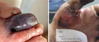
Iridocyclitis in dogs
Dogs also suffer from iridocyclitis, just like humans. The causes of the disease in dogs can be the following:
- influence of infectious parasites;
- inflammation occurring in the cornea of the eye;
- autoimmune and allergic processes.
Signs of iridocyclitis in dogs
The development of iridocyclitis in a dog can be determined by the following existing symptoms:
- swelling of the iris;
- photophobia – if light hits the iris of the eye, the pet will begin to experience discomfort;
- the vision pattern becomes blurred;
- infiltrates together with hemorrhage in the iris and anterior chamber of the eye;
- the vitreous body loses its transparency;
- Posterior adhesions begin to form.
Diagnosis and treatment
In order to make the most accurate diagnosis, the veterinarian examines the patient through ophthalmoscopy. After making an accurate diagnosis, the specialist does the following:
- Dilates the dog's pupils. This is done in order to reduce pain. To expand the crustaceans, solutions of atropine, glucocorticoids, and adrenaline are used;
- Treats infection. To cure an animal of this disease, vitamins and antihistamines are prescribed. Antibiotics may be used.
Possible complications in dogs
If the treatment is structured correctly, then the probability of encountering complications is close to 0. But still, diseases that could be caused by iridocyclitis are:
- cataract;
- vitreous abscesses;
- atrophy of the eye.
For prevention, it is necessary to get rid of foci of infection in the dog’s body in a timely manner and constantly monitor its condition.
Classification
The duration of the disease is classified as acute, subacute or chronic. By nature, iridocyclitis is defined as exudative, serous, fibrinous-plastic, hemorrhagic. Moreover, the fibrinous-plastic form of the pathology is the most dangerous, because provokes adhesions and a decrease in the transparency of eye tissue.
Acute iridocyclitis manifests itself sharply with pain. It is localized in the eye area, but sometimes spreads to the entire head. At night, the discomfort intensifies significantly, depriving patients of sleep.
Chronic iridocyclitis is a consequence of other pathologies - influenza, tuberculosis or herpes virus infection.
Serous iridocyclitis is a rare form of pathology. In this case, inflammatory phenomena are accompanied by the formation of a large amount of purulent mucus. This process leads to increased pressure inside the eye.
Fibrinous plastic iridocyclitis results from serious eye injuries. Often this pathology develops into a slowly progressive chronic form.
Depending on the etiological factor, the following types of iridocyclitis are distinguished:
- infectious or infectious-allergic;
- allergic non-infectious;
- systemic;
- post-traumatic;
- metabolic (developing as a result of metabolic dysfunctions);
- unknown origin.
Choreoretinitis
Choreoretinitis is an inflammation of the posterior uvea involving the retina. It can occur in acute or chronic form.
Symptoms of the disease
Choreoretinitis can be perepapillary (focus of inflammation near the optic nerve head), central (in the macular zone), peripheral (near the dentate line), equatorial (in the equator zone). Symptoms vary depending on the location of the inflammation.
The peripheral form usually has no symptoms, and the disease is discovered accidentally during an eye examination.
Central choreoretinitis by :
- “flashes”, dark spots and sparks before the eyes;
- “fog” and deterioration of visual acuity;
- distortion of the size and shape of objects;
- severe weakening of twilight vision, “night blindness”.
These symptoms portend a serious deterioration in vision; you should urgently consult an ophthalmologist!
1 Diseases of the choroid. Diagnosis and treatment
2 Diseases of the choroid. Diagnosis and treatment
3 Diseases of the choroid. Diagnosis and treatment
Symptoms
The intensity of the pathology depends on the type and duration of exposure to the pathogenic factor. Unilateral iris disease is usually found. The earliest signs of an acute form of pathology:
- soreness and hyperemia of tissues;
- pain that increases with pressure on the eye;
- fear of bright light;
- increased secretion of tears;
- decreased quality of vision;
- a veil before the eyes.
A change in the structure of the affected iris is determined. It changes its color to red or greenish. Corneal syndrome of varying severity appears. It is possible to form effusion of varying contents in the eye tissues. If purulent effusion accumulates at the bottom of the anterior chamber, a hypopyon grows in it - a gray or greenish strip.
Precipitates of different colors grow on the posterior surface of the cornea. They contain liquid. Due to the fact that the lens comes into contact with the swollen tissues of the iris, synechiae are formed, leading to irreversible changes in the pupil. A circular adhesion is formed if the anterior surface of the eye lens and the iris are fused.
If the course of iridocyclitis is unfavorable, there is a high risk of developing blindness due to a blocked pupil.
With a chronic type of pathology, pain in the eyes is not felt or it is very weak.
Photo
Chronic iridocyclitis
Chronic iridocyclitis is not an independent disease - it is just a consequence of the development of other pathologies. These include herpes, influenza and tuberculosis. The pain is not so severe, the conjunctiva does not redden strongly and sharply, and the disease proceeds slowly, just as it develops.
With this type of disease, atrophic changes are more noticeable: the adhesions of the iris and lens have become rough, which leads to overgrowth of the pupil, clouding of the vitreous, etc.
{banner_horizontalnyy2}
Diagnostics
Diagnosis of the patient includes examination, laboratory tests, and fluoroscopy.
First, the doctor examines the eyeball, palpates it, and analyzes the anamnesis in detail. The diagnosis is clarified by checking visual function and measuring eye pressure using tonometry. The diagnosis is confirmed by ultrasound of the eye.
To determine the etiology of iridocyclitis, an extensive blood and urine test is used. To determine the presence of systemic pathologies, rheumatic tests are prescribed. In order to confirm or refute the allergic genesis of iridocyclitis, allergy tests and tests for the presence of antigens are recommended.
In order to determine the state of immunity, patients examine the content of serum immunoglobulins.
Consultation with other specialists is required:
- phthisiatrician;
- rheumatologist;
- dentist;
- ENT doctor;
- allergy specialist;
- skin doctor.
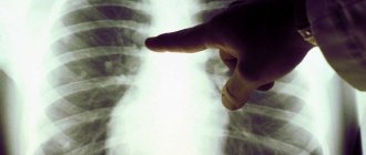
The diagnosis is clarified by performing an X-ray examination of the lungs and paranasal sinuses.
Pathological anatomy
For obvious reasons, fresh cases of diffuse iridocyclitis can only become the object of pathological examination as a rare exception. Therefore, changes in them can be judged only on the basis of experimental observations and on the basis of similar changes in traumatic iridocyclitis and iridocyclitis due to corneal ulcers.
In fresh cases, we are dealing with fibrinous exudation into the tissue of the iris and ciliary body and infiltration of the stroma with lymphocytes and polynuclear cells. Infiltration is mostly diffuse in nature, but it also occurs focally, mainly in the sphincter region in front of the pigment epithelium and at the root of the iris near Schlemm's canal. In the ciliary body, exudate is located mainly along its surface, and both layers of the epithelium are abundantly penetrated by emigrating cells.
In older cases, the organization of exudate into connective tissue is noted. Depending on where the organization occurs, there is a circular fusion of the pupillary edge of the iris with the anterior capsule of the lens and fusion of the pupil, connective tissue films on the surface of the iris, on the surface of the ciliary body and behind the lens. Wrinkling of these films can lead to secondary changes; on the iris to inversion of its pigment layer onto the anterior surface, on the ciliary body to detachment of the ciliary body and retina.
In the stroma of the iris and ciliary body, during old processes, atrophy is noted, the disappearance of chromatophores, their transformation into clumsy shapeless cells, depletion of pigment, and the development of coarse fibrous connective tissue. Precipitates consist of lymphocytes stuck together, with pigment grains included in them.
Pathological changes in tuberculous iridocyclitis manifest themselves in two ways. On the one hand, with nodular forms, the development of typical tuberculous nodules is observed, which in confluent forms give the formation of more or less extensive tuberculous granulomas. Tuberculosis nodules develop in the tissue of the iris and ciliary body, but deposition of specific tuberculosis products on the surface of these membranes is often observed, from where they can be transferred with a fluid current to various places in the eye, thus giving rise to a strain of tuberculosis for vaccination.
In chronic diffuse tuberculous iridocyclitis and in old cases that have led to eye atrophy, changes often have nothing characteristic of tuberculosis and consist of diffuse infiltration of tissue with lymphocytes and individual foci of round cell infiltration, mainly behind the sphincter at the pigment epithelium, in the region of the iris root and under epithelium of the ciliary body in the recesses between the processes.
With syphilitic iridocyclitis, in addition to diffuse infiltration of tissue with lymphocytes and epithelioid cells, the formation of perivascular nodules with typical vascular changes is observed - infiltration of adventitia with cells and proliferation of the intima, that is, the phenomena of peri- and endovasculitis.
Treatment
Therapeutic measures are aimed at preventing the influence of the pathological factor.
Non-operative measures are used to prevent the formation of adhesions and reduce the likelihood of progression of complications. At the initial stages of the disease, medications are administered to increase the diameter of the pupil, medications to relieve the inflammatory process, hormonal, and antihistamine medications.
Treatment measures are carried out in a hospital. The basis of therapeutic measures is the introduction into the body of various disinfectants, antimicrobial or antiviral agents. They are used in the form:
- drops;
- injections into a muscle or vein;
- intraocular or conjunctival injections of drugs.
Glucocorticosteroid medications are used to get rid of iridocyclitis of autoimmune etiology.
Detoxification treatment is prescribed. In case of intense inflammatory phenomena, blood purification and plasmapheresis, administration of mydriatics are used (these drugs do not allow the growth of adhesions and the development of a persistent decrease in pupil diameter). Depending on the presence of the underlying disease, immunostimulating agents and proteolytics are used. Physiotherapeutic procedures include electrophoresis, laser and magnetic therapy.
To reduce the intensity of pain, local analgesics are prescribed - Inocaine, Naklof, Alcaine, Lidocaine.
For iridocyclitis provoked by Koch microbes, antibacterial treatment is indicated.
If it is necessary to treat adhesions, the patient is prescribed surgery. In this case, synechiae are removed. Surgery is performed urgently when glaucoma progresses. In difficult to treat cases, the diseased eye is removed.
Origin and spread
Simultaneous damage to the iris and ciliary body is due to the fact that there is a blood relationship between them. They are connected to the central nervous system by a common nerve that causes excitation of the fibers. In addition, blood to both elements comes from a single vessel.
At the acute stage, the disease is accompanied by pain in the eyes, severe redness and swelling appear. Increased lacrimation and correction of the pupil opening provoke a change in the shade of the iris. As a result, visual acuity decreases, deposits from cellular elements accumulate on the cornea, and festering fluid collects in the frontal cell.
| The acute period lasts about a month and a half, the chronic period can last for six months. |
Complications
Diseases caused by iridocyclitis contribute to blindness and pose a threat to the preservation of the organ of vision:
- chorioretinitis;
- pupil deformation;
- increase in intraocular pressure;
- decreased transparency of the lens;
- clouding of the eye structures;
- retinal rupture and detachment;
- development of an inflammatory purulent focus;
- inflammation of the eye tissues;
- atrophy of the organ of vision.
In order to prevent the development of complications, it is necessary to abandon the so-called traditional methods of treatment. This disease is dangerous and requires early medical attention and effective therapy.
Iridocyclitis - what kind of disease is this?
This pathology is also called anterior uveitis due to the fact that inflammation affects not only the iris, but also part of the vascular network of the eye - the ciliary, or ciliary body. The iris is a thin, movable diaphragm with a hole in the center - the pupil. This structure of the eye regulates the amount of light rays entering it, which is achieved by contracting the pupillary area. Depending on the light source, it expands or contracts. The iris also ensures a constant temperature of the fluid located between it and the cornea and participates in its outflow.
The ciliary body is part of the choroid/tunica media. It supports the lens and is involved in the process of accommodation. In addition, the ciliary body is the connecting link between the iris and the vascular network. They have a common blood supply, and therefore the pathological process affecting one of the listed structures subsequently passes to the other. What causes this inflammation? Let's consider the main reasons for its occurrence. The form of the course, and therefore the treatment process, depends on them.
Forecast
Provided early therapy is started and conscious compliance with all the advice of the ophthalmologist, the prognosis of the disease is favorable. Recovery occurs in about a fifth of patients. In other patients, inflammation has a subacute course with mild relapses. More often, the disease returns in case of exacerbation of rheumatism or gout.
In case of ineffective and delayed treatment, iridocyclitis transforms into a treatment-tolerant stage with a steady decrease in vision.
Prognosis of iridocyclitis
The prognosis of iridocyclitis largely depends on its form and the adequacy of the treatment undertaken. As a rule, if the cause of the disease can be eliminated, then iridocyclitis is cured. In the case where iridocyclitis is a symptom of a severe systemic disease, every effort must be made to prevent the occurrence of complications and the spread of inflammation to the rest of the tissues of the eye. In general, the prognosis for iridocyclitis is favorable, subject to treatment and observation by an ophthalmologist.
Video from YouTube on the topic of the article:
Prevention
No specific prevention of pathology has been developed. It is advisable to eliminate all pathological and bacterial foci in the body. Common systemic pathologies need to be cured completely or transferred to a stage of stable remission. Compliance with the rules of a healthy lifestyle is of great importance.
The following preventive recommendations should be followed:
- adhere to personal hygiene requirements;
- monitor the intensity of visual work;
- avoid excessively bright light, dust;
- do not be in a draft;
- avoid being near air conditioners;
- eat food with sufficient amounts of vitamins and microelements that are good for the eyes;
- adhere to the established vaccination schedule (you need to be vaccinated against influenza annually);
- At the first signs of an eye disease, immediately contact an ophthalmologist.
Course of the disease
The course of iridocyclitis varies. Acute cases usually require at least 5-6 weeks to complete. Iridocyclitis of rheumatic, gouty, gonorrheal origin is acute, but is very prone to relapses, which sometimes torment patients very often, repeating several times a year.
Sometimes iridocyclitis, especially tuberculosis, takes a very slow, sluggish course from the very beginning. In this case, the phenomena of irritation are sometimes completely absent, and only visual disturbances attract the patients’ attention to the eye. The disease lasts for months, sometimes years, sometimes exacerbating and sometimes subsiding.
In most cases, especially with chronic and recurrent iridocyclitis, the disease affects both eyes.
Useful video
More interesting and useful information about iridocyclitis can be found in the video at the following links:
Iridocyclitis is a disease that requires immediate medical intervention. The preservation of normal vision depends on how early the patient receives help. Self-medication of this ophthalmological disease is extremely dangerous, because. can lead to irreversible changes in the tissues of the eye.
Author's rating
Author of the article
Alexandrova O.M.
Articles written
2031
about the author
Was the article helpful?
Rate the material on a five-point scale!
( 2 ratings, average: 3.00 out of 5)
If you have any questions or want to share your opinion or experience, write a comment below.
Etiology and pathogenesis
The causes of iridocyclitis are very diverse. These include:
- Injuries to the visual analyzer - penetrating wounds, bruises, foreign bodies, consequences of surgical treatment,
- Inflammation of the cornea or sclera,
- Viral infection - influenza, measles, herpes, cytomegalovirus,
- Pathogenic and opportunistic bacteria - staphylococci, streptococci, Koch's bacillus, gonococci, Treponema pallidum, chlamydia, toxoplasma,
- Pathogenic fungi - candida, actinomycetes,
- Various helminthiases and parasitoses,
- ENT diseases - otitis, sinusitis, tonsillitis,
- Dental diseases - caries, stomatitis, hilar cysts,
- Autoimmune diseases - rheumatoid arthritis, scleroderma, sarcoidosis, spondyloarthrosis,
- Allergies to food, medications,
- Endocrinopathies – diabetes mellitus, hyperfunction of the thyroid gland.
Despite such a variety of etiological factors of the disease, the direct cause of iridocyclitis is infection. Under the influence of external and internal etiopathogenetic factors, the blood-ophthalmic barrier is disrupted, which contributes to the deposition of immune complexes in the uveal tract of the eye. Cells of the choroid have increased susceptibility to antigens and CECs that penetrate the eye from infectious foci. Antigen-antibody complexes are formed in the blood, immunological inflammation of the vascular network of the eye and its damage by inflammatory mediators develops. In the tissues of the eye, phenomena of immunocytolysis occur, immunopathological reactions, vasculopathy, dysfermentosis, dyscirculatory processes, cicatricial and dystrophic changes develop. The result of such phenomena is a swollen and thickened iris. The patient develops a clinical picture of iridocyclitis.
Provoking factors of this pathology:
- immunodeficiency,
- neuropsychic exhaustion, stress,
- intense physical activity,
- unbalanced diet.
Pathogenetic links of the main morphological forms of iridocyclitis:
- The fibrinous-plastic form is characterized by the presence of fibrinous exudate in the anterior chamber of the eye with its partial organization and manifests itself with dangerous symptoms. A complication of this form is irreversible pupillary fusion and blindness.
- The purulent form develops a couple of days after a traumatic injury to the eye or is a complication of purulent tonsillitis, furunculosis, or abscess. The disease is severe. Pus accumulates in the anterior chamber of the eye. The process grows rapidly, the picture of panuveitis and endophthalmitis develops.
- The hemorrhagic form is a consequence of damage to the vascular walls by viruses and is characterized by the accumulation of bloody exudate in the anterior chamber of the eye.
- Mixed iridocyclitis is characterized by the appearance of white precipitates and pigmentation on the cornea, synechiae, and signs of focal chorioretinitis.