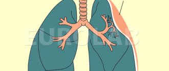
Pulmonary emphysema is a disorder of the anatomical structure of the lung tissue, characterized by a pathological expansion of the air spaces that are located beyond the terminal bronchioles, accompanied by destructive changes in the alveolar walls. For the treatment of patients suffering from pulmonary emphysema, all conditions have been created at the Yusupov Hospital:
- Chambers with a European level of comfort, equipped with forced-air ventilation and air conditioning;
- Providing patients with high-quality dietary nutrition and personal hygiene products;
- Use of the latest diagnostic equipment from leading global manufacturers;
- Treatment with modern medications that are effective and have a minimal range of side effects.
Doctors take an individual approach to choosing a treatment regimen for each patient. Severe cases of pulmonary emphysema are discussed at a meeting of the Expert Council with the participation of professors, associate professors, and doctors of the highest category. Leading specialists in the field of pulmonology collectively develop a patient management scheme.
Along with chronic obstructive bronchitis and bronchial asthma, emphysema belongs to the group of chronic obstructive pulmonary diseases (COPD). All these diseases are accompanied by impaired bronchial obstruction. This is due to some similarity in their clinical picture. Each form of COPD has its own specific features. Correct timely diagnosis of these diseases allows pulmonologists at the Yusupov Hospital to conduct rational therapy and targeted prevention of complications of pulmonary emphysema.
Causes of emphysema
The causes of this pathology are divided into two groups.
- Impaired elasticity and strength of lung tissue:
- Congenital structural features of lung tissue. Pressure in the alveoli increases due to collapse of bronchioles due to birth defects.
- Hormonal imbalance. The smooth muscles of the bronchioles lose their ability to contract due to an imbalance between estrogens and androgens. The consequence of this is stretching of the bronchioles and the formation of voids in the lung parenchyma.
- Inhalation of polluted air with impurities of tobacco smoke, coal dust, smog, toxins. The most dangerous impurities are oxides of sulfur and nitrogen - by-products of processing automobile fuel and emissions from thermal power plants. Microparticles of these compounds are deposited on the walls of bronchioles. They affect the pulmonary vessels that supply the alveoli, damage the ciliated epithelium, and activate alveolar macrophages. Additionally, the level of neutrophils and proteolytic enzymes increases, leading to the destruction of the walls of the alveoli.
- Congenital alpha-1 antitrypsin deficiency. This pathology leads to the fact that proteolytic enzymes acquire functions unusual for them - instead of destroying bacteria, they destroy the walls of the alveoli. Normally, alpha-1 antitrypsin should neutralize these manifestations immediately after they occur.
- Age-related changes. The blood circulation of an elderly person undergoes changes for the worse, and sensitivity to air toxins increases. In older people, lung tissue recovers more slowly after pneumonia.
- Respiratory tract infections. When pneumonia or bronchitis occurs, the immune system stimulates the activity of protective cells: macrophages and lymphocytes. A side effect of this process is the dissolution of the protein in the walls of the alveoli. Additionally, sputum clots do not allow air to pass from the alveoli to the outlet, which leads to tissue stretching and overfilling of the alveolar sacs.
- Increased pressure in the lungs:
- Occupational hazards. The costs of the profession of wind instrument musicians and glassblowers are increased air pressure in the lungs. Long-term exposure to these harmful substances leads to impaired blood circulation in the walls of the bronchi. Due to the weakness of the smooth muscles, some of the air remains in the bronchi, and the next portion is added to it when inhaling. This leads to the appearance of cavities.
- Chronic obstructive bronchitis. With this pathology, the patency of the bronchioles is impaired. When you exhale, the air does not completely leave the lungs. Because of this, both the alveoli and small bronchi stretch, and over time, cavities appear in the lung tissue.
- Blockage of the bronchial lumen by a foreign body. Causes an acute form of emphysema because air cannot escape from this segment of the lung.
The exact cause of the appearance and development of this pathology has not yet been established. According to scientists, the appearance of emphysema is influenced by several factors.
Signs and symptoms of emphysema
- Cyanosis – the tip of the nose, earlobes, and nails become bluish. As the disease progresses, the skin and mucous membranes become pale. The reason is that small capillaries are not filled with blood, oxygen starvation is recorded.
- Shortness of breath of an expiratory nature (with difficulty exhaling). Insignificant and unnoticeable at the beginning of the disease, it progresses later. It is characterized by difficult, stepwise exhalation and gentle inhalation. Due to the accumulation of mucus, the exhalation is elongated and puffing. Differentiation from shortness of breath in heart failure - does not worsen in the supine position.
- Intensive work of the muscles that provide breathing. To ensure the functioning of the lungs during inhalation, the muscles that lower the diaphragm and raise the ribs intensively tense. As you exhale, the patient tenses the abdominal muscles that lift the diaphragm.
- Swelling of the neck veins. Occurs due to increased intrathoracic pressure during coughing and exhalation. With emphysema complicated by heart failure, the neck veins also swell when inhaling.
- Pinkening of the complexion during a coughing attack. Thanks to this symptom, emphysema patients are nicknamed “pink puffers.” The amount of discharge when coughing is small.
- Weight loss. The symptom is associated with excessive activity of the muscles that provide breathing.
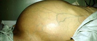
- Increased liver size and prolapse. Occurs due to stagnation of blood in the vessels of the liver and prolapse of the diaphragm.
- Changes in appearance. Appear in patients with long-term chronic emphysema. Signs: short neck, bulging supraclavicular fossa, barrel-shaped chest, sagging belly, intercostal spaces drawn in during inspiration.
Briefly about the main thing
Emphysema is a respiratory disease that is accompanied by a dry cough, impaired respiratory function, and increased intrathoracic pressure. Smoking citizens, as well as people with chronic pulmonary pathologies, are susceptible to developing the disease. Emphysema can be treated at home with regular check-ups with your doctor. For complex therapy, medications and traditional methods of treatment are used: herbal infusions, breathing exercises and oxygen therapy. In advanced cases, they resort to surgery.
Share with your friends
Do something useful, it won't take much time
Types of emphysema
Emphysema is classified into several categories.
According to the nature of the flow:
- Spicy. It can be caused by significant physical activity, an attack of bronchial asthma, or the entry of a foreign object into the bronchial network. There is swelling of the lung and overstretching of the alveoli. The condition of acute emphysema is reversible, but requires emergency treatment.
- Chronic. Changes in the lungs occur gradually; at an early stage, a complete cure can be achieved. Without treatment it leads to disability.
By origin:
- Primary emphysema. The origin is associated with the innate characteristics of the body. It is an independent disease and can be diagnosed even in newborns and infants. It is difficult to treat and progresses at an accelerated pace.
- Secondary emphysema. The origin is associated with the presence of obstructive pulmonary diseases in a chronic form. The onset of the disease may go unnoticed, and increased symptoms lead to loss of ability to work. If the disease is not treated, the size of the cavities that appear can be significant, occupying entire lobes of the lungs.
By prevalence:
- Diffuse form. Tissue damage and destruction of the alveoli occurs throughout the lung tissue. Severe forms of the disease may result in donor organ transplantation.
- Focal form. Changes in the parenchyma are diagnosed around foci of tuberculosis, scars, and the site of bronchial blockage. Symptoms of emphysema are less severe.
According to anatomical features, in relation to the acinus:
- Panacinar (vesicular, hypertrophic) form. Diagnosed in patients with severe emphysema. There is no inflammation, there is respiratory failure. There is no healthy tissue between the damaged and swollen acini.
- Centrilobular form. Destructive processes affect the central part of the acinus. Due to the expansion of the lumen of the bronchi and alveoli, an inflammatory process develops, and mucus is released in large quantities. Fibrous degeneration of the walls of damaged acini occurs. The intact lung parenchyma between the areas that have undergone destruction performs its functions without changes.
- Periacinar (parasepital, distal, perilobular) form. Develops during tuberculosis; in this form, the extreme parts of the acinus near the pleura are affected. It may result in a complication - rupture of the affected area of the lung (pneumothorax).
- Peri-scar form. It is characterized by minor symptoms and appears near fibrotic foci and scars in the lungs.
- Bullous (bubble) form. Bullae (bubbles) with a diameter of 0.5-20 cm are formed near the pleura or throughout the parenchyma. They arise at the site of damaged alveoli. They can rupture, become infected, and put pressure on surrounding tissues.
- Interstitial (subcutaneous) form. Due to the rupture of the alveoli, air bubbles form under the skin. They move through the lymphatic pathways and gaps between tissues under the skin of the head and neck. Spontaneous pneumothorax may occur due to the rupture of bubbles remaining in the lungs.
Due to the occurrence:
- Senile emphysema. Occurs due to age-related changes in blood vessels, violations of the elasticity of the walls of the alveoli.
- Lobar emphysema. Observed in newborns, it appears due to obstruction of one of the bronchi.
Bullous emphysema
Bullous pulmonary emphysema is understood as a critical disorder of the structure of the lung tissue, in which destruction of the interalveolar septa occurs. This creates one large cavity filled with air. Bullous emphysema can occur against the background of general pulmonary emphysema, as one of the extreme degrees of its development, or it can also develop against the background of healthy surrounding lung tissue. Such bullous transformation is facilitated by past inflammatory and suppurative processes in the lungs, especially with a chronic course (chronic abscess, bronchiectasis, tuberculous lesions). The mechanism of its appearance initially has a vicarious nature of emphysema, which over time transforms into a bulla.
If bullous emphysema is represented by single bullae on the surface of the lungs, a person usually does not know about its existence. It is not accessible to diagnosis even with x-ray examination. The situation is completely different with multiple bullae over the entire surface of the lung tissue. Such patients have all the symptoms of pulmonary emphysema, including signs of respiratory failure of varying degrees.
The danger of bullous emphysema occurs with severe thinning of the superficial membrane of the bulla. In this case, there is an extremely high risk of rupture. This is possible with sudden changes in pressure in the chest (cough, physical stress). When a bulla ruptures, air from the lungs rapidly enters the pleural cavity. A dangerous condition called pneumothorax occurs. In this case, the air accumulated in the pleural cavity creates high pressure, which compresses the affected lung. If the defect in the lung tissue is large enough, it is not able to close on its own, which leads to a continuous flow of air into the pleural cavity. When its level becomes critical, it begins to enter the mediastinum and subcutaneous tissue, which causes the development of subcutaneous and mediastinal emphysema. This is very dangerous, as it can result in decompensated respiratory failure and cardiac arrest.
Diagnosis of emphysema
Examination by a doctor
At the first symptoms or suspicion of pulmonary emphysema, the patient is examined by a pulmonologist or therapist.
The examination takes place according to the following scheme:
- The first stage is collecting anamnesis. Sample topics for questions to ask the patient:
- How long does the cough last?
- Does the patient smoke? If yes, then for how long, how many cigarettes does he use per day?
- Is there shortness of breath?
- How does the patient feel during increased physical activity?
- Percussion is a special technique of tapping the chest with the fingers of the right hand through the palm of the left placed on the chest. Possible symptoms:
- Limited lung mobility;
- “boxy” sound over areas of increased airiness;
- Drooping of the lower edge of the lungs;
- Difficulty determining the boundaries of the heart.
- Auscultation - listening to the chest using a phonendoscope. Possible manifestations of the disease:
- Increased exhalation;
- Muffled heart sounds due to sound absorption by the air-filled lung parenchyma;
- Decreased breathing;
- When bronchitis occurs - dry wheezing;
- Tachycardia is an attempt by the heart to compensate for oxygen starvation by increasing heart rate;
- Strengthening the second heart sound as a result of increased blood pressure in the pulmonary circulation, as a sign of damage to the right half of the heart;
- Rapid breathing with a frequency of 25 or more breaths per minute, as a sign of overstrain of the respiratory muscles and respiratory failure.
Instrumental methods for diagnosing pulmonary emphysema
- Radiography is a study of the lungs by obtaining their image on a special film using X-rays. The picture is taken in a direct projection, when the study is carried out with the patient facing the device. When analyzing the image, the doctor identifies lung pathologies and the stage of spread of the process. If it is necessary to clarify the diagnosis, magnetic resonance and computed tomography and spirometry are prescribed.
Indications for the study:
- Annual medical examination
- Dyspnea,
- Weak breathing
- Wheezing, pleural friction noise when listening to the chest,
- Prolonged cough
- Suspicion of tuberculosis or pneumonia, bronchitis, emphysema;
- Pneumothorax.
Contraindications: lactation and pregnancy.
Possible symptoms:
- Enlargement of the lungs, their overlap, compression of the mediastinum;
- Transparency of the affected areas;
- Widened intercostal spaces;
- Changes in the vascular system of the lungs;
- Drooping of the lower edge of the lungs and diaphragm;
- Detection of bullae and air pockets.
Indications:
- Symptoms indicate the presence of cysts, tumors, but the x-ray did not show them;
Contraindications:
- Mental illnesses that prevent maintaining a long-term immobile position;
- Fear of closed spaces;
- Severe obesity;
- Presence of implants, pacemaker, unremoved fragments.
Symptoms of emphysema determined by MRI:
- Bullae and cavities of various sizes;
- Enlarged lung;
- Compression of healthy tissue;
- Increased amount of fluid in the pleura;
- Damage to the alveoli and their capillaries;
- Impaired blood supply;
- Lowering the diaphragm.
Indications:
- Clarification of X-ray examination data;
Contraindications:
- Individual intolerance to contrast agent;
- Severe diabetes mellitus;
- Pregnancy;
- Severe obesity;
- Extreme weakness;
- Kidney failure.
Symptoms of emphysema:
- Identification of the area of expanded areas;
- Fixing the size and location of bullae;
- Vasodilation at the root of the lung;
- The appearance of airy areas.
Indications:
- Diagnosis of blood vessels at an early stage of emphysema development;
Pregnancy is an absolute contraindication to the examination.
Symptoms of emphysema:
- Blood flow disorders;
- The appearance of areas of compression of the lung tissue.
Indications:
- Prolonged cough;
Contraindications:
- Tuberculosis;
- Hypertension;
- Condition after stroke and heart attack, operations on the chest and peritoneum;
- Pneumothorax;
- Bloody sputum.
Symptoms of the disease:
- Changes in vital and residual lung capacity;
- Reduced ventilation and speed indicators;
- Increased airway resistance;
- Reduced extensibility of the lung parenchyma.
Indications:
- Signs of lack of oxygen (cyanosis);
Symptoms:
- Blood oxygen less than 15%;
- Oxygen tension is less than 60-80 mmHg;
- Carbon dioxide voltage is more than 50 mm Hg.
Deviations from the norm for emphysema:
- increased red blood cell count over 5 1012/l
Treatment of emphysema
Directions of treatment:
- Combating further development of the disease;
- Prevention of severe complications (respiratory and heart failure);
- Improving the quality of life of patients.
Mandatory treatment measures:
- Conservative therapy for easier breathing, better lung function;
- To give up smoking;
- Performing a complex of therapeutic exercises for lung ventilation;
- Treatment of the main cause of the disease.
Treatment of emphysema (medicines)
| Group of drugs | Drug names | Mechanism of therapeutic action | Mode of application |
| α1-antitrypsin inhibitors | Prolastin | The introduction of this protein reduces the level of enzymes that destroy the connective fibers of lung tissue. | Intravenous injection at the rate of 60 mg/kg body weight. 1 time per week. |
| Mucolytic drugs | Acetylcysteine (ACC) | Improves the removal of mucus from the bronchi, has antioxidant properties - reduces the production of free radicals. Protects the lungs from bacterial infection. | Take 200-300 mg orally 2 times a day. |
| Lazolvan | Liquefies mucus. Improves its removal from the bronchi. Reduces cough. | Used orally or inhaled. Orally during meals, 30 mg 2-3 times a day. In the form of inhalations using a nebulizer, 15-22.5 mg, 1-2 times a day. | |
| Antioxidants | Vitamin E | Improves metabolism and nutrition in lung tissues. Slows down the process of destruction of the walls of the alveoli. Regulates the synthesis of proteins and elastic fibers. | Take 1 capsule per day orally. Take courses for 2-4 weeks. |
| Bronchodilators (bronchodilators) Phosphodiesterase inhibitors | Teopek | Relaxes the smooth muscles of the bronchi, helps to expand their lumen. Reduces swelling of the bronchial mucosa. | The first two days take half a tablet 1-2 times a day. Subsequently, the dose is increased - 1 tablet (0.3 g) 2 times a day after 12 hours. Take after meals. The course is 2-3 months. |
| Anticholinergics | Atrovent | Blocks acetylcholine receptors in the bronchial muscles and prevents their spasm. Improves external respiration indicators. | In the form of inhalations, 1-2 ml 3 times a day. For inhalation in a nebulizer, the drug is mixed with saline solution. |
| Theophyllines | Long-acting theophylline | It has a bronchodilator effect, reducing systemic pulmonary hypertension. Increases diuresis. Reduces fatigue of the respiratory muscles. | The initial dose is 400 mg/day. Every 3 days it can be increased by 100 mg until the required therapeutic effect appears. The maximum dose is 900 mg/day. |
| Glucocorticosteroids | Prednisolone | Has a strong anti-inflammatory effect on the lungs. Promotes the expansion of bronchi. | Used when bronchodilator therapy is ineffective. At a dose of 15–20 mg per day. Course 3-4 days. |
Therapeutic measures for emphysema
- Electrical stimulation through the skin of the intercostal muscles and diaphragm. It is carried out with pulsed currents with a frequency of 5-150 Hz, individually selected for each patient. The procedure is aimed at facilitating exhalation, improving lymph and blood circulation, and providing muscles with energy. It effectively prevents muscle fatigue and further respiratory failure. During electrical stimulation, tiny muscle contractions occur that are not accompanied by pain. A course of 10-15 sessions is conducted.
- Oxygen inhalations. A long procedure (up to 18 hours in a row) of breathing through an oxygen mask. In severe cases, oxygen-helium mixtures are used.
- Breathing exercises. A set of specially selected exercises to strengthen the respiratory muscles is carried out for 15 minutes 4 times a day.
The complex includes slowly exhaling into the water through a cocktail straw, an exercise for diaphragmatic breathing with drawing in and inflating the abdomen, as well as a bench press with abdominal tension.
Medicines
Photo: forumdaily.com
Medications used include bronchodilators. These include β2-agonists and anticholinergic drugs. The principle of action of β2-agonists is to relax the smooth muscles of the bronchi due to the stimulating effect on β2-adrenergic receptors. Short-acting drugs, the effect of which does not exceed 6 hours, include salbutamol. This drug is used to relieve an attack of bronchial obstruction. In other cases, preference is given to drugs with long-acting effects (formoterol, salmeterol). Their use can reduce the severity of shortness of breath and improve the quality of life.
A representative of anticholinergic drugs is Atrovent. This drug has a good bronchodilator effect and, due to its weak systemic effect on the body, causes virtually no side effects. Only some people report a bitter metallic taste in their mouth.
In case of progression of the condition, when bronchodilators do not cope with the task, glucocorticosteroids are prescribed, which can be used in inhalation form, as well as in injections and tablets. Budesonide is a representative of inhaled glucocorticosteroids. Long-term use of the drug can cause some side effects. The most common symptoms are sore throat, dry or irritated mouth, and cough; candidiasis of the oral mucosa is much less common. Prednisolone is used in tablet and injection forms. This drug has more side effects, as it acts systemically on the body. Therefore, the prescription of glucocorticosteroids is considered in each individual case, and in no case is the starting therapy.
Methylxanthines (aminophylline, theophylline) have a dubious effect. Their purpose still causes numerous disputes between various specialists. It is believed that methylxanthines can only reduce the number of exacerbations of bronchial obstruction, but do not affect the improvement of lung function. Therefore, it is recommended to use these drugs exclusively in combination with bronchodilators, but not to prescribe them as monotherapy.
Surgical treatment of emphysema
Surgical treatment is prescribed in rare cases, when medications are ineffective and there is a significant area of lung damage.
Indications for surgery:
- Multiple bullae (more than a third of the chest area);
- Severe shortness of breath;
- Complications of the disease: pneumothorax, oncological process, bloody sputum, infection.
- Frequent hospitalizations;
- Transition of the disease to a severe form.
Contraindications to surgery may include severe exhaustion, old age, chest deformation, asthma, pneumonia, or severe bronchitis.
Types of operations for emphysema
- Lung transplantation (lobes, together with the heart), replacement with a donor organ. It is carried out in case of extensive organ damage, multiple bullae. Complications include rejection of the donor organ.
- Reducing the volume of the lung to a quarter with removing damaged areas by opening the chest. After removing the affected lobe of the lung, sealing sutures are applied.
- Minimally invasive method (thoracoscopy) to remove the affected area of the lung. It is carried out under the control of a video camera by making three incisions: for the camera and for the surgeon’s instruments.
- Bronchoscopy. It is carried out through the oral cavity, provided that the affected area is located close to the large bronchi.
As a result of surgery, ventilation of the lung is restored, it is not compressed by pathologically enlarged areas. After 3 months, the patient feels a significant improvement in his condition. Shortness of breath may return 7 years after surgery.
Complications of the disease
This disease sometimes leads to various complications. Among them:
- Infectious complications
. Pneumonia often develops, and lung abscesses occur. - Inadequate breathing. Because there is a disruption in the exchange process between oxygen and carbon dioxide in the lungs.
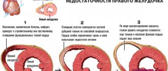
- Heart failure
. In severe cases of the disease, an increase in pulmonary pressure is observed. In this regard, there is an increase in the right ventricle and atrium. All parts of the heart gradually change. Therefore, there is a disruption in the blood supply to the heart. - Complications of the surgical plan
. If the cavity, which is located near a large bronchus, ruptures, then air can enter it. Pneumothorax forms. If the septum between the alveoli is damaged, bleeding will occur.
Is hospitalization necessary to treat emphysema?
Subject to the doctor's recommendations, optimal diet and medication regimen, outpatient treatment of the disease is possible.
Reasons for inpatient treatment:
- clarification of the diagnosis;
- intensification of symptoms, appearance of new signs (cyanosis of the skin and mucous membranes, weakness, shortness of breath without exertion, sputum production with blood);
- concurrent serious illnesses;
- the appearance of arrhythmia;
- ineffectiveness of outpatient treatment (deterioration of peak flow measurements).
Nutrition for emphysema (diet)
Diets No. 11 and No. 15 are aimed at strengthening the immune system, detoxifying the body and replenishing the patient’s energy reserves.
Principles of dietary nutrition:
- The calorie content of the daily diet is not lower than 3500 kcal. Diet: 4-6 times a day, little by little.
- Fat intake is at least 80-90 g. This can be vegetable oil, butter, and high-fat dairy products. The ratio of the proportion of animal fats to vegetable fats is 2:1.
- Proteins are consumed in quantities of up to 120 g per day. There should be at least half of animal products (eggs, meat of all types, sausages, sea and river fish, seafood, liver). Fried meat is excluded.
- The amount of carbohydrates in the diet is from 350 to 400 g. These are cereals, bread, jam, honey, pasta.
- Providing vitamins through the consumption of fresh fruits and vegetables, and the introduction of bran into food.
- Any drinks are allowed: juices, kumis, rosehip compote.
- Limiting salt to 6 g to prevent edema and cardiac complications.
The diet of patients with emphysema should not contain alcohol, cooking fats, or high-fat confectionery products.
ethnoscience
The patient can use folk remedies to expand the bronchial lumens, remove mucus, improve respiratory function and generally strengthen the body.
First of all, it is recommended to use decoctions and infusions of herbs. They are taken orally or inhaled.
Black radish with honey
In the treatment of pulmonary emphysema, you can use the following folk remedies:
- Infusion of wild rosemary. Add 1 teaspoon of dried and crushed herbal preparation to 500 ml of boiling water and leave for an hour. Drink 150 ml of warm tincture twice a day.
- Black radish juice. Fresh vegetables are washed and peeled. Grate it and squeeze out the juice. 50 ml of juice is mixed with 2 tablespoons of honey. Take 2 tablespoons of the drug twice a day. It is advisable to do this before eating.
- Infusion of horsetail and fennel. Pour boiling water into a half-liter jar with folk remedies taken in equal proportions (1 tablespoon each). The infusion is kept for an hour. Drink 100 ml three times a day.
- Milk with carrot juice. Add 1 tablespoon of carrot juice to a glass of warmed fat milk. The drink is consumed on an empty stomach for three weeks.
- Tea made from mint, sage and thyme. One and a half teaspoons of dried and crushed herbs mixed in equal proportions are poured into a thermos and poured with a glass of boiling water. Drink 70 ml after breakfast, lunch and dinner.
In the process of treating pulmonary emphysema with folk remedies, you can also use onion and garlic juice, propolis, aloe juice and Kalanchoe, etc.
It is important to treat non-traditional methods of combating pulmonary emphysema without fanaticism. It must be remembered that an unsuccessful attempt at self-medication can result in serious consequences, and sometimes even cost life.
Disease prognosis
Pulmonary emphysema is a complication of bronchopulmonary diseases. This means that the changes in the lung tissue that arose in this case are irreversible. All that remains is to slow down the progression of the disease and reduce signs of respiratory failure by improving bronchial patency.
Therefore, the prognosis for pulmonary emphysema depends on:
- Timeliness and adequacy of treatment of the underlying disease;
- An early and correct therapeutic approach to the treatment of emphysema;
- The patient’s compliance with all treatment and lifestyle recommendations;
- Duration of the disease.
In any case, it will not be possible to completely get rid of emphysema under any circumstances. But it is possible to influence the progression of the disease. If the underlying disease of the bronchopulmonary system, which caused emphysema, is characterized by a relatively stable course, then the prognosis for maintaining emphysema at its minimum level is quite favorable. If you follow all the recommendations of specialists, then the signs of respiratory failure will be insignificant and the person will be able to live in his usual rhythm.
The prognosis in the case of decompensated bronchial diseases with severe emphysema is in any case unfavorable. Such people are forced to take expensive medications for life that can only maintain basic vital breathing parameters. Significant improvements in quality of life occur extremely rarely. Life expectancy depends on the degree of compensation of the pathological process, age and regenerative resources of the body.
Fluimucil to thin sputum
Fluimucil is a mucolytic product. It thins mucus, reduces cough, and has an antioxidant effect on the human body. Fluimucil also has anti-inflammatory properties by suppressing the formation of oxygen-containing elements. Pulmonary emphysema, treatment with drugs and preparations that have a similar effect to Fluimucil:
| A drug | Active substance | Manufacturer | Price |
| Ambrobene | Ambroxol | Germany | 120 rubles |
| Lazolvan | Ambroxol | Germany, Poland | 150 rubles |
| Medox | Ambroxol | Czech | 120 rubles |
| Acetine | Acetylcysteine | India | 150 rubles |
| Acc | Acetylcysteine | Austria, Germany | 130 rubles |
Fluimucil is prohibited for use in cases of gastric ulcers, chronic gastritis, and anemia. It is also worth refraining from therapy with a mucolytic agent during pregnancy and lactation. As adverse reactions, patients may experience headaches, skin rashes, and a reflex urge to cough.