When doctors say “respiratory failure,” they mean that the respiratory system, which includes the oral and nasal cavities, larynx, trachea, bronchi and lungs, cannot supply the blood with the necessary amount of oxygen or is unable to remove excess carbon dioxide from the blood. This condition occurs when one or more of the mechanisms by which oxygen enters the blood or CO2 is removed from the blood is disrupted.
It's hard to breathe with Covid
With the development of pulmonary lesions against the background of COVID-19, signs of respiratory failure become one of the criteria for the severity of the patient’s condition and the reason for a CT scan or hospitalization. At home, you can find out about respiratory failure by:
- number of breaths per minute - more than 22 times per minute ,
- presence of pale or bluish skin ,
- according to the level of blood oxygen saturation (saturation) in the presence of a pulse oximeter - saturation less than 95% .
This is respiratory failure of the 1st degree (DN 1), it becomes a criterion for a condition of moderate severity and an absolute indication for a CT scan of the lungs.
- With such DN1, the patient may be hospitalized .
- But if there are no places in the hospital and the CT changes correspond to moderate severity, it can be treated at home .
Developing acute respiratory failure during coronavirus leads to oxygen starvation of organs and tissues. In addition, for a long time there are no other reasons for artificial ventilation of the lungs (loss of consciousness, weakness of the diaphragm, auxiliary respiratory muscles). Therefore, the first step in compensating for oxygen deficiency in most patients is oxygen therapy.
Attempts to carry out oxygen therapy at home for Covid using:
- oxygen cushion or
- even masks with a balloon are unacceptable frivolity.
The condition of a patient with covid pulmonary damage may suddenly deteriorate sharply due to concomitant microthrombosis, the addition of pulmonary embolism, disseminated intravascular coagulation syndrome and require urgent initiation of mechanical ventilation. Therefore, DN for pneumonia, which requires oxygen support, is carried out strictly in a hospital under the supervision of specialists, against the background of laboratory and hardware monitoring.
Diagnostics
You should start looking for the causes and treatment of shortness of breath immediately after you begin to notice such changes. The following methods are used to diagnose respiratory failure:
- Collection of anamnesis of the disease. The doctor carefully studies the patient’s medical history, his complaints, and learns his lifestyle.
- External examination of the patient. By studying the skin, chest muscles, and heart rhythm, the doctor can confirm or refute his guesses.
- Blood gas analysis is a reliable test. If there are deviations, a disease of the chest organs can be assumed.
The doctor makes the final diagnosis after a comprehensive diagnosis. It must be confirmed by laboratory tests.
What is respiratory failure
The human body is quite complex, and it has various ways in which it maintains those parameters necessary to ensure life (this includes the provision of oxygen). And if one path begins to “overlap,” the body immediately opens and expands a “bypass road” called “compensatory mechanisms.”
When they resolve the situation, chronic failure develops (in this case, chronic respiratory failure). When the disease develops so quickly that compensation does not even have time to form, the deficiency is called acute and poses an immediate threat to life.
Below we will look at how to understand, even before the doctor arrives, what is causing the respiratory failure and how to provide assistance. Because in many cases, saving a life is 80-90% dependent on the competent actions of the victim’s relatives.
Consequences
Complications concern the heart, central nervous system, lungs and bronchi themselves. Without treatment, a person is guaranteed to die. It is the matter of time.
In addition, the following problems are possible:
- Heart failure.
- Ischemic disease.
- Dementia.
- Decreased memory and concentration.
- General cognitive deficit.
- Pulmonary hypertension.
- Stroke.
- Heart attack.
- Left ventricular hypertrophy.
Pulmonary insufficiency is a severe and dangerous pathological process, which is accompanied by respiratory and cardiac disorders. The goal of treatment is to eliminate the primary disease and prevent complications.
Briefly about oxygen transport
In this section we will trace the path through which oxygen from the air enters the blood. The respiratory system is conventionally divided into 2 parts:
Dead space
This is the name of the large part of the respiratory system in which there is no contact of inhaled air with blood. It is conventionally divided into anatomically dead space, which includes:
- Nasal cavity. Serves for primary purification and warming of air. Even if it is completely blocked (due to edema or tumor), respiratory failure does not develop.
- Oral cavity and pharynx. The oral cavity is not intended for breathing, since the air is not warmed or purified here. But due to their direct communication in the form of the pharynx, it can also be used for breathing. At the border of the oral cavity and pharynx, as well as in the pharynx itself, there is a ring of lymphoid tissue (tonsils), which acts as a barrier to foreign substances entering with air and food. If inflammation develops in the tonsils or their surrounding tissue and they increase in volume. If the degree of their increase is so great, they block the path of air - respiratory failure develops.
- The larynx is the entrance to the trachea. Above it is a cartilaginous structure - the epiglottis, and in the larynx itself are the vocal cords. Inflammation, accompanied by swelling of the mucous membrane, as well as swelling of these structures block the path of air. Respiratory failure develops.
- The trachea is a cartilaginous tube between the larynx and bronchi. It rarely develops such significant edema that it blocks the path of inhaled air, but when a tumor develops here, chronic respiratory failure is formed.
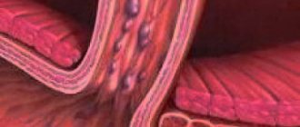
- The bronchial tree to the bronchioles are the tubes into which the trachea is divided. As they branch, they gradually become smaller and lose their cartilaginous base (bronchioles do not contain cartilage at all in the wall). The larger the bronchus where edema or swelling develops, the more air does not reach the lung, and the greater the severity of respiratory failure. There are processes, such as chronic bronchitis or bronchial asthma, that affect all bronchi at once, causing respiratory failure.
There is also functional dead space - areas of the respiratory system in which gas exchange also does not occur. This is anatomically dead space plus those areas of the lungs where air reaches, but in which there are no blood vessels. Normally there are few such areas.
Departments in which ventilation occurs
These are the final sections of the lungs - the alveoli. These are peculiar “bags” with a thin wall into which air enters. On the other side of the wall there is a blood vessel. Due to the difference in oxygen pressure, it penetrates through the wall of the alveoli and the wall of the vessel, entering directly into the blood. If the alveolar wall becomes edematous (pneumonia), it grows with connective tissue (pulmonary fibrosis) or the vessel wall undergoes pathological changes, and respiratory failure develops. The same process occurs when fluid appears between the alveolar wall and the vessel (interstitial pulmonary edema).
It turns out that the main gas exchange occurs at the level of the alveoli. Therefore, if, during respiratory failure, oxygen is introduced directly into the blood, bypassing all respiratory tracts, this will save a person’s life. The membrane oxygenation method allows this, but it requires special equipment, which is purchased by a small number of hospitals. It is used mainly for severe degrees of parenchymal respiratory failure (for example, pneumonia, pulmonary fibrosis, respiratory distress syndrome) - when the lung tissue does not perform its function.
Breath control
Although a person can speed up or slow down his own breathing with the power of thought, it is a self-regulating process. The center of breathing is located in the medulla oblongata, and if this accumulation of nerve cells does not give a command that an inhalation should occur (exhalation is considered a passive process that inevitably follows inhalation), no effort of will will help to do it.
The regulator of the respiratory center is not oxygen, but CO2. It is the increase in its concentration in the blood that activates more frequent breathing. What happens is this: the level of carbon dioxide increases in the blood and immediately increases in the cerebrospinal fluid. Cerebrospinal fluid bathes the entire brain - both the brain and the spinal cord. The signal that there is more CO2 is immediately picked up by special receptors in the medulla oblongata, and it gives the command to breathe more often. The command goes along the fibers that are part of the spinal cord and reaches its III-V segments. From there, the impulse is transmitted to the respiratory muscles: those located in the intercostal spaces (intercostal) and the diaphragm - the main respiratory muscle.
The diaphragm is a muscular plate stretched like a dome between the lower ribs and delimiting the chest cavity from the abdominal cavity. When it contracts, it moves the abdominal organs down and forward, as a result, negative pressure appears in the chest cavity, and it “pulls” the lungs with it, forcing them to straighten. The intercostal muscles help to further expand the chest: they pull the ribs down and forward, expanding the chest in the lateral and anteroposterior directions. But without the diaphragm, the efforts of the intercostal muscles alone will not result in normal oxygen saturation of the blood.
When the respiratory muscles contract, the size of the chest increases, and the resulting negative pressure pulls on the thin and elastic lungs, causing them to expand and fill with air. The lungs are “wrapped” in a thin “film” in 2 layers. This is the pleura. Normally, there should be nothing between its two layers - neither air nor liquid. When they get there, the lungs become compressed and can no longer expand normally. This is respiratory failure.
If air (pneumothorax) or liquid (hydrothorax) enters the pleural cavity in large quantities or continues, not only the lung is compressed: the overfilled “container” also puts pressure on the nearby heart and large vessels, preventing them from contracting normally. In this case, cardiovascular failure is added to respiratory failure.
Oxygen balance indicators
The oxygen content in the blood can be understood by:
- hemoglobin level: its norm is 120-140 g/l. It is estimated that each of its molecules binds 1.34 grams of oxygen. Determined by a general blood test;
- hemoglobin saturation with oxygen, that is, the ratio of the amount of oxygenated hemoglobin (oxyhemoglobin) to the total amount of these molecules. Normally, oxygen saturation is 95-100% and depends on the oxygen content in the inhaled gas mixture. So, if a person breathes 100% oxygen (this is only possible in hospitals or specialized ambulances and only when using special equipment), the saturation of hemoglobin with oxygen is higher. The oxygen content in atmospheric air is approximately 21%. If a person is in an enclosed space with low oxygen levels, the hemoglobin will be very poorly oxygenated. This indicator is determined by an analysis called “blood gases” and is taken from an artery and vein.
- the partial pressure of oxygen in arterial blood, that is, the individual pressure of this particular gas on the walls of the vessel. The higher the oxygen pressure, the better the blood is saturated with it. Normally, the partial pressure of oxygen in arterial blood is 80-100 mm Hg. If this indicator is reduced, a diagnosis of respiratory failure is made. The indicator is determined by analyzing blood gases.
To understand the processes occurring in the body, it is important for doctors to know not only how much oxygen is contained in arterial, that is, O2-saturated blood, but also:
- how it will be delivered to the tissues (this already depends on the cardiovascular system);
- how the tissues will use it (calculated from the oxygen content in the venous blood and data on the work of the heart).
Based on the deviation from the norm of the last two indicators, compensation for respiratory failure is judged (how quickly the heart will pump blood and tissues will “consume” oxygen from the blood more efficiently). It also happens that a person develops symptoms the same as with respiratory failure, but no pathology of the respiratory tract is detected. Then determining the delivery of oxygen to the tissues and its absorption by them is important for making a diagnosis.
Complications
Here are the complications that respiratory failure can lead to:
- From the side of the heart and blood vessels. Respiratory failure increases the load on the cardiovascular system, causing specific diseases such as ischemia, hypotension, heart attack, etc.
- The digestive organs (intestines, stomach) are also at risk due to respiratory failure. This disease can cause stomach ulcers, intestinal bleeding and irregular bowel movements.
- DN most clearly affects the brain and other organs of the central nervous system. The patient becomes irritable, lethargic, and unable to concentrate.
- Quite often, respiratory failure leads to inflammatory processes in the lungs (pneumonia, bronchitis).
As you can see, there are many causes of shortness of breath. Treatment should begin as soon as you notice warning signs in your body. Perhaps the hot weather or your fatigue are to blame. But respiratory failure syndrome is a serious disease that, if not treated sufficiently, can cause death in the patient. Therefore, it is important to diagnose acute and chronic DN in time and begin treatment without delay.
Causes of respiratory failure - acute and chronic
There is a classification that divides respiratory failure into 2 types:
- Ventilation. It occurs due to a huge number of reasons not related to damage to lung tissue.
- Pulmonary. It is associated with damage to the lungs in the normal state of the ventilation sections (functional dead space).
There is a second division of respiratory failure into:
- hypoxemic, which occurs when there is insufficient partial pressure of oxygen in the blood;
- hypercapnic, when there is a high pressure of carbon dioxide in the blood, that is, it is clear that CO2 is not removed sufficiently.
The first classification is used for treatment at the initial stage of providing specialized medical care. The second is to correct the state of blood gases a little later, after the diagnosis has been clarified and emergency measures have been taken in relation to the patient.
Let's consider what the main reasons can cause ventilation and pulmonary forms of respiratory failure.
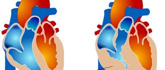
Mechanism of development of SLI
SLN often develops as a complication after diseases that lead to pulmonary hypertension and right-sided heart failure. The first version of the development of SLN begins with a pathology of the lungs, leading to an increase in the stiffness and density of the lung tissue, which is why the right ventricle requires more effort for systole. Subsequently, its wall hypertrophies, and the cavity expands, forming the so-called “pulmonary heart” (CP). Another variant of pathogenesis is associated with the progression of left ventricular heart failure to the last stage, as a result of which blood stagnates in the large and then small circle, increasing the pressure in the pulmonary vessels.
Causes of ventilation failure
This condition can be either hypoxemic or hypercapnic. It occurs due to a large number of reasons.
Violation of cerebral regulation of breathing. This may happen due to:
- insufficient blood supply to the respiratory center. This is a form of acute respiratory failure. It develops either by a sharp decrease in blood pressure (with blood loss, any type of shock), as well as with displacement of brain structures in the cranium (with a brain tumor, trauma or inflammation);
- lesions of the central nervous system without displacement of the brain into the natural openings of the skull. In this case, acute respiratory failure develops with meningitis, meningoencephalitis, strokes, and chronic - with brain tumors;
- traumatic brain injury. When it causes swelling of the brain with disruption of the respiratory center, this is acute respiratory failure (ARF). If 2-3 months have already passed after the injury, and disturbances in adequate oxygen metabolism occur, this is chronic respiratory failure (CRF);
- overdose of drugs that suppress the respiratory center: opiates, sleeping pills and sedatives. This is how ODN develops;
- primary insufficient air entry into the alveoli. This is a hypercapnic form of chronic renal failure, caused, for example, by extreme obesity (Pickwick syndrome): when a person is unable to breathe frequently and deeply.
Impaired conduction of impulses to the respiratory muscles due to:
a) spinal cord injuries. When it is injured or inflamed, ARF develops;
b) disorders of the spinal cord or nerve roots along which impulses travel to the respiratory muscles. Thus, with polyradiculoneuritis (damage to several spinal nerve roots), the development of inflammation of the spinal cord, inflammation of the nerves that go to the respiratory muscles, ARF develops. If a tumor grows slowly in the spinal cord - CDN.
Neuromuscular disorders due to:
- erroneous administration of drugs that relax all muscles, including respiratory ones (these drugs are called muscle relaxants and are used during anesthesia, after which the person is transferred to mechanical breathing). This is acute respiratory failure;
- poisoning with organophosphorus compounds (for example, Dichlorvos). This is also acute respiratory failure;
- myasthenia gravis – rapid fatigue of the striated muscles, which also includes the respiratory muscles. Myasthenia gravis causes ARF;
- myopathies are non-inflammatory diseases of muscles, including respiratory muscles, when their strength and motor activity decrease. This can develop both acute and chronic forms of respiratory failure;
- ruptures or excessive relaxation of the diaphragm. Calls ODN;
- botulism, when botulinum toxin entering with food is absorbed into the blood, and then into the nervous system, where it blocks the impulse from the nerves to the muscles. Botulinum toxin acts on all nerve endings, but causes ADN in severe cases associated with its entry into the body in large quantities. Sometimes ARF can develop when a person with botulism seeks medical help late;
- tetanus, when tetanus toxin entering (usually through a wound) causes paralysis of striated muscles, including respiratory muscles. Calls ODN.
Disturbances of the normal anatomy of the chest wall:
- with open pneumothorax, when air enters the pleural cavity through a wound in the chest wall, causing ARF;
- with floating rib fractures, when a rib fragment is formed that is not connected to the spine, and it usually moves freely in the direction opposite to the movement of the chest. If one rib is damaged, CDN occurs; if several ribs are damaged at once, ARF occurs;
- kyphosis (bending backward, towards the back) of the spine in its thoracic region, which limits the movement of the chest when inhaling and causes CDN;
- pleurisy - accumulation of inflammatory fluid and/or pus between the layers of the pleura, which, like pneumothorax, limits the expansion of the lungs. Acute pleurisy causes ARF;
- Chest deformities - congenital, resulting from rickets, trauma or surgery. This restricts the movement of the lungs, causing CDN.
Airway obstruction at the level of anatomically dead space (obstructive respiratory failure)
It occurs due to:
- laryngospasm - muscle contraction at the level of the larynx, which occurs in response to a lack of calcium in the body of a child under 3 years old, with the development of pneumonia, diseases of the larynx, trachea, pharynx, pleura, inhalation of toxic gases, fear. This causes ARF;
- entry of a foreign body into the trachea or bronchi. If blockage of the trachea or large bronchi causes acute respiratory failure, when assistance must be provided in a few minutes, then blockage of smaller bronchi may not be so acute;
- laryngostenosis - narrowing of the lumen of the larynx. It can develop against the background of infectious diseases or due to the entry of a foreign body into the larynx, in response to which a spasm of its muscles occurs, which does not allow the foreign object to pass further (ODN). Laryngostenosis can also cause CDN when it occurs as a result of tumors of the larynx or compression from the outside by an enlarged thyroid gland or tumors of the soft tissues of the neck;
- narrowing of the lumen of the bronchi during bronchial asthma, bronchitis, when swelling of the bronchial mucosa occurs. In acute bronchitis and exacerbation of bronchial asthma, in some cases ARF develops, while chronic bronchitis and the interictal period in bronchial asthma is the cause of CRF;
- narrowing of the lumen of the bronchi due to the accumulation of a large amount of mucus in them (for example, with cystic fibrosis). Provoke CDN;
- narrowing of the lumen of the bronchi as a result of bronchospasm, caused by allergens and infectious agents. This disease is accompanied by ARF;
- bronchiectasis, when, as a result of chronic inflammation or congenital characteristics of the bronchi, they expand and pus accumulates in them. Causes the formation of CDN.
Disorders of regulation of the respiratory center
Disturbances in the activity of the respiratory center can occur due to poisoning with narcotic analgesics, sleeping pills, and anesthetic agents. Skull injuries, cerebral hemorrhages, strokes, inflammatory processes of the brain and its membranes, comatose states of various etiologies can also damage the cells of the respiratory center or disrupt their function due to edematous processes in the brain. In these conditions, the respiratory center ceases to adequately respond to acidification of the blood and cerebrospinal fluid, an increase in carbon dioxide and a decrease in oxygen content in the arterial blood.
In such patients, external respiration is sharply weakened, it becomes superficial, sometimes pathological (Cheyne-Stokes, Biota); in severe cases, breathing stops. The concentration of carbon dioxide in their blood increases and the oxygen content decreases. Hypoxia and hypercapnia damage cells of the central nervous system, myocardium and other organs and systems, directly leading to cardiac arrest.
Toxic or hypoxic damage to the brain, in turn, causes (or deepens) coma and predetermines obstructive breathing disorders. Muscle tone decreases, and in patients the root of the tongue sinks, blocking the airways at the level of the throat. Without urgent measures to restore their patency, patients may die.
Often in comatose patients there is an accumulation of saliva in the mouth and throat, sputum in the tracheobronchial tree, which the conscious person is not able to cough up and swallow or spit out. The sputum becomes infected, and such patients soon develop inflammatory processes, purulent tracheoesophageal bronchitis, pneumonia, which is one of the most common causes of death in patients who were in a coma for a long time.
Respiratory disorders in fainting patients can be caused by passive leakage (regurgitation) of contents from the stomach into the oral cavity and its further flow (aspiration) into the trachea and bronchi. According to A.P. Zilber (1989), aspiration of 10-15 ml of acidic gastric contents is fatal in most cases.
Preventive and therapeutic measures for this pathology are classified as emergency. The nurse must be able to provide first aid even before the arrival of a specialized medical team.
Urgent measures:
- assess the level of depression of the central nervous system - the depth of coma;
- to prevent aspiration, the patient must be turned on his side, slightly lowering the upper half of the body (in case of probable regurgitation, gastric contents flow out through the mouth and do not flow into the trachea);
- open the patient’s mouth, use a gauze swab on the clamps (or a finger wrapped in a handkerchief) to clear the contents from the mouth and throat;
- straighten the patient's neck, placing a cushion under the shoulders so that the head leans back. In most cases, the root of the tongue moves forward, freeing the throat; the airways become open;
- remove the lower jaw. To do this, being behind the victim’s head, you should place the thumbs of both hands on his chin, with the others covering the lower jaw so that the little fingers are under its joints. Carefully move the lower jaw forward and upward so that its teeth are fixed with the teeth of the upper jaw (malocclusion position). Further support of the lower jaw is provided without much effort, even with one finger;
- if there is an air duct, insert it into the oral cavity. To do this, its distal part, curved upward, is inserted between the victim’s teeth, up to the palate. Then turn 180 ° and push forward, making sure that the back of the tongue is located above the concave part of the air duct;
- If the victim is unable to breathe on his own, he must undergo artificial ventilation using the mouth-to-mouth method or using a hand-held portable device.
As part of a specialized emergency care team, a nurse conducts an in-depth study of the patient’s condition (measures pulse rate, respiration rate, blood pressure, body temperature, establishes cardiac monitoring, records an electrocardiogram, collects biological fluids for laboratory tests), and assists the doctor in conducting intensive care .
Urgent actions of a specialized team:
• re-assess airway patency using direct laryngoscopy; • clear the upper respiratory tract of existing fluids (sputum, blood, gastric contents) using an electric vacuum suction;
• in case of ineffective breathing, perform artificial ventilation of the lungs using a mask with an Ambu bag or a stationary ventilator;
• establish the patient’s oxygenation system. To do this, connect an oxygen supply of 8-10 liters per minute to the ventilator;
• set up a system for infusion therapy;
• catheterize a central or peripheral vein;
• if the patient's own breathing is further ineffective, inject atropine sulfate (0.1% - 0.5 ml) intravenously and, under the control of direct laryngoscopy, perform tracheal intubation, transferring the patient to controlled breathing;
• insert a tube into the stomach and empty it of contents;
• create an individual observation card for the patient, where you note data on his condition and completed appointments;
• conduct a comprehensive instrumental and laboratory examination of the patient;
• carry out etiopathogenetic and symptomatic therapy.
Causes of pulmonary type DN
These types of respiratory failure are not caused by the accumulation of carbon dioxide in the blood, but by an insufficient supply of oxygen. Provoke the development of ARF. The main pulmonary causes are:
- Pneumonia, when inflammatory fluid accumulates in the alveoli of a separate area (areas), and individual walls of the alveoli swell, as a result of which oxygen cannot enter the bloodstream. Causes acute respiratory failure.
- Respiratory distress syndrome - damage to the lungs as a result of injury, pneumonia, inhalation (aspiration) of liquids, ingress of fatty tissue into the vessels of the lungs, inhalation of radioactive gases and aerosols. As a result, inflammatory fluid leaks into the lungs; after a while, some of the inflammatory changes are stopped, but in some parts of the lung tissue is replaced by connective tissue.
- Pulmonary fibrosis is the replacement of normal lung tissue with connective tissue. The more altered sections there are, the more severe the condition will be.
- Pulmonary edema is the leakage of fluid into the alveoli (alveolar edema) or into the lung tissue between the vessels and alveoli (interstitial edema), resulting in significantly reduced oxygen delivery to the blood.
- Lung injury. In this case, the penetration of oxygen from the alveoli into the blood becomes impossible in some area due to damage to the blood vessels and saturation of the lung tissue with blood.
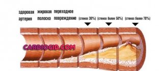
- Embolism of the branches of the pulmonary artery, that is, blockage of the branches of the artery that carries blood from the heart to the lungs, fat, air, blood clots, tumor cells, foreign bodies. As a result, larger or smaller areas of the lungs cease to receive blood supply, and accordingly, much less oxygen enters the blood.
- Atelectasis, that is, collapse of parts of the lungs and their exclusion from gas exchange. The reasons may be compression of the lung by fluid in the pleura, blockage of the bronchus, violation of the technique of artificial ventilation of the lungs, when some part of it is not ventilated.
Prevention and recommendations
Pulmonary insufficiency is a condition that has several causes (chronic obstructive pulmonary disease (COPD), pneumonia, acute pulmonary edema, asthma, obesity, kyphoscoliosis, overdose of drugs or drugs that depress the respiratory centers, tetanus, severe hypothyroidism).
Therefore, preventing these diseases or conditions is an effective means of reducing the likelihood of developing pulmonary failure.
Especially important:
- no smoking;
- adopt a healthy lifestyle and diet;
- perform regular physical activity;
- get a tetanus vaccination;
- don't use drugs.
Signs of ARF
Symptoms of acute respiratory failure are:
- increased breathing. In adults - more than 18 per minute, in children - above the age norm;
- inclusion of auxiliary respiratory muscles in the act of breathing. The retraction of the intercostal spaces, the area above the collarbone, becomes noticeable, the wings of the nose swell;
- increased heart rate above 90 beats per minute, due to intoxication, arrhythmia may begin;
- feeling of lack of air;
- you can notice asymmetrical movements of the chest;
- change in skin color: the skin becomes pale, and the lips and nasolabial triangle become bluish, the fingers acquire the same color;
- with severe respiratory failure, loss of consciousness is observed; before this, inappropriate behavior and delirium may be observed;
- feeling of panic, fear of death.
The severity of respiratory failure is determined by indicators such as respiratory rate, level of consciousness, and the level of partial pressure of O2 and CO2 in the arterial blood. To determine the partial pressures of gases, it is necessary to perform an analysis of gases from arterial blood, which requires time and appropriate equipment. Therefore, for faster diagnosis, the “saturation” indicator is used, which is determined using a pulse oximeter device. The sensor of this device is a clothespin, inside of which there is an infrared emitter. The sensor is placed on a person’s finger and in a matter of seconds allows one to judge the degree of oxygen saturation of capillary blood.
There are 4 degrees
- Breathing speeds up to 25 per minute, and the heart rate reaches 100-110 beats per minute. The person is conscious and adequate, feels short of breath, and may experience slight blueness of the lips. Oxygen saturation 90-92%, CO2 partial pressure 50-60 mm Hg. when breathing normal air.
- Respiration rate is 30-35 per minute, pulse is 120-140 per minute, blood pressure rises. The skin is bluish and covered with cold, sticky sweat. The person is restless or inhibited, and may be euphoric. Saturation is reduced to 90-85%, CO2 partial pressure is 60-80 mm Hg.
- Breathing is shallow, 35-40 per minute, pulse - 140-180 per minute, blood pressure is reduced. The skin is sallow, the lips are bluish. The person is inadequate, inhibited. Saturation is reduced to 80-75%, CO2 partial pressure is 80-100 mm Hg.
- Here a hypoxic coma develops, that is, a person is unconscious and cannot be awakened. Pulse - 140-180 per minute, breathing rate depends on brain damage: it can be more than 40 per minute or less than 10 per minute. Saturation is reduced to 75% and below, and the partial pressure of CO2 is more than 100 mm Hg.
Depending on the severity, doctors will provide assistance to the person. If in the first degree, while the examination and determination of the causes of ARF are ongoing, the person is allowed to breathe humidified oxygen using a face mask (the content of the supplied oxygen will be no more than 40%, while the air contains 20.8%). At stages 2-4, the patient is put under anesthesia in order to transfer him to mechanical breathing using an artificial ventilation device.
In addition to the symptoms of respiratory failure itself, a person develops signs that tell the doctor the reason for the development of this serious condition:
- if the symptoms of ARF develop after a traumatic brain injury, there is probably either a brain contusion or the formation of a hematoma in it;
- if before the development of ARF a person had a cold, and then complained of a headache and fever for some time, after which a disturbance of consciousness developed and shortness of breath appeared, it is probably meningitis or meningoencephalitis;
- when a person suffers from hypertension or he became very nervous, after which he suddenly lost consciousness and began to “breathe incorrectly” against this background, he probably suffered a hemorrhagic stroke;
- Poisoning with drugs that depress the respiratory center is indicated by scattered drugs, syringes, and inappropriate behavior some time before the illness. Examination of the pupils in this situation is not informative, since hypoxia/hypercapnia also changes the pupillary diameter;
- if signs of ARF appeared after eating canned food, dried or dried river fish, brawn, sausage, and the person initially complained of blurred vision, fog before the eyes or double vision, it may be botulism. If a person has not eaten canned food or fish, and he has the same symptoms, this indicates a stroke or a tumor in the brain stem;
- when a person suffered from a cold or diarrhea with an increase in temperature, and his legs gradually began to go numb, and then his arms and stomach, while his movement was impaired, this may be Guillain-Barre syndrome;
- if a person suddenly feels a sharp pain in the chest, or has a chest injury, after which it suddenly becomes worse to breathe, these are symptoms of pneumothorax;
- if the symptoms of ARF develop against the background of a cold with fever and cough, it is probably acute pneumonia, although it may also be acute bronchitis.
Symptoms
With pulmonary failure, the following symptoms appear:
- shortness of breath of varying degrees of intensity;
- in the morning, a sick person may experience headaches;
- insomnia;
- heart rate increases;
- nausea and vomiting;
- the skin acquires a bluish tint;
- auxiliary muscle structures are involved in the respiratory act;
- memory impairment;
- decrease in blood pressure;
- the frequency and depth of breathing changes;
- disturbance of consciousness.
What to do if you have ARF
First aid for respiratory failure should be provided after calling an ambulance. There is no talk of waiting for a local therapist in case of ARF.
The algorithm of actions is as follows:
- Call an ambulance.
- You can seat the person near the table so that he can put his hands on the table and raise his shoulders higher - closer to his chin. This will allow the auxiliary respiratory muscles to have more range of motion.
- Try to calm the victim.
- Free him from his outerwear, unfasten all the buttons and the belt of his trousers so that nothing interferes with his breathing.
- Provide fresh air flow from windows and vents.
- Constantly reassure the patient and be with him.
- If a person has asthma, help him take 1-2 breaths from his inhaler.
- If such symptoms occur after eating fish or canned food, give him “Activated carbon” or other sorbents.
If ARF develops as a result of a foreign body entering the throat, an urgent need to perform the Heimlich maneuver: stand behind the victim, clasp him with both hands. Clench one hand into a fist and place the palm of your other hand under it. Now, with a pushing movement directed upward, bending our elbows, we press on the abdominal area “under the stomach” until the victim’s airways are completely cleared.
If the development of ARF was preceded by a cold or a barking cough, it is recommended to inhale with a 0.05% naphthysine solution through a nebulizer before the ambulance arrives: 3-4 drops per 5 ml of saline solution.
When signs of ARF are present in a person who has been in a car accident, then it is possible to remove him from the car or shift him only after fixing his cervical spine with a Shants-type collar.
Treatment of ARF
It is carried out by the ambulance resuscitation team and continues in the hospital. The first action is to provide oxygen support (through a mask or with transfer to artificial ventilation). Further, it depends on the cause of ARF:
- for bronchial asthma and chronic bronchitis - this is the intravenous administration of Eufillin, inhalation administration of Berodual or Salbutamol;
- for pneumonia - administration of antibiotics;
- for pneumothorax - surgical treatment in the thoracic surgery department;
- for botulism and tetanus - administration of specific serums (antibotulinum or antitetanus);
- for Guillain-Barré syndrome - administration of intravenous immunoglobins;
- for stroke - treatment in a neurological department;
- with an intracerebral hematoma, surgical removal is possible;
- for myasthenia gravis – the prescription of specific drugs: “Proserina”, “Kalimina”;
- in case of opiate overdose - administration of antidotes;
- for pleurisy - treatment with antibiotics and rinsing the pleural cavity with antiseptics;
- for pulmonary edema - lowering blood pressure, administering antifoam drugs.
In severe pulmonary fibrosis and bilateral pneumonia, a person can only be saved with the help of extracorporeal membrane oxygenation.
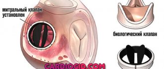
Treatment and observation of a patient with SLN
Cardiopulmonary failure is a chronic disease that requires lifelong treatment. Under the influence of correctly prescribed medications, symptoms decrease, which significantly improves the quality of life. It is important to treat not only SLN, but also its cause. For most patients, treatment consists of dietary adjustments, medications, and surgical interventions.
The main medications used to treat SLN:
- ACE inhibitors - dilate blood vessels, lower blood pressure, improve blood flow and reduce stress on the heart
- Angiotensin receptor blockers - the principle of action is similar to previous drugs. Prescribed for intolerance to ACE inhibitors
- Beta blockers – slow down heart rate
- Thrombolytic drugs - for conservative treatment of pulmonary embolism
- Glucocorticoids and cytostatic drugs - for the treatment of connective tissue pathology
- Anti-tuberculosis drugs - for appropriate pathology
- Mucolytic drugs - optimize the viscosity of sputum and promote its discharge
- Diuretics – remove fluid that forms swelling, thereby lowering blood pressure and improving breathing
- Aldosterone antagonists – act like diuretics
- Digoxin – strengthens heart contractions, reducing their frequency
- Nitroglycerin – improves blood flow in the myocardium
- Statins – used to treat atherosclerosis
- Anticoagulants - used to reduce blood clotting properties
The drugs are selected individually and require several visits to the doctor to calibrate (titrate) the effective dosage. It is very important to use them regularly and not stop taking them on your own.
Chronic respiratory failure
Chronic respiratory failure develops as a result of chronic pathologies of the respiratory tract (chronic bronchitis, bronchial asthma, tumors of the larynx, trachea or bronchi), deformation of the chest wall, myasthenia gravis. It manifests itself with symptoms such as:
- blue-violet tint of the skin on the face and fingers, increasing during physical activity;
- more frequent breathing (over 20 per minute);
- fluttering of the wings of the nose;
- fast fatiguability;
- change in the shape of fingers and nails. Fingers become like drumsticks, and nails become like watch glasses;
- frequent headaches;
- change in the shape of the chest (in some cases it becomes barrel-shaped).
In these cases, only treatment prescribed by a doctor after a thorough examination can help.
Author:
Krivega Maria Salavatovna resuscitator
conclusions
Not all diseases that lead to the development of cardiopulmonary failure are reversible, but the right treatment can reduce symptoms to a minimum. Lifestyle changes such as regular physical activity, healthy eating, reducing dietary salt, reducing stress and excess weight improve prognosis and quality of life.
The only way to prevent the development of SLN is to control the underlying disease and follow the instructions of the attending physician.