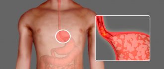
The esophagus is covered with a fairly dense network of vessels. There is a complex blood flow system here. Therefore, a person often develops esophageal varicose veins (EVV).
Varicose veins of the esophagus provoke disturbances in blood flow in the vessels. The disease appears with age and affects adults, most often older people over 50 years of age.
Men get sick 2 times more often than women. There is a hereditary form of varicose veins, which is much less common in medical practice. Children who are born with this condition also suffer from other diseases.
VRVP develops as a result of liver disease, most often cirrhosis. Therefore, patients experience changes in the color of the skin and whites of the eyes, they become yellowish. A person loses his appetite, his body weight drops sharply, his stomach hurts and he often feels sick.
This disease is accompanied by normal or minor internal bleeding.
About 30% of all bleeding from the upper alimentary tract occurs as a result of varicose veins.
Dilatation of the veins of the esophagus. ICD I85.0 (I85.9)
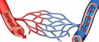
The walls of the esophageal veins are thin and easily stretchable, so an increase in pressure in them quickly leads to dilatation, deformation and bag-like bulging of part of the vessel. This is also facilitated by the fact that the veins of the esophagus are surrounded by loose, pliable connective tissue.
The anatomy of the venous system of the esophagus is quite complex. Blood from this organ flows to the three most important veins of the body. Impairment of blood flow in any of them can cause the appearance of phlebectasis in the esophagus. Most often, phlebectasis occurs in the lower parts of the tubular organ, as a result of disturbances in the v. system. portae (portal vein).
Various liver diseases (hepatitis, thrombosis, sclerosis, cirrhosis) lead to blood flow slowing down and pressure in the vein increasing (portal hypertension). The outflow of venous blood from internal organs, including the esophagus, slows down sharply, resulting in stagnation in the venous system.
The venous vessels of the esophagus, not adapted to such a volume of blood and pressure, stretch, their walls become thinner, the lumen of the veins becomes uneven, the veins lose their elasticity and bulge in a bag-like manner. Varicose nodes appear. The mucous membrane of the esophagus above the affected vessels becomes thinner and inflamed. All this creates conditions for damage to the integrity of the venous wall and the occurrence of bleeding - the most dangerous complication of varicose veins, threatening the life of the patient.
The danger of bleeding, ways to stop it
Acute (suddenly occurring) blood loss is dangerous due to the development of hemorrhagic shock. This is a condition that is characterized by a significant release of blood from the vascular bed. According to statistics, four out of five people who suffered venous bleeding of the esophagus died.
The release of blood occurs due to a tear in the mucous membrane lining the surface of the esophagus. Acute blood loss can also be caused by the following factors:
- eating hot or too dry food;
- a sharp increase in pressure;
- consumption of alcoholic beverages;
- intense physical activity;
- obsessive cough;
- entry of foreign bodies into the esophagus;
- exacerbation of reflux disease.
In addition, the most common cause of acute blood loss from the veins of the esophagus is attacks of uncontrollable vomiting in people suffering from chronic alcoholism.
At home, it is not possible to save a patient who has bleeding from the veins of the esophagus. Therefore, in such cases, it is necessary to immediately call an ambulance, having first placed the patient in a horizontal position on his side. The victim must be hospitalized in a ward of a specialized intensive care unit or resuscitation unit. Balloon dilatation is most often used to stop bleeding. The mechanism of this procedure is to install a special device into the lumen of the esophagus, followed by inflating the built-in ball, which compresses the damaged vessels.
After the patient’s critical condition has weakened, drugs that increase coagulation are introduced into his body. If the hemorrhage was significant, a decision is made to transfuse blood and its components. The patient must be constantly under the supervision of specialists with regular measurement of blood pressure and pulse.
VRVP classification
All phlebectasias of the esophagus are divided into congenital and acquired.
- Congenital phlebectasis of the esophagus is a rare pathology, accompanied by many other severe abnormalities. The disease is associated with genetic predisposition and disorders that occur during pregnancy at the time of organ formation.
- Acquired are the result of impaired blood flow in large veins and, as a consequence, phlebectasis in the veins of the esophagus. Esophageal varices are common in liver cirrhosis.
Depending on the size of varicose veins (varicose nodes) and the severity of the disease, four degrees of varicose veins are distinguished.
| Degree of disease development | Condition of veins and location of phlebectasias | X-ray characteristics | Condition of the esophagus | Clinical manifestations of pathology |
| 1st degree | Single dilated veins | Not defined | Satisfactory | Doesn't appear |
| 2nd degree | The veins of the esophagus dilate, become tortuous, and moderately enlarged varicose nodes appear. | An unclear venous contour is revealed. You can often see areas of stretched or tortuous veins. | The esophageal mucosa is not changed, its integrity is not compromised. | The clinic is not specific. Some patients experience discomfort when swallowing food. |
| 3rd degree | The lumen of the vessel narrows, the veins take on a serpentine shape, and the first angioectasia (dilation) appears. | In the middle of the esophagus and above, bulging vessels are detected. Distinct protrusion of venous nodes into the esophageal cavity. | The mucous membrane is thinned, erosions and foci of inflammation are detected on it. The mucous membrane is easily injured. | Belching and pain in the epigastrium and retrosternal space appear. Signs of micro- and sometimes macro-hemorrhage appear. |
| 4th degree | Veins are twisted, collected in nodes with numerous angioectasia | The esophagus is narrowed. Polypoid and cluster-shaped phlebectasias are identified. | The esophageal mucosa is thinned, with many erosions and inflammatory foci. | The patient has a clinical picture of severe esophagitis. A characteristic salty taste of blood appears in the mouth. The development of massive bleeding is very likely. |
External factors
In addition to pathological causes, there are a number of extraneous causes that can affect the appearance and development of the disease, these include:
In addition to pathological causes, there are a number of extraneous causes
- alcohol abuse;
- medications that negatively affect the condition of the liver;
- some diseases transmitted by inheritance;
- viral diseases affecting the liver (hepatitis).
Important! Pregnant women should pay increased attention to their health, since any viral infections suffered during this period can be transmitted to the fetus, increasing its risk of developing esophageal varices.
Causes of VRVP
The cause of VRVP is congestion in the esophageal veins and increased pressure in them. The main factor for this is disruption of blood flow and increased pressure in one of the three main veins.
Pathologies most often occur in the v. system. portae. This is due to the fact that almost any disturbance of blood flow in the system of this vein leads to portal hypertension. Moreover, an obstacle to blood flow can occur in any area v. portae:
- the lower ones are thrombosis of the splenic vein, congenital narrowing of the portal vein;
- hepatic – liver cirrhosis, active hepatitis, hepatocellular carcinoma, schistosomiasis;
- upper – constrictive pericarditis, right ventricular failure.
With portal hypertension at the initial stages of development of varicose veins, phlebectasias are localized in the distal part of the organ, and only as the disease progresses, the veins of the stomach and the veins of the middle part of the esophagus are involved in the process.
VRVP can form when compressed by the superior vena cava (hypertrophied organ, enlarged lymph nodes, tumor). In this case, varicose veins of the esophagus will develop in the upper part of the organ.
In rare cases, VRVP occurs as a result of disturbances in the entire circulatory system (severe degrees of heart failure). In this case, blood circulation in all organs is disrupted. As a result of a violation of the outflow of blood from the esophagus, deformation of the veins is observed along the entire length of the organ, although the size of the nodes will be significantly smaller than with portal hypertension.
Therapy with folk remedies
In combination with traditional therapy, traditional therapy methods can be used. In this case, consult your doctor before using them. In the treatment of folk methods, it is recommended to use medicinal compositions from rose hips and red rowan.
For the composition you need to take 1 tbsp. l. rowan berries and 1 tbsp. l. rose hips, add 500 mg of boiling water and simmer over low heat for 5 minutes. Next, the drink is filtered and cooled.
Take the composition ½ cup 4 times a day.
Dilated veins of the esophagus: symptoms
For a long time, the pathology does not manifest itself in any way. The first symptom of varicose veins is often bleeding. Sometimes hemorrhagic manifestations are preceded by unpleasant sensations in the retrosternal region (behind the sternum). More often, symptoms of esophagitis appear within a few days (sour belching, heartburn, difficulty swallowing solid food).
More often, hemorrhage is immediately preceded by overeating and sudden physical exertion. Although sometimes bleeding occurs during sleep.
The severity of hemorrhagic syndrome can be very different, from minor bleeding to massive blood loss that threatens the patient’s life. But even minor, constant bleeding can lead to serious consequences. Patients are constantly worried about:
- weakness;
- nausea;
- vomiting streaked with blood;
- melena.
The result of this is exhaustion, sudden weight loss, and iron deficiency anemia.
With massive blood loss, when blood flows out in a stream from damaged vessels, the condition immediately becomes critical, with rapidly increasing symptoms:
- suddenly the patient feels severe weakness, accompanied by a faint or faint state;
- severe sweating;
- nausea followed by vomiting large amounts of blood with clots;
- Blood pressure drops sharply, tachycardia appears, often accompanied by various kinds of arrhythmias, and the patient loses consciousness.
Complications and prognosis
The most severe and dangerous complication that pathology of the esophageal veins can lead to is bleeding. It leads to rapid and massive blood loss, hemorrhagic shock and, ultimately, death. In this case, even the presence of nearby medical workers and special equipment cannot always save the patient’s life.
According to statistical data, in patients who have suffered hemorrhage due to varicose veins, in 75% of cases a relapse occurs within a short period of time (1-2 years).
About 50% of patients die from blood loss. Moreover. the prognosis for life is extremely unfavorable: even if a person manages to control the veins of the esophageal gastrointestinal tract, complications from the liver may occur.
Diagnostics
Diagnostic measures are aimed at identifying the cause that caused varicose veins, as well as determining the stage of varicose veins, which determines the treatment strategy for the patient.
Mandatory diagnostic measures.
Laboratory research:
- study of peripheral blood (hemoglobin, red blood cells, hematocrit);
- study of venous blood (blood sugar, bilirubin and its fractions, AST and ALT, amylase, creatinine, total protein);
- coagulogram.
- Ultrasound examination of the liver
- X-ray examination with contrast of the esophagus (allows you to determine the nature of the dilation of veins and varicose nodes).
- fibroesophagoscopy (the main diagnostic method that allows you to determine how much the veins are changed, the presence of damage and hemorrhages, the condition of the venous nodes).
Methods for diagnosing the disease
If you suspect this pathology, you should contact a specialized specialist - a gastroenterologist. The doctor, after a face-to-face consultation and history taking, prescribes a number of diagnostic measures. A laboratory blood test is carried out, and, as a rule, a rapid decrease in hemoglobin can be detected, and acute anemia is observed.
In parallel, an ultrasound examination of the abdominal organs and an MRI of the liver are prescribed (for maximum information with a contrast agent). An X-ray examination of the esophagus with a contrast agent is also carried out, which makes it possible to identify a pathologically altered vein, wall deformation, and the condition of the organ mucosa.
The most informative method for examining the esophagus for varicose veins is esophagogastroduodenoscopy (gastroscopy).
This is an endoscopic examination of the digestive tract, which allows you to examine the esophagus, stomach and duodenum.
The camera equipped with the endoscope projects an image of the mucous membrane onto the screen, which allows the specialist to carefully examine the condition of the esophagus. Diagnostic imaging can be further improved by the use of fluoroendoscopic techniques.
In addition, endoscopy of the esophagus, if necessary, allows the collection of a biological sample of any suspicious lesion. Such biomaterial can be sent for further histological examination.
Also, the technical capabilities of esophagogastroduodenoscopy make it possible to remove polyps and eliminate sources of acute bleeding (peptic ulcer, esophageal varices, etc.).
It is necessary to pay attention to the preparatory stage before conducting the examination. The patient should not eat, drink liquids or smoke for at least 6 hours before the procedure. Before the procedure begins, a local anesthetic is sprayed into the patient’s mouth and throat to reduce the gag reflex. Therefore, the patient will not feel any significant discomfort.
Esophagogastroduodenoscopy is a valuable method for gastroenterological diagnostics for identifying pathologies, and in some cases for treating pathologies of the upper gastrointestinal tract. In addition, the endoscope can be used to remove foreign objects (mainly in children or psychiatric patients).
Treatment
If a diagnosis of varicose veins is established, then, depending on the degree of phlebectasis in the organ, appropriate treatment is immediately prescribed.
Basic principles of management of patients with esophageal varicose veins:
- treatment of the disease that caused varicose veins;
- delay the progression of varicose veins as long as possible;
- slow down the occurrence of hemorrhagic complications as much as possible;
- quickly stop the bleeding that has begun and rehabilitate the victim as fully as possible;
- use all means to prevent the recurrence of hemorrhages.
Varicose veins of the esophagus I degree
There are no clinical manifestations suggesting esophageal varicose veins at this stage of the disease. The veins are slightly dilated, their lumen is free. Nodes are just beginning to form, there may be 1-2 of them. The only method to detect the disease at this stage is esophagoscopy.
At this stage, the main method is conservative treatment, the patient is supervised by a gastroenterologist, therapy is mainly aimed at treating the disease that caused varicose veins. In addition, the patient is prescribed:
- diet therapy with strict adherence to the diet;
- develop a work and rest regime with the exception of heavy physical activity;
- warn about the need to eliminate bad habits;
- prevention of esophagitis: antacids (Gaviscon, phosphalugel), PPIs (omeprazole, rabeprazole), IGRs (famotidine, nizatidine), prokinetics (domeperidol, itopride).
Esophageal varicose veins, grade 2
In the second degree of pathology, the veins are dilated and tortuous, but their lumen is free. The mucous membrane is slightly changed or completely intact (not affected). At this stage, the disease can be detected not only by endoscopy, but also by contrast radiography of the esophagus. Clinical manifestations are determined mainly by the clinical picture of esophagitis:
- belching with a bitter or sour aftertaste, heartburn;
- discomfort in the retrosternal area;
- symptoms of dysphagia to varying degrees.
There are no bleedings with stage 2 VRVP.
A patient at this stage of the disease is usually prescribed:
- drugs that increase the pH of gastric juice;
- hemostatic drugs;
- vitamin complexes containing rutin and tocopherol to strengthen the vascular wall;
- iron-containing and vasoconstrictor drugs.
Grade 3 varicose veins of the esophagus
At this stage of phlebectasis, the veins are changed, serpentine in shape, their lumen is narrowed, the tone of the walls of the venous vessels is reduced, multiple nodes are clearly visible, and angioectasias are detected. The mucous membrane is changed, its integrity is damaged, multiple foci of inflammation and erosion appear on the surface. Gastroesophageal reflux is pronounced. With grade 3 VRVP, the risk of bleeding is very high. Hemorrhage can be caused by a variety of reasons:
- lifting weights;
- straining;
- feverish conditions;
- sudden rise in blood pressure.
The main goal of drug treatment is to prevent bleeding by:
- drug therapy for reflux esophagitis (diet, antacids, PPIs, IGRs, vitamins);
- transfusions of plasma, red blood cells, blood;
- administration of vasoconstrictor drugs.
If bleeding begins, it is necessary to stop it as soon as possible and carry out restorative therapy:
- tamponade of bleeding esophageal veins using a double-balloon Blackmore probe is a temporary measure, more often used in case of massive bleeding; inflated balloons press against the bleeding vessel, as a result the bleeding stops;
- ligation of varicose veins of the esophagus is ligation of bleeding veins (sometimes electrocoagulation of bleeding vessels is performed);
- restoration of blood volume, patients are immediately prescribed a transfusion of blood substitutes, and, if necessary, blood;
- measures are taken to prevent infection of the damaged mucosa and blood vessels.
After the bleeding has stopped and hemodynamic parameters have become stable, surgical treatment is prescribed to prevent relapses.
There are many methods of surgical treatment of varicose veins of the esophagus:
- anastomosis to reduce pressure in the affected vessel;
- prosthetics of venous vessels;
- suturing and removal of affected veins of the esophagus.
The method of surgical treatment and the extent of the operation are individually selected for each patient.
Stopping massive bleeding, and even more so preventing recurrent bleeding, is a very difficult task. Everything becomes more complicated if the cause of varicose veins is cirrhosis of the liver, a severe, constantly progressive disease. It is impossible to stop its progression with the current level of medical knowledge, which means it is impossible to reduce the level of constantly progressing portal hypertension. Even such a radical method as a liver transplant does not always lead to the desired results. And with the progression of portal hypertension, varicose veins progress, which is why patients with cirrhosis of the liver often die from bleeding from the esophagus.
Prevention
There are no specific preventive measures for this disease. The only thing that patients need to do is to promptly eliminate those diseases that can lead to the formation of such an illness. To do this, you need to undergo regular examination by a gastroenterologist.
The prognosis for varicose veins of the esophagus is unfavorable, since such a disease is incurable. Patients should take measures to prevent the development of the pathological process and prevent the formation of hemorrhages. The average life expectancy is about five years. After bleeding occurs, a relapse of the disease occurs approximately two years later.
Prognosis of VRVP in liver cirrhosis
The prognosis for VRVP is very serious, however, if the disease is detected in the early stages (stage I, and in some cases stage II), then it becomes possible not only to prolong the patient’s life, but, provided the patient follows all medical recommendations and proper treatment, for a long time maintain ability to work. In cases where the pathology is detected at stages III-IV, the prognosis is disappointing, especially in cases where hemorrhagic complications have begun. The main reason for this is that the diseases that cause varicose veins of the esophagus, especially liver pathology, are currently poorly treated, which means that varicose veins of the esophagus will progress all the time.
Which doctor should I contact?
If the diagnosis is known, you need to contact a phlebologist. If you have problems with the digestive system, you will need to consult a gastroenterologist.
If the clinic does not have a phlebologist, you can contact an angiologist. This specialist has a broader specialization. An angiologist deals not only with veins, but also with all capillaries, arteries, and any vessels.
If you are not sure about varicose veins, you should first contact your local physician. When the diagnosis is confirmed by the therapist, he gives a referral to a gastroenterologist, phlebologist or angiologist.
The surgery is performed by a vascular surgeon and gastroenterologist.