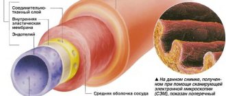
The widest and thickest-walled vessel extending from the heart and forming a large circle of blood circulation is the aorta. It plays a key role in maintaining hemodynamics and blood supply to almost all vital organs. The vessel is located in the thoracic part, directly above the apex of the heart and continues to the lumbar region: its beginning is represented by a bulb connected to the left ventricle of the heart, and the end is a branching into two iliac arteries. The anatomy corresponds to the structure of all large vessels of the elastic type, and most pathologies can be surgically corrected if detected early.
Development mechanism
It is based on a decrease in elasticity or organic, cellular changes in the structures of the aorta. The tissues soften and are easily destroyed. Usually we are talking about congenital factors; in 20% of situations there is an acquired cause of the pathological process.
The disorder develops gradually. There is no rapid gap observed. First, blood penetrates between the inner and middle linings (dissection). Then the space separating the muscle layer from the outer shell is involved.
The pressure on the serous structure is growing rapidly. It is not designed for such loads. A rupture occurs and massive bleeding occurs.
Often the patient does not have time to understand the essence. The last thing a person feels is a sharp pain at the level of the sternum. Then consciousness is lost and death occurs.
Disease prognosis
Without surgical intervention, the expansion constantly progresses, which is why the course of an aortic aneurysm is dangerous. For aneurysms with a diameter of less than 4 centimeters, the risk of rupture is low. If it is detected in elderly patients (over 70 years), then the intervention of a vascular surgeon is not required; dynamic observation is sufficient. With a diameter greater than 5 cm, the risk of rupture is very serious, so the indications for surgical treatment must be more active. Thromboembolism in most cases does not depend on the size of the aneurysmal sac, so when they appear, it is always necessary to raise the question of surgical treatment.
Classification
Typification of the pathological process is carried out for many reasons. They have meaning for practitioners and theorists, but they mean little to the patient. Still, it's worth checking out some common options. There are several criteria.
Localization of the violation. Difficult to understand. The main one is the De Bakey classification. According to it, there are three forms of violation:
- First type. The dissection begins in the ascending aorta and spreads to the abdominal and thoracic aorta. It is considered perhaps the most severe type, since the area of destruction is large. The symptoms are characteristic. They appear early.
- Second type. The localization is identical. But only the arch and the ascending section are involved. The area is sufficiently delimited, so the surgical field is clearly visible, which allows achieving high results of therapy.
- It is possible for the pathological process to spread only to descending structures. Signs are almost undetectable even at an advanced stage. Until it's too late. This is the third type.
The unit's technique is used by surgeons to identify the exact location, extent of the disorder, and plan intervention.
There is a simplified Stanford classification. She only names dissection of the ascending aorta and disruption of the integrity of the descending aorta.
Classification according to the duration of the pathological process is also used:
- Acute dissection. From the moment the anomalous transformations begin until the end of the second week. It is not accompanied by any symptoms, which makes diagnosis almost impossible. There are no complaints, and there is no reason to see a doctor.
- Subacute form. From 2 weeks to 2 months. Signs are minimal and nonspecific. Like bloating in the chest, weakness, instability of blood pressure. At any moment, an emergency condition and death of the patient may occur.
- Chronic type. Up to six months. The clinic is minimal.
Attention:
Few people survive the first 20-30 days. Usually 70% of patients die within the first 10 days from the start. The peak occurs during the week.
The violation can be classified depending on the stage:
- Aortic dissection is the penetration of blood between the inner lining and the muscle layer.
- The second stage of the process is accompanied by the entry of liquid tissue at the border of the muscular structures and the serous membrane.
- The last phase is rupture of the vessel and massive bleeding.
Folk remedies
Photo: sad-dom.com
Treatment of aortic aneurysm using folk remedies
Aortic dissection and aneurysm rupture require immediate surgical intervention. At an early stage of the disease, if it proceeds without dangerous complications, prevention and treatment of abdominal aortic aneurysm with folk remedies will be effective.
Effective folk remedies
Alternative treatment for aortic aneurysm will help normalize a person’s well-being and strengthen blood vessels. Herbal infusions are very effective and tonic.
- Hawthorn is the most accessible and effective remedy. Since ancient times, mankind has known the amazing properties of this plant. The fruits and leaves of hawthorn contain many important vitamins and are also capable of removing bad substances from the body (salts, heavy metals, etc.). Hawthorn is most effective for cardiac disorders. Decoctions and infusions will help improve blood circulation and normalize blood pressure. To prepare a simple medicinal infusion, you need to pour crushed dry hawthorn berries (4 tbsp) with boiling water (3 cups) and let it brew thoroughly.
- Viburnum infusion has anti-inflammatory properties, fights shortness of breath, and is also useful for vascular spasms and hypertension. The fruits of this plant contain a huge amount of vitamin C, which is necessary for the body, especially during illness. Therefore, for such a disorder as abdominal aortic aneurysm, treatment with folk remedies must necessarily include this miraculous infusion. Of course, viburnum is not a panacea, but with complex treatment it will only bring benefits. To prepare the infusion, dry berries are poured with boiling water and infused for 3.5 hours.
- Celandine - helps well in the fight against the most common cause of aneurysm development - atherosclerosis. The leaves, stems and flowers of this plant are dried and then infused in boiling water. It is recommended to drink 50 grams of infusion daily.
- Dill infusion is no less useful. Dill helps lower blood pressure, eliminates headaches and has a beneficial effect on heart function. You can use both herbs and seeds for infusion. 1 tbsp. dill is poured with boiling water (about 200 ml) and left for an hour. Treatment of aortic aneurysm with folk remedies must be combined with a healthy lifestyle and a balanced diet. Physical as well as psychological stress should be avoided.
Before starting treatment with these methods, you should consult a doctor.
The information is for reference only and is not a guide to action. Do not self-medicate. At the first symptoms of the disease, consult a doctor.
Symptoms depending on the form
Signs differ only when assessing the acute and chronic types. Localization does not provide specifics.
Emergency condition
Accompanied by pronounced clinical symptoms.
- Unbearable pain in the chest, just below the neck or in the area between the shoulder blades. Accompanied by pressing sensations, lumbago, swelling in the affected area. It appears suddenly, in one moment.
- Weak pulse, lightheadedness, increased sweating and pale skin, cyanosis of the nasolabial triangle. Similar reactions are typical for heart diseases, so it is impossible to immediately understand what happened. Collapse leads to the terminal phase, the death of the patient. If you don't provide first aid.
- Rapid jump in blood pressure. By 30-50 mm Hg in a short time. For a few minutes. This is dangerous in itself. There is a risk of stroke or heart attack. Then, as the disorder progresses, hypertension is replaced by a deep, critical drop in blood pressure.
- Dyspnea. Expressed.
- Tachycardia. Which, as the collapse approaches, is replaced by a reverse process.
These are typical signs. There are also more rare symptoms of aortic dissection, which occur in 2-20% of patients.
Among them:
- Peripheral blood flow disorders. Legs and arms suffer. Accompanied by pain, poor skin, a crawling sensation, as if the limbs were numb.
- Kidney failure. Lack of urine or a rapid decrease in the amount of diuresis, pain in the lumbar region.
- Abdominal discomfort, flatulence, diarrhea or constipation (much more often) with the development of false painful urge to evacuate (tenesmus). The reason is a decrease in the speed and efficiency of blood flow in the digestive tract. An extremely rare occurrence, but it is possible.
- Cerebrovascular insufficiency. Not necessarily a stroke. Transient ischemia is likely. Accompanied by headache, spatial disorientation, decreased reflexes, and focal neurological symptoms.
- Infarction or temporary disturbances of myocardial trophism. Chest pain, increased heart rate, other signs.
- Fainting often occurs. They can go into a coma, from which the patient can no longer be brought out.
Chronic form
The clinical picture is sluggish, because it increases gradually. The duration of development of the full symptomatic complex is about a day or two.
The manifestations are:
- Stomach ache. At the level of the epigastric region, slightly lower. May be diffuse, without clear localization. Accompanied by flatulence, nausea, diarrhea. Constipation (stool disorders). Tenesmus is detected. Uncomfortable urge to defecate without results.
- Chest pain. In this case, the localization of discomfort depends on the location of the dissection area. The ascending part is the place just below the neck, the arch and below - between the shoulder blades, etc. There may be discomfort in the abdominal cavity. A typical sign of aortic dissection.
- Dysphagia, disturbances in the speaking process, up to the complete disappearance of the voice. They are caused by compression of the nerves involved in conducting impulses from the brain.
- Angina pain. They arise abruptly, are highly forceful, and pulsate in time with the heartbeat.
- Darkening in the eyes, shortness of breath, disturbances of consciousness, syncope. They are caused by the same reason - acute weakening of blood circulation in the brain, trophism (nutrition) of cardiac structures through the coronary arteries.
- Edema. Usually peripheral. The lower extremities suffer.
- Paleness of the skin. Also fingertips, nails, mucous membranes.
Regardless of the clinical picture, the probability of death is always approximately the same. The location of the hearth plays a role.
The maximum risk is observed in the first 7 days (60% of fatal outcomes). Over the next month - about 80%. About 40% of people die before diagnosis.
There is no chance of spontaneous recovery. Lethal outcome is the only possible one. Surgery is required.
Normal size indicators
In a healthy adult, the aorta has a diameter of about 3 cm, tapering to 1.75 cm as it branches into the common iliac arteries opposite the 4 lumbar vertebrae.
Normal aortic size
Over time, the diameter and length of the aorta change. Starting from 50-60 years, the diameter of the vessel increases by about 1 mm per year, as elasticity is lost. Longitudinal growth of the aorta also occurs, especially in patients suffering from an aneurysm.
Longitudinal growth is also part of the normal growth process, but it is accelerated in some patients who are predisposed to aneurysmal disease. The ratio of the ascending to descending aorta, which is usually 3:2, also changes over time, as after age 55 the descending aorta enlarges more than the ascending aorta.
Dimensions of the main vessel:
| Ascending aorta | 2 to 3.7 cm in diameter |
| Total size of the arch In the isthmus region | About 2.5-3cm |
| The aorta narrows by approximately 3 mm | |
| Descending thoracic aorta | Ranges from 2 to 2.5 cm |
However, these measurements will be reduced in female patients because women have smaller aortas than men, even if they have the same body weight and surface area.
Causes
As mentioned, the basis is congenital pathologies. Which ones exactly:
- Coarctation of the aorta. Narrowing of an artery in a certain area. It is accompanied by a host of other symptoms and requires treatment on its own. The aorta does not always begin to dissect.
- Aortic valve stenosis. Leads to an increase in pressure in the largest vessel of the human body. Depending on other factors, it may result in an aneurysm, spontaneous rupture, or the condition in question.
There are also more rare deviations. Genetic, hereditary nature. Ehlers and Morphan syndromes.
Acquired reasons are numerous:
- Pregnancy, severe toxicosis. Usually late gestation, with first child. Refers to a possible complication in “old-parous” women. The probability is not higher than 3-5%.
- Iatrogenic factor. Associated with the actions of doctors during examination. Usually after inserting a catheter into the aorta.
- Chest injuries. Fractures, severe bruises. The probability is also not high, because the skeleton reliably covers the soft tissues.
- Aortitis. Inflammation of the walls of the artery of infectious, fungal, and much less often autoimmune origin. Occurs infrequently. In approximately 3% of all reported cases.
- Operations performed. As an unlikely complication.
- Hypertonic disease. Current for a long time, with high. Stable blood pressure levels.
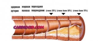
- Atherosclerosis of the aorta. Also long-lasting, against the background of stenosis or blockage of the vessel by cholesterol plaque, its calcification. There is no correlation between the duration of the pathological process and the likelihood of an emergency condition as such.
Causes are used by doctors to identify the etiological factor, i.e. origin of the disease. Without eliminating this point, there can be no full-fledged treatment. A relapse or early complication after the operation is possible.
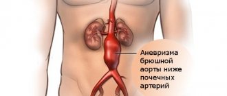
Aneurysm of the aorta of the heart - what is it?
An aortic aneurysm is a pathological enlargement of one of the sections of the largest artery in the human body. The cause of the development of pathology is weakness of the arterial wall. The process of formation of an aortic aneurysm is irreversible, so this disease requires surgical intervention.
The thoracic aorta most often suffers from an aneurysm. It accounts for about 2/3 of all cases. According to postmortem autopsies, aortic aneurysm is found in 0.6-6.5% of patients. At the same time, an aortic aneurysm can be considered any expansion of the lumen of the vessel that exceeds its normal diameter by 2 times. As for the age of patients with aortic aneurysm, it mainly develops in older people against the background of hypertension and atherosclerosis.
Aortic aneurysm is a deadly disease. A person with such a diagnosis risks his life every minute. An aneurysm can rupture at any time, causing severe internal bleeding. Due to such a serious complication, up to 40% of patients die, despite the fact that they are delivered to the hospital in a timely manner.
Content:
Diagnostics
It is carried out urgently. There is no time for long thoughts. The main problem is the lack of competence and awareness of doctors.
A vascular surgeon can qualitatively detect the pathological process. There is a huge shortage of such in provincial clinics in Russia and the countries of the former Union. And general doctors do not have sufficient experience and qualifications.
It is recommended to contact a surgeon and describe your complaints in as much detail as possible. This will allow the doctor to form a clear clinical picture in his head.
Anamnesis collection is mandatory.
Other methods:
- Blood pressure measurement (a difference of more than 10 mmHg in two arms is detected).
- Auscultation. Listening to heart sounds. Sinus murmurs are detected.
- Chest X-ray. With targeted visualization of the aorta. A high level of professionalism of the doctor and his assistant is also required.
- Ultrasonography. Basic diagnostic method.
- An MRI is required. The opportunity must be found in a short time. Allows you to visualize every millimeter of examined tissue.
There is no time to resort to other methods. This is the required minimum. You can do it literally in a day. Therefore, the examination should ideally be carried out in a hospital setting.
But neither the patient nor the doctor knows about the need for hospitalization. Precious time is lost, risks increase.
Since the possibility of high-tech research is few and far between, we have to limit ourselves to ultrasound and x-rays. Which is usually enough to make a diagnosis if doctors pay enough attention.
Vessel functions
The aorta's job is to supply blood to the entire body. To perform this function correctly, blood must be transported against gravity in the head and, with great effort, enter the last muscle cell. The strong pressure created by the heart helps here.
This pressure is called blood pressure, which in a healthy adult should be 120/80 mmHg. To withstand the pressure created by the heart, the aorta has an expandable wall. This allows the vessel to create a pressure reservoir in milliseconds when the heart is not pumping. The elasticity of the main artery is a very important property that ensures full functioning.
Treatment
Therapy is strictly surgical. Urgent. You can't hesitate. In rare cases, it is possible to stabilize the condition without such intervention and manage the patient in this way for years (chronic forms). Still, the radical method remains the gold standard.
Conducted open access. The point is to replace the affected area of the dissected aorta along the entire length of the structure.
This is a difficult intervention, which is why it is not carried out everywhere and requires a high level of professionalism from doctors.
Upon completion, drug therapy is indicated. Because the root cause does not go away. It is possible to provoke a relapse or aortic dissection in another location.
Etiotropic therapy depends on the provoking pathological process.
Monitoring of blood pressure is mandatory (beta blockers, calcium antagonists, ACE inhibitors are used to reduce the rate), heart rate (in the presence of functional and organic disorders, cardioprotectors are used like Mildronate, glycosides - Digoxin, antiarrhythmics according to indications - Quinidine).
In addition, statins are prescribed to remove cholesterol and dissolve already formed plaques - Atoris, others. Antiplatelet agents to normalize blood flow (Aspirin Cardio, Heparin).
Some pathologies themselves require surgical treatment: defects, advanced forms of atherosclerosis.
The question of whether surgical correction is carried out in parallel with the elimination of the dissection or whether a separate act is planned is decided by doctors.
Upon completion, diet correction is required (minimum salt, up to 7 grams per day, animal fats, fried, smoked, semi-finished and canned foods).
Sleep at least 7 hours per night, adequate physical activity, at the level of walking in the fresh air.
No smoking. Alcohol. Especially drugs. Self-administration of any medications is prohibited. By indication only.
How can you detect an aortic aneurysm?
Diagnosis of this deadly defect of the aortic wall is quite difficult. Therefore, if there is a risk of aneurysm formation (identified atherosclerosis, hypertension, old age, etc.), it is necessary to undergo regular examinations.
Ultrasound allows you to detect an aneurysm in the early stages of its formation and even when it is not there yet, but it should appear soon.
Abdominal aortic aneurysm is easily visualized on x-ray. The fact is that calcium deposits on the aneurysm, which gives a shadow during the study.
If an aneurysm is detected during an ultrasound, an MRI or CT scan is prescribed. These techniques allow you to collect as much information as possible about the existing defect.
Forecast
The issue has already been addressed. Death occurs most often in the first two days. Within a week, the initial peak period ends.
Only 15% of patients manage to live for more than a year; this is great luck; one cannot speak of a pattern. Pure chance.
After surgical therapy, the survival rate is 89-90%, other situations include complications, but death occurs early and relatively rarely.
As for maintaining ability to work, physical professional activity is excluded. We need to reconsider the scope of our efforts.
The ability to take care of oneself at home is limited only in the early period of rehabilitation (about 6 months after surgery).
Prevention
Prevention, thanks to which it is possible to prevent the occurrence of aortic dissection in healthy people, is nonspecific (i.e., effective not only in the case of this pathology) and includes:
- complete cessation of smoking;
- reducing alcohol standards to the level of “only for holidays”, or better yet, a complete refusal;
- physical education and sports;
- elimination of factors that cause a rise in blood pressure (stress, kidney disease);
- treatment and prevention of diseases that contribute to aortic dissection (atherosclerosis);
- immediate alertness in the event of a sudden, at first glance inexplicable appearance of interruptions in the functioning of the heart, gastrointestinal tract and respiratory system and immediate examination by specialized specialists;
- regular, high-quality, not just for show, medical examinations with a vascular surgeon and cardiologist.
Complications
A complication of aortic dissection is its complete rupture. The mortality rate from aortic rupture is up to 90%. 65-75% of patients die before they arrive at the hospital, and the rest before they reach the operating room. The walls of the aorta are an elastic structure that requires complete integrity. A rupture occurs when its strength is lost. This can happen when internal or external pressure is more than the walls can withstand.
Pressure occurs as the tumor progresses. Bleeding can be retroperitoneal or intraperitoneal and can create a fistula between the aorta and intestine.