Sputum is a pathological secretion of the lungs and respiratory tract (bronchi, trachea, larynx), which is separated when coughing.
Healthy people do not produce sputum. Normally, the glands of the large bronchi and trachea constantly produce a secretion in amounts of up to 100 ml/day, which is swallowed when secreted.
Tracheobronchial secretion is a mucus that contains glycoproteins, immunoglobulins, bactericidal proteins, cellular elements (macrophages, lymphocytes, desquamated bronchial epithelial cells) and some other substances.
This secretion has a bactericidal effect, promotes the removal of inhaled small particles and cleanses the bronchi. In diseases of the trachea, bronchi and lungs, the formation of mucus increases, which is expectorated in the form of sputum.
Smokers without signs of respiratory diseases also produce copious amounts of sputum.
A general sputum analysis is a laboratory test that allows you to evaluate the nature, general properties and microscopic features of sputum.
Based on a general analysis of sputum, the inflammatory process in the respiratory organs is judged, and in some cases a diagnosis is made.
During a general (clinical) examination of sputum, the following is carried out:
- examination of discharge;
- establishing their quantity;
- determination of color, transparency and viscosity;
- microscopy;
- bacterioscopy;
- bacteriological culture;
- study of cellular composition.
A general (clinical) examination determines the presence of microorganisms (bacteria, fungi), as well as parasites.
A general clinical analysis of sputum detects, first of all, pathogenic microorganisms, the most significant of which is mycobacterium or the causative agent of tuberculosis.
Malignant tumor cells, an admixture of pus or blood may also be detected, which also indicates certain diseases.
If pathogenic microorganisms are detected, their sensitivity to antibiotics and bacteriophages is immediately established, which allows the doctor to prescribe the most effective treatment.
Sputum for general clinical examination is collected for the following diseases:
- bronchitis – inflammatory lesion of the bronchial wall;
- pneumonia – inflammation of the lung tissue;
- bronchial asthma - an allergic reaction of the bronchi with bronchospasm;
- pulmonary tuberculosis is an infectious disease caused by Koch's bacillus;
- whooping cough is predominantly a childhood infection caused by Bordetella;
- bronchiectasis - local expansion of the bronchial wall (sputum stagnates in the formed cavity);
- tumors of the respiratory system;
- helminthic infestation – echinococcosis, etc.;
- fungal infection - actinomycosis, etc.;
- silicosis is an occupational pathology that develops with prolonged inhalation of silicon compounds.
The study is mandatory if there is a cough with sputum, as well as if there is suspicious X-ray data or pathological data on auscultation of the lungs.
Where does sputum come from?
Sputum usually forms in the lower parts of the respiratory tract (trachea, bronchi, bronchioles) and, accumulating, irritates the receptors, causes a cough reflex and comes out through the mouth.
Some patients call phlegm nasal discharge that flows down the back wall of the nasopharynx ( sputum is not only snot ), as well as discharge from chronic pathologies of the oropharynx and larynx.
That is why sputum production can be associated with various pathological processes.
In case of such complaints, it is necessary to conduct a full examination of the patient, find out the details of the medical history and conduct a series of tests.
Why are all these various studies needed?
The main purpose of the described study is to clarify the diagnosis. Under normal circumstances, no sputum is produced. Goblet cells of the ciliated epithelium secrete from 10 to 100 ml of liquid, which is swallowed by a person.
The progression of the pathological process in the bronchi or lungs leads to a change in the activity of the corresponding structures with an increase in cough, shortness of breath, and chest pain. The amount of liquid mucus increases, and bacterial microflora may join. The result is sputum production with coughing.
Based on the expected diagnosis and the results of a visual assessment of the secretion of the bronchial glands, the doctor prescribes the appropriate type of study. The use of different options for sputum analysis makes it possible to assess the physicochemical properties of the fluid, cytological changes (presence of cancer cells), and the presence of bacterial invasion.
Types of sputum tests (studies)
Sputum examination can be carried out either with the naked eye or with the help of specialized equipment.
Depending on the disease that the doctor suspects, the following types of diagnostics may be used:
- General sputum analysis . The doctor evaluates the physical characteristics of the mucus that appears after coughing;
- Cytological (microscopic) examination . To carry out appropriate diagnostics, the doctor needs a microscope. Using image magnification, the liquid is analyzed. The technique allows you to see the presence of pathological cells that appear in certain diseases;
- Chemical Research . Changes occurring in the metabolism of alveolocytes and ciliated epithelium of the bronchi are assessed;
- Bacteriological method or sputum culture . The essence of the study is based on sowing bacteria obtained from the contents of the respiratory tract on a nutrient medium. The growth of colonies confirms the presence of the pathogen in the respiratory tract. An important advantage of culture remains the ability to test the sensitivity of bacteria to specific antimicrobial drugs in the laboratory.
In severe cases, for timely diagnosis of the pathology of the patient’s respiratory system, all three research options are simultaneously prescribed. Based on the results obtained, appropriate therapy is selected.
General sputum analysis
Fact! General or macroscopic analysis allows you to evaluate sputum immediately after it is obtained. This research option has been used by doctors for hundreds of years. Even before the invention of the microscope and modern analyzers, doctors made diagnoses based on the appearance of the expectorated liquid.
Below we will describe the key aspects that the doctor pays attention to during diagnosis.
Quantity
The daily amount of mucus secreted ranges from 50-100 ml to 1.5 liters, depending on the underlying pathology, which disrupts the normal secretion process of goblet cells. Respiratory diseases such as bronchitis or pneumonia are accompanied by the release of up to 200 ml of fluid (daily amount).
A sharp increase in this indicator occurs when pus or blood accumulates with further release through natural pathways. Bronchiectasis, drained abscess, and gangrene of the lung occur with the release of up to 1.5 liters of fluid.
Character
Depending on the nature of the liquid that is expectorated during a cough, pulmonologists distinguish the following types of sputum:
- Mucous . Favorable scenario. Diseases in which it occurs: bronchial asthma, chronic bronchitis, tracheitis;
- Mucopurulent . Additionally, a bacterial infection is added. In addition to coughing and mucus, liquid is released, which is the waste products of microorganisms and bacteria “digested” by immune cells. Diseases – lung abscess, bacterial pneumonia, gangrene;
- Purulent . The reasons for the occurrence are the same as in the previous case. The difference is a higher percentage of pus and tissue breakdown products. The patient's condition worsens;
- Bloody . When individual red blood cells or portions of blood enter the liquid expectorated during a cough, it acquires a characteristic color. The symptom indicates vascular damage. Possible causes are cancer, trauma, pulmonary infarction, actinomycosis.
Assessing the nature of liquid discharge when coughing helps to understand the pathological process that develops in the patient’s respiratory system and select adequate treatment.
Color
The color palette of sputum released during a cough depends on its nature.
Possible combinations:
- Mucous membrane – grayish or transparent;
- Mucopurulent – gray with yellow or purulent patches;
- Purulent - the fluid may be dark yellow, green or brown;
- Bloody - various shades of red. It is important to remember that a “rusty” color indicates the presence of modified red blood cells. If a vessel is damaged, the blood is scarlet or pink (depending on the intensity of blood loss).
Interesting! Anthracosis is an occupational disease of miners caused by inhalation of dust rich in coal. The corresponding pathology is accompanied by the release of mucous sputum, but due to coal impurities it is black in color.
Smell
Sputum in 75% of cases has no characteristic odor. The exception is the release of purulent contents. Dead tissue particles cause a putrid odor. When a lung cyst in which echinococcus (helminth) has developed breaks through, a fruity aroma appears.
Layering
The mucus released when coughing is predominantly homogeneous.
The separation of sputum into layers is characteristic of the following pathologies:
- Lung abscess . In this case, 2 layers are formed - serous and putrefactive;
- Gangrene of the lung . In this case, a third (upper) foamy layer is additionally formed, which is caused by the vital activity of the corresponding microorganisms that produce gas bubbles.
Visual assessment of mucus allows you to quickly establish a diagnosis without performing auxiliary tests.
Impurities
Impurities in sputum include red blood cells, pus, or serous fluid. The presence of the described inclusions allows the doctor to assess the degree of damage to the lung tissue and understand which pathological process is primary for a particular clinical case.
Chemical research
Chemical analysis of the fluid secreted during coughing allows us to determine the severity of the pathological process. Depending on the results of the study, the doctor selects appropriate medications aimed at stabilizing the function of the ciliated epithelium.
Reaction
Normally, the pH of sputum ranges from 7 to 11. The progression of the processes of decomposition of lung tissue leads to the oxidation of the corresponding reaction (indicator below 6). The reason for the change in pH value is based on disturbances in the metabolic processes of salts and minerals.
Protein
Protein is always present in the liquid released when coughing. The norm is up to 0.3%. A slight increase in the corresponding figure to 1-2% may indicate the progression of tuberculosis. A significant increase in the indicator (10-20%) is a sign of the development of lobar pneumonia. Laboratory examination of mucus with protein determination makes it possible to differentiate these pathologies against the background of an analysis of the clinical picture (cough, shortness of breath, chest pain) and the results of other diagnostic procedures.
Bile pigments
Bile pigments, or more precisely, cholesterol microparticles, are secreted with mucus during coughing in the following pathologies:
- abscess;
- formation of echinococcal cyst;
- malignant tumors of the respiratory tract.
Microscopic examination
Microscopic analysis of sputum allows, using an appropriate optical apparatus, to detect the presence of cells or microorganisms that should not normally be present in mucus secreted by coughing.
Epithelial cells
Epithelium in sputum is a normal variant. On microscopic examination, attention is drawn to a sharp increase in the concentration of cells or the formation of epithelial casts. This picture indicates damage to the respiratory tract and inner lining.
Alveolar macrophages
The function of alveolar macrophages is to provide local immune protection. A small number of cells may be present in the mucus. A sharp increase in the concentration of macrophages indicates a chronic inflammatory process (bronchitis, bronchiectasis, asthma, tracheitis).
Leukocytes
The appearance of leukocytes indicates the presence of acute inflammation, which can occur against the background of a bacterial infection. Possible pathologies are abscess, pneumonia, bronchiectasis.
Red blood cells
Blood cells appear in sputum when small or large vessels rupture. The doctor judges the nature of the bleeding by the number of red blood cells. Separately, it is worth highlighting the appearance of modified cells that penetrate through the dilated walls of blood vessels without rupturing the latter. A typical example of the disease is lobar pneumonia.
Tumor cells
Atypical cells in sputum are a sign of a developing cancer process. To clarify the location and type of pathology, additional tests are required.
Fact! With the help of microscopic examination, cell differentiation is also established. The less similar the structures are to the original tissue, the worse the prognosis for the patient.
Elastic fibers
The appearance of elastic fibers in the mucus released when coughing is a sign of serious lung damage with tissue breakdown. Examples of diseases are gangrene, late stage bronchiectasis, tuberculosis and cancer, accompanied by destruction of organ parenchyma.
Detection of Mycobacterium tuberculosis
Microbiological analysis of sputum is one of the important methods for verifying the presence of tuberculosis. The causative agent of the disease is mycobacterium (Koch's bacillus).
Determining the presence of a microorganism is possible using the bacterioscopic method using a microscope. To visualize the pathogen, it is necessary to stain the material under study using the Ziehl-Neelsen method. If Koch's bacillus is detected in the sputum after a cough, the phthisiatrician must indicate BC (+) in the documentation, which indicates the release of the pathogen. Such patients require isolation. CD (-) – the patient does not spread the bacteria.
Fact! Sputum analysis for tuberculosis is also carried out by inoculating the test fluid on a nutrient medium. The advantage of the method is 100% accuracy. If the bacterium is present in the body, it will grow in the laboratory. The main disadvantage of the described diagnostics is the length of time it takes to obtain analysis results (sometimes more than a month).
Bacteriological examination for infectious lung diseases
Bacteriological research for inflammatory lesions of the respiratory tract is usually used to verify community-acquired forms of infection (pneumonia, actinomycosis, etc.).
The analysis is carried out in three stages:
- sputum collection for research;
- sowing the liquid onto a previously prepared nutrient medium;
- reseeding the required colony with the study of the chemical and physical characteristics of the pathogen.
If it is necessary to establish susceptibility to antimicrobial agents, a sensitivity test is additionally performed. Paper mugs treated with antibiotics are placed in a Petri dish where a colony of microorganisms has grown. Those drugs around which the zone of colony destruction is maximum are recommended for use in a particular patient.
Carrying out
You need to take a sputum test to detect pathogenic microorganisms and tubercle bacilli.
It is carried out in several ways:
- general analysis;
- fluorescence microscopy;
- bacteriological (cultural) method;
- bacterioscopic study of biomaterial.
General sputum analysis
This is the most basic and effective method that allows you to evaluate the quantitative and qualitative characteristics of a biomaterial.
His goal:
- identification of a pathogenic process in the respiratory system;
- assessment of the nature of the disease;
- monitoring the course of chronic pathology;
- analysis of the effectiveness of prescribed treatment.
This method takes into account the following factors:
- quantity;
- character;
- smell;
- layering;
- color;
- presence of impurities.
General analysis allows you to get a picture immediately after its examination. This method has been used for many decades and provides reliable information about diseases. More in-depth data can be obtained by conducting a number of other analyses.
Bacterioscopic examination
This method is also called Ziehl-Neelsen staining. This is one of the main research methods for suspected tuberculosis development. With its help, AFBs are identified, indicating the presence of severe pathology.
Diagnosis of sputum is carried out according to the following algorithm:
- treatment of biomaterial with carbol fuchsin;
- decolorization of sputum with a 3% solution of hydrochloric acid alcohol or a 5% solution of sulfuric acid;
- subsequent staining of the material with a 0.25% solution of methylene blue.
After the manipulations, the resulting biomaterial is assessed using an immersion system. The presence of AFB is indicated by the appearance of red and blue colors in the area of pathogenic organisms.
The result is viewed as standard in 100 fields of view. If AFBs are not detected, scanning is carried out in 200 fields of view.
The analysis is free, efficiency is high. The only drawback of the method is low sensitivity.
Bacteriological (cultural) method
The sputum is cultured on a nutrient medium. To detect the tuberculosis bacillus, only a few vital cells are enough.
The following types of culture media are used in the analysis:
- liquid semi-synthetic and synthetic;
- agar-based semi-liquid;
- dense, egg-based.
The primary causative agent of tuberculosis is determined, as well as the sensitivity of the pathogenic organism to antibacterial dosage forms. This is an excellent method that allows you to select the most effective medicine in each individual case.
The negative side of the method is its duration. The intensity of MBT growth is determined from 21 to 90 days, but with the use of modern computer technology this period has decreased to 3-4 days.
Fluorescence microscopy
This method is 30% more effective than bacterioscopic examination. Organic fluorochromes are used as coloring materials. Using a luminescent device under ultraviolet illumination, they begin to glow blue or violet. The presence of AFB, BC, VK is indicated by a golden color on a dark background.
When using fluorescent microscopy, not only the presence of pathogenic microorganisms is detected, but also their quantitative characteristics. This allows you to assess the epidemic danger to others and the stage of the disease.
Diseases for which a doctor may prescribe a general sputum test
Collecting sputum for general analysis can be prescribed for almost any disease that is accompanied by expectoration after coughing. However, appropriate diagnostics are rarely used for seasonal viral infections because they are unnecessary. In these cases, cough and other symptoms regress with plenty of fluids and bed rest.
Pathologies requiring sputum analysis:
- tuberculosis;
- lung abscess;
- malignant neoplasms;
- gangrene of the lung;
- bronchial asthma;
- Chronical bronchitis;
- pneumoconiosis is an occupational disease of the bronchopulmonary system.
Confirmation of the diagnosis is carried out using laboratory, physical, and instrumental methods.
Nature of the discharge
Mucous secretion is released during acute or chronic bronchitis, bronchial asthma, pneumonia, lung cancer, bronchiectasis, pulmonary echinococcosis accompanied by suppuration, actinomycosis.
Sputum mixed with pus is observed with lung abscess, echinococcosis and bronchiectasis.
Mucus mixed with blood or consisting entirely of blood is characteristic of tuberculosis. The appearance of blood may indicate the presence of oncology, bronchiectasis, or suppuration of the lung. This phenomenon is also observed in middle lobe syndrome, pulmonary infarction, trauma, actinomycosis and syphilitic lesions. Blood can also be released during lobar and focal pneumonia, congestive processes, cardiac asthma and pulmonary edema.
Serous sputum is observed with pulmonary edema.
How to prepare for the test in order to pass it correctly?
Preparing a patient to collect sputum for analysis is a responsible process on which the quality of diagnosis may depend. If simple rules are ignored, additional impurities appear in the mucus, preventing the laboratory assistant from establishing the root cause of cough and respiratory pathology in general.
Recommendations:
- Preparing the container . Containers sold in pharmacies remain optimal. If such a bottle is not available, even a half-liter jar or small plastic tank (no more than 1 liter) will do. However, it must be taken into account that such containers are extremely inconvenient and can only be used in atypical circumstances when there is no access to normal containers;
- Two hours before the examination, you need to brush your teeth and rinse your mouth. Removing food particles and saliva improves diagnostic accuracy;
- Consult your doctor . The doctor will explain in detail how to properly collect sputum for analysis.
If a person is giving bronchial mucus for the first time, then it often takes several attempts to complete the procedure correctly.
Features of performing bacteriological culture
You can improve the accuracy of your readings by following simple recommendations from your doctor. Before taking a sputum test, it is necessary to rinse your mouth with antiseptics to prevent pathogenic microflora of the oral cavity from entering the sample. The resulting mucus is immediately placed in a nutrient medium (Petri dish). After a few days (5-14 days), the concentration of leukocytes can be assessed and bacteria such as legionella, staphylococcus, CD and others can be identified.
After determining the type of cocci, a study is carried out to determine the resistance of the pathogenic flora to various types of antibiotics. Thus, the most effective drug for the treatment of tuberculosis and pneumonia is selected.
How to submit sputum for analysis?
In addition to the preparation nuances described above, the rules for collecting sputum provide for the use of a morning portion of mucus. The reason is the accumulation of secretions from the night, which greatly facilitates expectoration after coughing. You can take bronchial mucus at other times of the day, however, the quantity and quality of the material being studied is reduced.
Algorithm for collecting sputum for general analysis:
- take a deep breath and hold the air for 10 seconds;
- exhale smoothly;
- repeat 2 breaths;
- on the third exhalation, the air must be forcefully pushed out of the chest, after which you need to cough;
- bring the container to your lower lip and spit out the mucus.
This algorithm allows you to collect the required amount of test material (2-5 ml). If difficulties arise, it is recommended to lean forward and lie on your side. To speed up the removal of mucus, you can additionally do moisturizing inhalation with steam or using an expectorant.
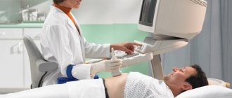
Collecting sputum in the described manner does not exclude saliva from entering the test sample during coughing. An alternative to this algorithm of action remains the collection of secretions from the respiratory tract during bronchoscopy. The doctor uses an endoscope to examine the condition of the ciliated epithelium and can take the required amount of fluid for appropriate analysis.
Material collection rules
Patients often have difficulty collecting sputum. To facilitate this process and ensure the reliability of the results, several rules should be followed:
- Sputum is always collected in the morning, since it accumulates in sufficient quantities at night.
- The minimum amount to be obtained is 1 ml. Ideally - 3 ml.
- Before collection, the patient sits in front of an open window.
- First, it is recommended to take two slow deep breaths with a slight breath hold.
- On the third inhalation, the patient stands up to maximize the expansion of the lungs and exhales sharply. If the diaphragm connects to the lungs as a result of these movements, it will provoke a cough impulse, and phlegm will come out. It is immediately spat out into the prepared container.
- Dishes for collecting material must be sterilized (usually by boiling, but there are also ready-made vessels).
- No saliva allowed. If there is not enough material, you can make several cough pushes. The lid of the container is opened only at the moment of spitting; the rest of the time it should be closed.
- The following methods stimulate sputum production: taking expectorants, irritating inhalations, drinking plenty of warm water, and physical exercise.
- If the patient is very weakened or sputum needs to be collected from a small child, you need to touch the root of the tongue with a sterile napkin, thereby causing a cough. In this case, part of the secretion from the bronchi gets onto the napkin. It is quickly transferred from a napkin to a glass slide and immediately sent to the laboratory.
To collect the material, choose a transparent container with a wide neck (for ease of spitting) and an airtight lid. The material should be delivered for examination no later than 2 hours.
Only then will the results not be distorted (extra microorganisms may have time to multiply in the sputum, and the result will become false positive). The collected material should only be stored in the refrigerator.
Interpretation of sputum tests, normal indicators, examples
Normally, a pulmonologist or phthisiatrician will interpret the sputum analysis. Below is a table showing the characteristics of mucus released after a cough in the absence of pathology.
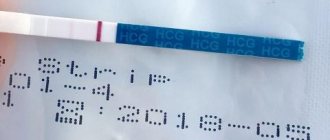
After examining the sputum, the laboratory assistant fills out the appropriate form (click to enlarge).
The specified document may look slightly different. It all depends on the specific laboratory. Below are options for study forms with proposed diagnoses.
Interpretation: pink color of mucous sputum in combination with the presence of mycobacterium tuberculosis (MT+) indicates the presence of a corresponding pathology.
Explanation: given the presence of leukocytes, mucopurulent consistency and a large amount of coccal flora, the most likely diagnosis remains chronic bacterial bronchitis.
Explanation: first of all, attention should be paid to a large amount of sputum (50 ml). In combination with the abundance of leukocytes, which cover the entire field of view of the microscope, and the presence of elastic fibers, one can judge the presence of an abscess that has broken into the bronchus.