Epidemiology of the disease
The most common trichophytosis in the Russian Federation is caused by anthropophilic fungi Trichophyton tonsurans and Trichophyton violaceum (the most common). The fungus causes sporadic outbreaks of mycosis in families, schools, kindergartens and boarding schools. More often children get sick who become infected from sick adults (mothers, grandmothers) suffering from chronic forms of the disease. When infected, a superficial form of the disease develops.
Zoophilic trichophytons Trichophyton mentagrophytes v.gypseum and Trichophyton verrucosum are the cause of the development of infiltrative-suppurative trichophytosis. The source of pathogenic fungi is animals.
Sources of infection
- Anthropophilic trichophytosis: a sick person.
- Zooanthroponotic trichophytosis: the source of Trichophyton verrucosum is large domestic animals (cattle, horses, calves, donkeys and goats, as well as wild animals), Trichophyton mentagrophytes is small animals (mouse-like rodents, gophers). Transmission of infection from a sick person is possible.
Transmission factors
- Anthropophilic trichophytosis: hair of the patient, skin scales and crumbling nail plates of the sick person.
- Zooanthroponotic trichophytosis: hay, straw, dust (in autumn during field work), wool of sick animals.
Transmission routes
- Anthropophilic trichophytosis: the patient’s clothes, his personal belongings (towels, combs, razors, scissors, hats, scarves, bed linen, etc.). Wrestling athletes are affected by contact from patients with trichophytosis of smooth skin (lichen of the trunk of gladiators).
- Zooanthroponotic trichophytosis: by contact through objects contaminated with sick animals (brushes, bedding, feeders, etc.).
Incubation period
- Anthropophilic trichophytosis (superficial) - 1 week.
- Zooanthroponotic trichophytosis (infiltrative-suppurative) - 1-2 weeks - 2 months.
Rice. 2. Calves are often the source of Trichophyton verrucosum.
Routes of infection
Trichophytosis refers to contagious skin diseases, being an exception to the total number of diseases associated with fungi. Every day, a healthy adult sheds millions of dead and dead cells from the upper parts of the epidermis, they are separated from the skin through the process of natural desquamation and enter the environment.
In a patient with trichophytosis, trichophyton microspores also enter the mass of exfoliated cells. Soon they can get on the skin:
- child;
- an elderly person;
- pet;
- a patient with a weakened immune system.
It is by this principle that ringworm spreads.
Characteristics of the main pathogens of trichophytosis
Trichophyton tonsurans
Trichophyton tonsurans belongs to the group of anthropophiles. Causes superficial trichophytosis. Distributed everywhere. Affects hair and smooth skin. Large-spored. The tissue form is endothrix. There is no glow in the rays of a Wood's lamp.
Colonies on Sabouraud's nutrient medium grow slowly, young colonies are velvety and whitish in color. With age, they become mealy, dense, dry, with many cracks, the crater in the center is brain-shaped and striated, the color of the colonies is from white to yellow, in the center - to brown.
The mycelium is septate, many microconidia, macroconidia are rare.
Rice. 3. Trichophyton tonsurans. In the photo on the left is a view of the fungus through a microscope, on the right is a view of the colonies when growing on a nutrient medium.
Rice. 4. Trichophyton tonsurans hair damage. The tissue form is endothrix.
Trichophyton violaceum
Trichophyton violaceum or purple trichophyton is an anthropophile. Causes superficial trichophytosis.
When sown on Sabouraud's medium, colonies grow slowly - they appear on days 4 - 5 in the form of a yellowish tubercle, and grow on days 25 - 30. Their surface is dense, leathery, radially striated, scalloped, wrinkled, dry, powdery, the edges are clearly defined, the color ranges from pale lilac to dark purple, rarely colorless.
The mycelium is branching, smooth, septate. Microconidia and macroconidia (exospores) are not found. Chlamydospores are detected only in old cultures.
The tissue form is endothrix (spores are located inside the hair) in chains, sometimes arranged randomly.
Rice. 5. Trichophyton violaceum. Microscopic picture (photo on the left), appearance of colonies when growing on a nutrient medium (photo on the right).
Rice. 6. Hair affected by Trichophyton violaceum. Spores are found in large numbers inside the hair, arranged in chains.
Trichophyton mentagrophytes
Trichophyton mentagrophytes is the main zoophilous trichophyton. There are 2 subspecies of this type of fungus: Trichophyton mentagrophytes var.gypseum and Trichophyton mentagrophytes var.interdigitale. They are the cause of infiltrative-suppurative trichophytosis.
Colonies of pathogens when sown on Sabouraud's medium begin to grow already on the 2nd - 3rd day. They have a powdery surface, often with pronounced graininess and wrinkles. The color is white to cream, with brownish-red coloration on the underside. The edges are often radiant.
The mycelium is branching, has partitions, and is often twisted in a spiral. Microconidia are round or pear-shaped, arranged in clusters.
The tissue form is ectothrix (spores are located on the outside of the hair).
Rice. 7. View of Trichophyton mentagrophytes colonies (photo on the left). Type of fungus under microscopy.
Rice. 8. Trichophyton mentagrophytes hair damage. The tissue form is ectothrix.
Rice. 9. Histological specimen. The mycelium threads of Trichophyton mentagrophytes var.interdigitale are visible in the stratum corneum.
Trichophyton verrucosum
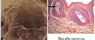
Trichophyton verrucosum is a cosmopolitan mushroom. It parasitizes the skin of cattle, calves, goats, donkeys and horses. It is the cause of infiltrative-suppurative trichophytosis. When infected, a person’s scalp, body, mustache and beard area are affected.
Colonies with a smooth or velvety surface. The reverse side is white or yellow-ocher in color.
The mycelium is septate. Microconidia are elongated, drop-shaped or spherical in the form of strings of beads. Chlamydospores are numerous.
The tissue form is ectothrix. The spores are located on the outside of the hair.
Rice. 10. Microscopic picture of Trichophyton verrucosum (photo on the left). Chlamydospores (photo on the right).
Rice. 11. View of Trichophyton verrucosum colonies when growing on different nutrient media.
Rice. 12. Fungal spores surround the outside of the hair.
Rice. 13. Cattle are the main source of infection.
Main forms of the disease
There are superficial and chronic forms of trichophytosis, caused by anthropophilic fungi of the endothrix type Trichophyton tonsurans and Trichophyton violaceum, and deep infiltrative-suppurative form, caused by zoophilic fungi of the exothrix type Trichophyton mentagrophytes v.gypseum, Trichophyton mentagrophytes var. interdigitale and Trichophyton verrucosum. The disease affects the scalp, smooth skin (superficial and deep layers), nails (in the chronic form of the disease) and internal organs.
The incubation period for the superficial form of the disease is 1 week, for the infiltrative-suppurative form - from 1 week to 2 months.
Skin damage, weak immunity, prolonged contact with infection and the presence of endocrine pathology contribute to the development of mycosis.
Rice. 14. Trichophytosis on the face of a child.
Prevention
Measures to prevent trichophytosis include preventing the spread of fungal infection and timely treatment of animals with ringworm - calves, cows, sheep.
Prevention of trichophytosis includes deratization of residential and industrial premises, as well as mandatory veterinary examination of domestic cats, dogs, hamsters, and rabbits.
To prevent trichophytosis, it is necessary to limit children’s contact with stray animals and use only personal combs and hats.
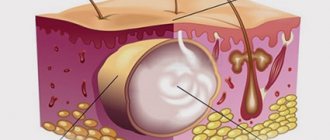
Hair damage due to trichophytosis
Based on the nature of hair damage, trichophytons can be divided into 2 groups: Trichophyton ectothrix and Trichophyton endotlrix.
Trichophyton ectothrix
The group Trichophyton ectothrix (ecto - outside) includes fungi whose spores surround the hair on the outside. Their spores are large (5 - 7 µm) and small (3 - 5 µm). At the base of the hair they are arranged in several layers, forming a wide sheath (in large-spored ones) or a narrower one (in small-spored ones). Along with spores, short or long chains of rounded spores are found in the peripheral region of the hair. Usually these are zooanthropophilic trichophytons. They are the cause of infiltrative-suppurative processes, in which inflammatory infiltrates are formed around the hair follicles, followed by purulent melting.
Trichophyton endothrix
The group Trichophyton endothrix (endo - inside) includes fungi whose spores are found inside the hair. They are round in shape, large (5 - 7 microns), densely fill the hair inside without going beyond its limits, are arranged either in the form of chains, or fill the hair like a bag of nuts. These are usually anthropophilic trichophytons. They are the cause of superficial forms of the disease.
Rice. 15.Trichophyton ectothrix: pathogen spores envelop the outside of the hair, like a muff (photo on the left). Trichophyton endothrix: The inside of the hair is filled with spores (photo on the right).
At-risk groups
First of all, this group should include people exposed to certain negative professional factors, such as:
- work in rooms with high temperature and humidity;
- long-term wearing of clothes made of air-tight or rubber fabrics;
- frequent contact with soil, animals and biologically contaminated surfaces.
Patients with endocrine and other chronic diseases, which often entail:
- weight gain;
- increased sweating;
- increase in normal body temperature.
It is these factors that make the skin vulnerable to the appearance of microcracks and abrasions, through which trichophytons can freely penetrate inside.
Unfortunately, ringworm is not a rare disease among children attending preschool institutions and staying in infant homes, orphanages and boarding schools. Both owners of pets (cats and dogs) and owners of livestock are at particular risk for the development of trichophytosis. Both of these categories of people can become infected with trichophytosis from their pets.
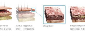
Signs and symptoms of superficial trichophytosis
The disease most often occurs in children aged 4–15 years, rarely in infants and adults. By puberty, self-healing occurs in most cases, mainly in boys. In some patients (usually women), mycosis becomes chronic and lasts until old age. Children often become infected from sick adults in the family, as well as in children's groups from sick children. Infection occurs during care, through hats, combs, brushes, clothing, underwear, and hairdressing tools. Fungi affect the scalp and smooth skin. Nails are not affected. The incubation period is up to 7 days.
Trichophytosis of the scalp
Outbreaks. When the disease occurs, small lesions (up to 2 cm in diameter) (small-focal form) or large lesions (large-focal form) appear on the scalp. New lesions gradually appear, and old ones, due to peripheral growth, increase in size and merge with each other. The skin becomes covered with scales (pityriasis desquamation). Their grayish-white color gives the lesion a whitish appearance. The inflammatory component in the lesion is weakly expressed.
Hair. The affected hair within the lesions breaks off at a low height (1 - 3 mm), sometimes at the very root (the appearance of “black dots”), loses color, shine, elasticity, thickens, some of them curl and take on the appearance of a comma. There is no glow under Wood's lamp. Along with broken hair, healthy hair growth is observed.
Course of the disease. Superficial trichophytosis in children lasts a long time. During puberty, self-healing occurs (often recorded in boys). Otherwise, the disease becomes chronic (often in women).
Rice. 16. Damage to the scalp. Broken hair looks like black dots (photo on the left). Pityriasis-like peeling in the lesion (photo on the right).
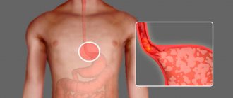
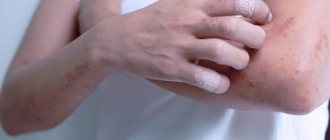
Trichophytosis of smooth skin
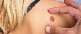
In the superficial form of the disease, the skin can be affected independently, or occur simultaneously with damage to the hair. The lesions are localized mainly in open areas of the body - the face, neck, forearms and torso. The disease occurs at any age, equally often in women and men.
When infected, one or more round spots of bright red color with clear boundaries appear on the skin. The center of the spot is covered with scales. Along the periphery there is an inflammatory ridge rising above the surface of the skin, covered with blisters and crusts. The spot increases in size due to peripheral growth. Reverse development occurs from the center, due to which the area of inflammation takes on the appearance of a ring. Itching is mild or absent.
Sometimes the lesions are weakly expressed (must be distinguished from erythrasma and pityriasis versicolor). Sometimes the inflammation is severe. The formed bubbles merge with each other and surround the central part like a shaft.
When hair follicles are damaged, fungal folliculitis is formed.
Rice. 17. Lesion on the skin of the face.
Rice. 18. Superficial trichophytosis. Lesions on the skin of the chest and hand.
Rice. 19. Focus of inflammation on the forearm and shoulder.
Rice. 20. Superficial form of trichophytosis. A focus of inflammation on the forearm.
Signs and symptoms of infiltrative-suppurative trichophytosis
The cause of the disease is zooanthropophilic fungi of the ectothrix type. The incubation period ranges from 1 week to 2 months. Adults and children living in rural areas and caring for animals are affected. In children, the disease most often affects the scalp, in adults - the area where the mustache and beard grow.
There are several forms of the disease that transform into each other: superficial, infiltrative and suppurative. The suppurative form of trichophytosis, in which pathogens are transmitted directly from animals, occurs with a pronounced inflammatory reaction. When pathogens are transmitted from person to person, the clinical picture of inflammation is similar to superficial trichophytosis.
Trichophytosis of hair
Zooanthropophilic trichophytons infect hair according to the ectotrix type: large spores surround the outside of the hair and a small number of them are detected in the form of chains inside the hair. Hair breaks off at low heights. Fungi cause the development of purulent inflammation of the follicles. With timely treatment, hair does not fall out.
Rice. 21. Superficial form of zooanthropophilic trichophytosis.
Damage to smooth skin with superficial zooanthroponotic trichophytosis
Initially, one or several round-shaped lesions appear on the skin, pale pink in color, with clear boundaries. Along the edges there is an inflammatory ridge covered with blisters and crusts. Gradually, the lesions increase in size, rise above the skin level, forming bizarre figures when merging. The top is covered with blisters, purulent crusts and plaques. The inflammatory process affects vellus hair. With a long course of the disease, flat plaques, sometimes quite large, form on smooth skin.
Rice. 22. Infiltrative form of zooanthroponotic trichophytosis. The smooth skin of the body is affected.
Infiltrative-suppurative form of zooanthroponotic trichophytosis
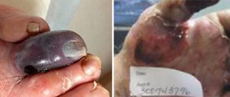
When a secondary infection occurs and there is no specific treatment, purulent inflammation develops involving the follicles. The lesions on the head take on the appearance of tumor-like formations of a hemispherical shape, initially dense, lumpy to the touch, then of a dough-like consistency, bright red in color, completely covered with purulent crusts. When pressed, pus and affected hair are released from the openings of the follicles. Lesions in the mustache and beard area resemble wine berries. Soreness appears. The overall picture resembles a honeycomb (Kerion). There is a musty smell coming from the fires. Increasing in size, inflammatory infiltrates reach the area of the palm or more. Hair falls out in the affected area.
General health worsens. Body temperature rises. Regional lymph nodes are enlarged.
In some cases, an allergic rash appears on the skin of the torso and limbs - trichophytids, reminiscent of a rash caused by measles or scarlet fever. Allergization of the body occurs as a result of the entry of fungal toxins into the lymphatic and blood vessels.
Spontaneous healing sometimes occurs within 2 to 3 months. At the site of inflammatory infiltrates, cicatricial atrophy of the skin is formed.
Immunity is strong. Repeated cases of the disease do not occur.
Rice. 23. Infiltrative form of trichophytosis.
Rice. 24. Infiltrative-suppurative form of the disease.
Rice. 25. Purulent inflammation of the follicles in the head area.
Rice. 26. Suppurative form of zooanthroponotic trichophytosis (kerion).
Rice. 27. Kerion is a suppurative form of the disease.
Rice. 28. Trichophytosis in the mustache and beard area.
Rice. 29. Suppurative form of the disease in the mustache and beard area (purulent folliculitis).
Rice. 30. Consequences of zooanthroponotic trichophytosis in a child: cicatricial atrophy with baldness.
Diagnosis
To confirm the diagnosis of “classic” ringworm, a routine clinical examination is sufficient. But sometimes it becomes difficult to distinguish the infiltrative-suppurative form of trichophytosis from a bacterial skin infection, and then a dermatologist and laboratory diagnostics come to the aid of the clinician.
There are several ways to determine the causative agent of trichophytosis - Microscopic examination.
- To carry it out, it is necessary to collect biological material from areas of skin lesions - scraping from the scalp, collecting hair “stumps”, scales, and crusts from the periphery of the lesions.
- Under a microscope, it is possible to clearly distinguish between a violation of the normal structure of the hair and the presence of numerous fungal spores inside it. In whitish scales and other fragments of skin scrapings, particles of fungal mycelium or gift can be detected.
- Sowing biological material on special media helps to establish the specific “autograph” of the pathogen and determine its type.
Signs and symptoms of chronic trichophytosis
The chronic form of the disease is recorded most often in women. The disease begins in childhood in the form of superficial trichophytosis and does not go away during puberty. Women (80% of cases) suffering from endocrine insufficiency, caused by decreased ovarian function, are especially susceptible to chronic trichophytosis. The disease lasts a long time, the manifestations of mycosis are scanty. Adults become a source of infection for children. The disease occurs with damage to the scalp, smooth skin and nail plates.
Damage to the scalp
The skin on the head is most often affected in the area of the back of the head and temples, where small areas of whitish peeling appear. Broken hair resembles black dots (black dot trichophytosis) - the only clinical sign of the disease. Atrophic scars are visible in places.
Rice. 31. Damage to the scalp with chronic trichophytosis.
Damage to smooth skin
Smooth skin during the disease is affected in the area of the forearms, elbows and the back of the hands, legs and knees, less often - the buttocks, face and torso, where pink-violet spots of 1 cm or more appear, prone to merging, without clear boundaries, without signs inflammation. Hyperkeratosis and lamellar peeling are observed on the skin of the soles and palms, and cracks sometimes form.
Rice. 32. Damage to the skin of the trunk.
Follicular form of chronic trichophytosis
The follicular (furuncle-like) form or Majocchi granuloma is rarely recorded in chronic trichophytosis, mainly in individuals with immune deficiency. Dermatophyte fungi affect the deep layers of the skin and hair follicles. Around them, infiltrates form on the affected areas of the skin, which over time become deep, similar to boil-like nodes, called trichophytosis granulomas (first described in 1883 by Majocchi). With suppuration, purulent melting of the follicles develops. Pus is released.
Cases of infection of lymph nodes by fungi of the genus Trichophyton are described.
Rice. 33. Majoka granuloma on the skin of the face and head.
Rice. 34. Folicular (boil-like) form of the disease or Majocchi granuloma.
Signs of damage to the nail plates
In 30% of cases, the disease affects the fingernails. The infection spreads when fungi are transferred from smooth skin or scalp. In some cases, nail mycosis may be the only sign of the disease. Infection from such patients is often transmitted to children when washing their hair or cutting their hair.
Initially, a cloudy spot appears on the free edge of the nails, the area of which increases over time. The nails become dirty gray in color, thicken, become lumpy and begin to crumble. The course of the disease is long. Nail trichophytosis should be distinguished from onychodystrophy and onychomycosis, which is caused by Trichophyton rubrum.
Rice. 35. The photo shows nail damage due to chronic trichophytosis.
What does the skin look like?
Smooth in texture, skin that is affected by chronic trichophytosis is usually bluish in color with the presence of gray and thin scales. The skin in the buttocks area is often affected, as well as on the inner thighs, elbows and forearms. Affected areas are found on the upper part of the body, but much less frequently, and the spread of trichophytosis over the entire surface of the body is very rare. The pattern on the skin is usually pronounced; there may be a thickening of the stratum corneum of the skin. The latter leads to the appearance of furrows on the feet and palms, namely at the site of skin folds. These grooves will become cracks over time. Blistering was never observed in cases involving the palms and soles. Nodules, blisters and crusts will also not be typical for the chronic type of trichophytosis.
Diagnosis of the disease
Diagnosis of trichophytosis is carried out on the basis of the clinic, microscopic and microbiological examination data. Skin flakes, nail plates and affected hair are subject to examination. Serological studies are carried out in the chronic form of the disease. Intradermal tests are performed with allergens such as trichophytin. There is no glow in the rays of a Wood's lamp (with microsporia the glow is emerald in color).
Rice. 36. Trichophyton verrucosum under a microscope (photo on the left). Growth of a Trichophyton mentagrophytes colony on a nutrient medium.
Rice. 37. Microscopy of hair determines the tissue form of trichophyton. Endothrix (photo on the left), when fungal spores are located inside the hair. Ectotrix (photo on the right), when fungal spores are located on the surface of the hair.
Treatment of trichophytosis
Treatment of trichophytosis is similar to the treatment of microsporia: see the article “Ringworm (microsporia) in humans.” Treatment uses systemic antifungal therapy. Local treatment is used simultaneously with oral medications. External treatment of lesions not only shortens the healing time, but also reduces the likelihood of transmitting infection to others. For single lesions, monotherapy is carried out using ointments, creams and gels with antimiotics.
Systemic treatment of trichophytosis
The main antifungal drug for the treatment of ringworm is Griseofulvin. The drug can be adsorbed in the horny substance of the epidermis, hair and nails. Drugs containing terbinafine and ketoconazole are also used. Fluconazole and Intraconazole are used. The choice of drug, determination of the dose and duration of treatment is carried out only by a doctor after establishing an accurate diagnosis.
Local therapy
Local therapy is carried out simultaneously with oral antifungal drugs. Antiseptics and keratolytics are used in treatment.
- Among the antiseptics used are 2 - 5% alcohol solution of iodine, rivanol, furatsilin, potassium permanganate, etc.
- Keratolytics include preparations containing birch tar, sulfur, salicylic and glycoic acids.
- To remove hair from damaged areas and detach the stratum corneum, milk-salicylic-resorcinol collodion or salicylic ointment is used, after which manual hair removal of vellus hair is carried out. Removal of “black spots” in chronic trichophytosis is carried out according to Arievich’s method.
- If there is significant inflammation, medications containing hormones are prescribed, for example, Travocort cream or Triderm.
- For suppurative trichophytosis, initially, in order to remove purulent crusts, bandages with 2% salicylic ointment are applied to the affected areas. Next, the crusts and vellus hair are removed, followed by the use of lotions with antiseptics. Antimyotes are then used locally. Antifungal drugs are used orally from the first days of treatment.
- Keratoplasty agents are used to resolve deep inflammatory infiltrates. By taking away water, drugs in this group help slow down the processes of fermentation and putrefaction in deep inflammatory infiltrates. The use of 20% Ichthyol ointment or pure Ichthyol is indicated.
Rice. 38. Suppurative form of zooanthroponotic trichophytosis (kerion).