Human echinococcosis is a zoonosis (a disease transmitted from animals to humans) caused by parasites, namely tapeworms of the genus Echinococcus. There are four forms of echinococcosis:
- cystic echinococcosis, also known as hydatid disease, or hydatidosis, which develops as a result of infection with the species Echinococcus granulosus;
- alveolar echinococcosis, which develops as a result of infection with E. multilocularis;
- two forms of neotropical echinococcosis: polycystic echinococcosis, which develops as a result of infection with E. vogeli; And
- monocystic echinococcosis caused by E. oligarthrus.
The two main forms of medical and public health significance are cystic echinococcosis (CE) and alveolar echinococcosis (AE).
Transmission of infection
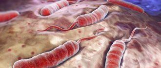
The intermediate hosts of Echinococcus are some herbivores and omnivores. These animals acquire the infection by ingesting parasite eggs contained in contaminated food and water, and the parasite then passes through the larval stages of development in their internal organs.
The definitive hosts of the parasite are carnivores, whose intestines contain sexually mature worms. Infection of the definitive hosts occurs when they eat the internal organs of intermediate hosts, which contain parasite larvae.
Humans are so-called accidental intermediate hosts in the sense that they become infected in the same way as other intermediate hosts, but do not participate in transmission of infection to the definitive hosts.
Several different genotypes of E. granulosus are known, some of which have distinct preferences for intermediate hosts. Some genotypes are considered species other than E. granulosus. Not all genotypes cause infection in humans. The genotype that causes the vast majority of cystic echinococcosis infections in humans is primarily maintained in the dog-sheep-dog cycle, although other domestic animals can be affected, including goats, pigs, cattle, camels and yaks.
Alveolar echinococcosis is commonly found in wild animals and is maintained in cycles between foxes or other carnivores and small mammals (mostly rodents) acting as intermediate hosts. Definitive hosts may also include domestic dogs and cats.
Immunological methods for studying echinococcosis
- Indirect hemagglutination reaction (IRHA) is a method for identifying antigens and antibodies, based on the ability of erythrocytes (red blood cells), on the surface of which antigens or antibodies are pre-adsorbed (collected), to agglutinate (stick together and precipitate) in the presence of homologous sera (obtained from sera of immunized (vaccinated, that is, having antibodies against a specific pathogen (in this case we are talking about the causative agent of echinococcosis)) people (people who have been vaccinated with certain known antibodies)) or corresponding antigens;
- latex agglutination reaction - agglutination of latex particles, on the surface of which the antigen is collected, occurs with antibodies;
- double gel diffusion - the method consists of placing the antigen and antibody into wells made in a thin layer of gel at a short distance from each other. The precipitation zone (the zone of sediment formation that occurs during the test) is permeable to antigens and antibodies that do not interact with those that formed the precipitate. As a result, several precipitation zones can form, each of which corresponds to an individual antigen and antibodies to it;
- immunoelectrophoresis (IEF) and counter immunoelectrophoresis (CIEF) are methods for studying the antigenic composition of biological materials;
Fluorescent antibody reaction (FA) - is based on the reaction of an antigen with antibodies associated with fluorescent (converting absorbed light into visible radiation) dyes, which give a characteristic glow when irradiated with short-wave light;
- scolex precipitation reaction - as a result of the interaction of the scolex (head of echinococcus) with the antibody, precipitation occurs in the form of so-called precipitates;
- enzyme-linked immunosorbent assay (ELISA) – detection of pathogen antigens, as well as specific IgG antibodies (a class of antibodies that provide protection against microorganisms and toxins (their waste products)) in blood serum.
Signs and symptoms
Cystic echinococcosis/hydatidosis
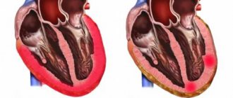
Infection of humans with E. granulosus results in the development of one or more hydatid cysts, most often localized in the liver and lungs and less commonly in the bones, kidneys, spleen, muscles and central nervous system.
The asymptomatic incubation period of the disease can last many years until hydatid cysts develop to a size at which clinical signs appear, with approximately half of all patients receiving medical treatment for the infection beginning to receive it several years after initial infection with the parasite .
If hydatides are localized in the liver, abdominal pain, nausea and vomiting are often observed. If the lungs are affected, clinical signs include chronic cough, chest pain, and shortness of breath. Other signs depend on the location of the hydatid(s) and the pressure exerted on adjacent tissues. Nonspecific signs include anorexia, weight loss, and weakness.
Alveolar echinococcosis
Alveolar echinococcosis is characterized by an asymptomatic incubation period of 5-15 years and the slow development of a primary tumor-like lesion, usually localized in the liver. Clinical signs include weight loss, abdominal pain, general malaise, and signs of liver failure.
After dissemination of the parasite through the circulatory and lymphatic systems, larval metastases can spread to either organs adjacent to the liver (eg, the spleen) or to distant sites (eg, the lungs or brain). If left untreated, alveolar echinococcosis progresses and leads to death.
Stage of the disease
In clinical practice, there are several conventional stages of echinococcosis:
- Latent - develops from the moment of invasion into the body until the first appearance of nonspecific subjective symptoms (poor health, fatigue);
- Mild changes—more pronounced subjective disorders;
- Progression stage—sharply expressed objective signs (rash, hepatomegaly);
- The stage of complications is suppuration and breakthrough of the cyst with further generalization of the process (the appearance of secondary foci) and the development of intoxication, up to toxic shock.
Spreading
Cystic echinococcosis is common throughout the world and is found on all continents except Antarctica. The distribution of alveolar echinococcosis is limited to the northern hemisphere, in particular to some areas of China, the Russian Federation and countries of continental Europe and North America.
In endemic areas, human incidence rates of cystic echinococcosis may exceed 50 per 100,000 people per year, and in parts of Argentina, Peru, East Africa, Central Asia and China, prevalence rates may be as high as 5–10%. Among farm animals, the prevalence of cystic echinococcosis detected in slaughterhouses in hyperendemic areas of South America varies from 20 to 95% of animals slaughtered.
The highest prevalence rates are observed in rural areas where older animals are slaughtered. Depending on the specific animal species infected, livestock production losses due to cystic echinococcosis are caused by liver culling, as well as decreased carcass weight, decreased hide value, decreased milk production, and decreased reproductive performance.
Laboratory methods for studying echinococcosis
- Complete blood count: eosinophilia is detected (increased concentration of eosinophils (blood cells that contribute to the development of an allergic reaction) in the blood);
- biochemical blood test: dysproteinemia (disturbance in the ratio of blood proteins) is detected - a decrease in albumin (simple, water-soluble proteins), prothrombin (a complex protein that is an important indicator of the function of the blood coagulation system) and an increase in gamma globulins (antibodies (cells used by the immune system) to detect and neutralize foreign objects)). This indicates a violation of the protein synthetic function (protein production) of the liver;
Echinococcus. The Kasoni serological test is a diagnostic allergy test for diagnosing echinococcosis, during which an echinococcal antigen (a genetically foreign substance that causes a reaction from the human immune system) is injected intradermally. A positive test will indicate the presence in the human body of antibodies (cells used by the immune system to detect and neutralize foreign objects) against the causative agent of echinococcosis;
- microscopic examination of sputum for the presence of the causative agent of echinococcosis;
- microscopic examination of urine for the presence of the pathogen echinococcus.
Diagnostics
The best technique for diagnosing both cystic and alveolar echinococcosis in humans is ultrasonographic imaging. This technique is usually complemented or confirmed by computed tomography (CT) and/or magnetic resonance imaging (MRI).
Sometimes cysts can be discovered accidentally during an x-ray. Specific antibodies are detected using various serological tests and can confirm the diagnosis. Early detection of E. granulosus and E. multilocularis infections is still needed to assist in clinical treatment options, especially in resource-poor areas.
Development of echinococcus
The embryos that enter the lungs grow and gradually transform into bubbles. A well-developed hydatid cyst consists of an inner membrane called germinative (membrana germinativa) and an outer plaque-like membrane, resembling a folded chicken protein in appearance. The fluid in the cysts is light watery or amber yellow. This liquid is isotonic, neutral or slightly acidic.
A small cyst contains several tens of milliliters of fluid, a large one contains much more. The literature contains descriptions of giant cysts with a capacity of up to 5 liters. The fluid pressure in the cyst can be high and reach 50 cm of water column. In 7% of cases, vesicles develop on the inner membrane of an echinococcal cyst, the number of which can reach a dozen. Some of the bubbles break off from the inner membrane and float freely in the fluid of the cyst.
Each vesicle contains a series of oval structures corresponding to the heads of the tapeworm, called scolices. Floating bubbles form the so-called. echinococcal sand (le sable hydatique). Primary pulmonary echinococcus can be single or multiple.
Treatment
Treatment of both cystic and alveolar echinococcosis is often expensive and complex and may require extensive surgery and/or long-term drug therapy. There are four treatment options for cystic echinococcosis:
- percutaneous treatment of hydatid cysts using the PAIR technique (puncture, aspiration, injection, reaspiration);
- surgical intervention;
- therapy with anti-infective drugs;
- observation.
The choice should be based primarily on the results of ultrasound echography of the cyst. The specific stage of the disease must be taken into account, as well as the available medical infrastructure and available human resources.
In the case of alveolar echinococcosis, the key components of treatment remain early diagnosis and radical (as in the case of a tumor) surgery followed by a course of anti-infective prophylaxis using albendazole. In cases of limited damage, radical surgery may be curative. Unfortunately, in many patients this disease is detected in late stages. As a consequence, if palliative surgery is not accompanied by a full course of effective anti-infective therapy, relapses often occur.
List of sources
- Sergiev V.P., Legonkov Yu.A., Musaev G.Kh., Poletaeva O.G. Cystic echinococcosis (unilocular): clinical picture, diagnosis, treatment, prevention. M.: Vector-Best; 2008.
- Shodmonov I.Sh., Razikov Sh.Sh. EPIDEMIC SIGNIFICANCE OF ECHINOCOCCOSIS // Modern problems of science and education. – 2015. – No. 2-1.
- Deineka I. Ya. Human echinococcosis. - M.: Medicine, 1968. - 376 p.
- Trishin, M.V. Epidemiology and diagnosis of echinococcosis in the child population of the Orenburg region in 1994-2012 / M.V. Trishin, P.V. Gureeva, I.A. Sim // Bulletin of the Orenburg Scientific Center of the Ural Branch of the Russian Academy of Sciences (electronic journal). - 2014. - No. 1. — P. 4-7.
- Dadvani S.A., Strelyaeva A.V., Gostishchev V.K. and others. Minimally invasive surgical interventions and chemotherapy for echinococcosis // Annals of Surgery. - 2000. - No. 4. - P. 38-41.
Health and economic burden
Cystic and alveolar echinococcosis represent a significant proportion of the disease burden. At any given time, the number of people suffering from these diseases in the world may exceed one million. Many of these people may have severe clinical syndromes that, if left untreated, can be life-threatening. Even with treatment, people's quality of life often decreases.
With regard to cystic echinococcosis, the postoperative mortality rate of surgical patients averages 2.2%, and in 6.5% of cases, relapses are observed after surgery, requiring a long recovery period.
Globally, echinococcosis is responsible for 19,300 deaths and approximately 871,000 disability-adjusted life years (DALYs) annually, the WHO Foodborne Disease Burden Epidemiology Reference Group (FERG) established in 2015.1.
The annual cost of treating patients and losses in livestock production due to cystic echinococcosis is estimated at US$3 billion.
Diet
Diet for liver cysts
- Efficacy: no data
- Terms: 1-6 months/lifetime
- Cost of products: 1500-1600 rubles. in Week
Diet 0 table
- Efficacy: therapeutic effect after 21 days
- Terms: 3-5 months
- Cost of products: 1200-1300 rubles. in Week
For echinococcal liver cysts outside the surgical period, the Diet for Liver Cysts . In the preoperative period (on the eve of the operation) dietary Table No. 15 ; after surgery – Diet 0 table ; in the early postoperative period, Dietary Table 1A , 1B , and in the late postoperative period - Treatment Table No. 5 . For echinococcal cysts in the lungs outside the surgical period - Diet 15 , in the postoperative period on days 1-2 Diet No. 0a , and from day 3 - No. 1 - surgical, from day 5 dietary Table No. 11 or 13 .
Surveillance, prevention and control
Reliable surveillance data are urgently needed to reflect the burden of echinococcosis and assess the progress and success of disease control programs. However, as with other neglected diseases that predominantly affect underserved and remote populations, there is a critical lack of data and attention must be given to the issue if echinococcosis control programs are to be successfully implemented and measured. more attention.
Cystic echinococcosis/hydatidosis
Surveillance of cystic echinococcosis in animals is difficult because infection in livestock and dogs is asymptomatic. In addition, local communities and veterinary services do not recognize the importance of surveillance and do not prioritize it.
Cystic echinococcosis is preventable, since the final and intermediate hosts of the parasite are domestic animal species. Periodic deworming of dogs using praziquantel (at least four times a year), improved slaughter hygiene (including proper disposal of infectious waste) and public education campaigns have been found to reduce, and in high-income countries prevent, – transmission of infection and reducing the burden of disease in humans.
Vaccination of sheep with recombinant E. granulosus antigen (EG95) offers promising prospects for the prevention and control of echinococcosis. Currently, commercial production of the vaccine has been established, which is registered in China and Argentina. Trials in Argentina demonstrated the benefits of vaccinating sheep, and the vaccine has been widely adopted in China.
A sheep vaccination program combined with dog deworming and culling of older sheep could lead to the elimination of cystic echinococcosis in humans in less than 10 years.
Alveolar echinococcosis
It is more difficult to prevent and control alveolar echinococcosis, since the cycle includes wild animal species both as definitive and intermediate hosts. Regular deworming of domestic carnivores with access to wild rodents may help reduce the risk of infection in humans.
Studies conducted in Europe and Japan have shown that deworming wild and stray animals, which are the definitive hosts of the parasite, using anthelmintic baits helps to significantly reduce the prevalence of alveolar echinococcosis. Trapping foxes and stray dogs appears to be extremely ineffective. The sustainability and cost-effectiveness of such campaigns are controversial.
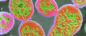
Parasite - Echinococcus
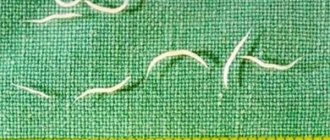
Echinococcus is the larva of Taenia echinococcus, which lives in the digestive tract of dogs, wolves, jackals, foxes, and sometimes cats. The Echinococcus tapeworm is small in size. Its length does not exceed 6 mm, thickness 0.3 mm. The parasite has a head and 3-4 segments. The head contains a mouth opening, 4 suckers, and a double corolla of hooks. The last segment is the largest, contains a uterus with 600-800 eggs (embryophores) with a diameter of about 30 mm.
In the middle of each egg there is an embryo armed with 6-8 hooks (the so-called oncosphere). The last segment of the Echinococcus tapeworm breaks off after some time and is excreted in the dog’s feces. Thus, Echinococcus eggs contaminate courtyards, streets, vegetable gardens and meadows.
Eggs are very resistant to environmental influences. Dried eggs live in the sun for 12 days, in water for 2 weeks, and at a temperature of 0° for 116 days.
WHO activities in countries
Strengthening the prevention and control of echinococcosis
In 1985, informal working groups on echinococcosis were established under the auspices of WHO. For 10 years, these groups, led by Professor J. Eckert (University of Zurich, Switzerland), organized expert meetings and promoted international scientific exchange and cooperation in the field of echinococcosis research. In 1995, WHO changed the structure of these groups and transformed them into a single group, the WHO Informal Working Group on Echinococcosis (WHO Informal Working Group on Echinococcosis). The mission of the WHO IWGE is to strengthen the prevention and control of echinococcosis through effective collaboration with strategic partners and relevant sectors. The current Chairman of the WHO IWGE is Professor Thomas Junghans (Heidelberg University, Germany), and the Co-Chair is Professor Okan Akhan (Hacettepe University, Turkey).
In 1995, the WHO NRGE developed a standardized classification of cystic echinococcosis (CE) that can be applied in all settings. In 2009, a consensus document on the diagnosis and treatment of TBE and alveolar echinococcosis (AE) reached by the WHO GWG (Brunetti et al, 2010) was published, providing updated guidelines for diagnosis and treatment.
The WHO IWGE is currently reviewing the diagnosis and appropriate clinical management of echinococcosis and developing technical guidelines of practical applicability. To address various aspects of these diseases, several working groups have been created and are working on the preparation of these documents. The group also facilitates the collection and mapping of epidemiological data.
Building capacity to improve early diagnosis and clinical management of TBE
Endemic countries have requested WHO support for early diagnosis and clinical management of cystic echinococcosis. WHO is providing capacity-building support through training courses for medical and paramedical personnel, with a focus on the clinical management of cystic echinococcosis in rural areas of affected countries. This is an integral component of efforts to achieve universal health coverage.
In Morocco, a project is being implemented focusing on the decentralization of diagnostic and therapeutic technologies and the promotion of the PAIR (puncture, aspiration, injection, re-aspiration) strategy in rural and hyperendemic areas.
Mongolia has recognized echinococcosis as a public health problem and, at the request of its Ministry of Health, WHO conducted a preliminary analysis of the situation in 2013. The analysis focused on early diagnosis and establishing a basic surveillance system covering both humans and animals needed to establish the true burden of the disease.
A cross-sectional study conducted in Bulgaria, Romania and Turkey in 2014–2015 found that the true burden of TBE is poorly understood and that many cases are asymptomatic and remain without appropriate medical diagnosis and treatment. This study assessed the prevalence of the disease among rural populations in three countries.
In the Region of the Americas in 2021, the Pan American Health Organization/WHO Regional Office of the Americas (AMRO) has prepared guidelines for the control of cystic echinococcosis. It was published in Spanish.
Working with veterinary and food safety authorities to facilitate the development of echinococcosis control programs
The transmission cycle of cystic echinococcosis (CE) involves dogs and intermediate hosts, usually sheep. To break the cycle of transmission, these animals must be taken into account when taking action. Echinococcosis control measures for dogs and sheep based on the One Health concept include deworming dogs with praziquantel at least 4 times a year and vaccinating sheep with EG95 vaccine.
As part of the One Health approach, WHO and its partner, the World Organization for Animal Health (OIE), are supporting the development of echinococcosis control programmes, including interventions for animals. Joint meetings are regularly held and technical support is provided, for example, in the countries of Central Asia and the Caucasus.
WHO is helping countries develop and implement pilot projects leading to the establishment of effective strategies for the control of cystic echinococcosis. Collaboration with veterinary and food safety authorities and other sectors is essential to achieve long-term results in reducing the burden of disease and maintaining the food value chain.
WHO is supporting individual countries, such as Mongolia, in developing programs to combat TBE. A multidisciplinary stakeholder meeting was convened in Ulaanbaatar in 2021 to develop a national action plan for echinococcosis control. Due to the lack of significant investment in the disease, progress has been slow, but WHO continues to bring together stakeholders and further action has been agreed upon in 2021. In addition, WHO supported the validation of diagnostic tests used to detect echinococcosis in dogs, which is important for surveillance, and the establishment of a core system in Bayankhongor Province, Mongolia.
China is including the prevention, treatment and control of echinococcosis in its economic development plans to bring increased attention to this significant problem in the country, especially in the Tibetan Plateau areas as well as in Central Asian areas.
WHO promotes One Health approaches, such as the approach developed by Dr Larriu in Patagonia, Argentina, which involves community health workers, dog deworming and sheep vaccination.
Improving the quality of CE data
Surveillance data is key to understanding the epidemiological situation and taking action in areas at risk, as well as setting priorities. In addition, data are needed to monitor progress in implementing interventions and assess the results achieved in disease control.
Indicators are specific variables that help analyze data and provide tools for health authorities and those involved in disease control. WHO has developed a new set of CE indicators at country and global levels and is developing reporting systems that will guide and support countries in data collection and reporting.
At the global level, indicator 1, the number of countries endemic for TBE, and indicator 2, the number of countries with enhanced control measures in hyperendemic areas, are used. A hyperendemic area was defined as an area with an annual incidence of 5 human cases per 100,000 people.
At the country level, epidemiological indicators and indicators of progress in disease control are used. The latter indicators include impact and outcome indicators.
(1) One DALY (disability-adjusted life year) can be considered one year of “healthy” life lost. The total number of DALYs in a population, or disease burden, can be used to measure the gap between current health status and an ideal health status in which all people live into old age free of disease and disability.
Analysis for echinococcus
A long stay in countries where this disease occurs is an argument for making a positive diagnosis. Eosinophilia in peripheral blood is of the same importance. The following immunological reactions are produced:
- Complement fixation reaction with sheep echinococcus fluid (Weinberg and Ghedini reaction),
- Intradermal reaction of Casoni with fluid from a human hydatid cyst.
The Weinberg reaction is positive in 50% of echinococcal diseases. The Casoni reaction consists of an intradermal injection of 0.1-0.2 ml of hydatid cyst fluid. If the reaction is positive, itching, redness and swelling of the skin appear. This reaction occurs immediately (early reaction) or after 2-3 hours (late reaction).
The reaction is considered positive only when the swelling and redness last for at least 3-4 hours. The intradermal reaction is positive in 80% of cases of echinococcus. The detection of a spherical or nearly spherical shadow on an x-ray in the lungs of a person arriving from a country in which echinococcus occurs endemically, eosinophilia, positive Weinberg and Casoni reactions make it possible to diagnose echinococcus.