Ornithosis (psittacosis) is an infectious disease that affects the human respiratory system and central nervous system, which is characterized by general intoxication and fever.
Has a natural focality. Belongs to the group of zoonoses. Not only wild or domestic birds, but also other animals can spread the infection. Certain strains of bacteria infect cows, goats, and sheep.
The causative agent of the disease is isolated by birds in secretions from the respiratory tract, as well as in feces. The main routes of transmission of infection are airborne dust and airborne droplets. Dry particles of droppings enter the human body through breathing. The greatest changes are found in the lungs. The entire lobe can be affected by patches of red-gray shades. The adrenal glands, spleen, heart muscle, liver and even the brain suffer. Lymph nodes enlarge.
Pathogen
The causative agent - Chlamydophila psittaci is an obligate intracellular parasite and has some structural features that determine the course of the disease:
It multiplies inside the affected cells.
Capable of forming L-forms - a pathogen that is partially or completely devoid of a cell wall, which allows it not to die and maintain its virulence for a long time, even when exposed to damaging factors (temperature, antibiotics, phagocytosis, etc.).
The pathogen can be in two forms - elementary and reticular bodies, this is important when prescribing treatment: elementary bodies are a spore-like form due to the presence of a strong shell that makes the pathogen insensitive to antibiotic therapy, and in this life cycle the pathogen is outside the cell, and when under unfavorable conditions, division is suspended with a decrease in the synthesis of the main membrane protein and an increase in the synthesis of heat shock protein (for this pathogen this protein is an exotoxin) - it causes the synthesis of anti-inflammatory cytokines, which in turn plays a role in the formation of chronic infection and long-term persistence (stay) of the pathogen with the formation of infertility in women. Reticular bodies are an intracellular multiplying form that is sensitive to antibiotics. It is with the reticular form that the pathogen divides inside the infected cells, resulting in the formation of intracellular inclusions - microcolonies of chlamydia.
Presence of exotoxin (heat shock protein) and endotoxin (membrane lipopolysaccharide).
Tropism (selectivity of damage) to the cells of the cylindrical epithelium of the respiratory and urogenital tracts (mainly), alveolocytes, vascular endothelium, endocardium, SMF (mononuclear phagocyte system - a physiologically protective system of cells, which includes: connective tissue histiocytes, liver Kupffer cells (stellate reticuloendotheliocytes) , alveolar macrophages of the lungs, macrophages of the lymph nodes, spleen, bone marrow, pleural and peritoneal macrophages, osteoclasts of bone tissue, microglia of nervous tissue, synoviocytes of synovial membranes, Langergais cells of the skin, pigment-free granular dendrocytes). When this system (SMF) is damaged, an IDS (immunodeficiency state) is formed.
The pathogen is relatively stable in the external environment:
- So at room temperature, the pathogen persists on average up to 2 days, on the shell - 3 days, in bird feces - up to 4 months.
- tolerates sub-zero temperatures well: at “-20°C” it lasts for half a year, at “-75°C” for a year or more;
- low temperatures have a detrimental effect: at 60°C the pathogen dies within 10 minutes, at lower temperatures the death of chlamydia occurs almost instantly.
- Disinfectants, both physical and chemical, have an inactivating effect: UVR, ether, formaldehyde, 0.5% phenol, 2% Lysol, 0.1% potassium iodite, 0.5% potassium permanganate, 6% peroxide solution - they destroy the pathogen in 2 day; Within 10 minutes, the pathogen dies under the influence of a 0.5% chloramine solution, and a 2% chloramine solution destroys the pathogen within a minute.
Susceptibility is high, without age or gender restrictions, but the tendency of the disease is greater among middle and older ages, but children are no exception. The prevalence is widespread, incidence is recorded in the form of sporadic cases, as well as group, industrial and family outbreaks.
What happens after infection?
So, chlamydia overcame the protective barriers and entered the body through the mucous membranes of the upper respiratory tract. After this, they quickly penetrate the bronchioles and small bronchi, often reaching the alveoli, which provokes inflammatory processes. Reproduction of pathogens occurs in cells. If, when diagnosed with psittacosis, the symptoms were interpreted incorrectly and treatment was delayed, chlamydia will have time to penetrate the blood, causing intoxication and damage to various organs and systems - from the adrenal glands to the central nervous system. Poisoning by toxins is explained by the influence of both the pathogen itself and its waste products.
Sometimes the infection enters the body through the lining of the digestive tract rather than through the upper respiratory tract. In this case, the development of ornithosis occurs according to the scenario described above, but without pneumonia, which is characteristic only of aerogenic infection. We also note that after an infection, immunity is developed, but it is short-term and unstable, so cases of repeated diseases are possible. Only comprehensive prevention of psittacosis and regular examinations of workers at poultry farms and other enterprises working with birds can help here.
Folk remedies
Photo: krrot.net
In the absence of suitable antibacterial therapy, psittacosis is dangerous due to possible complications: without treatment, chlamydia spreads to other organ systems and causes their degenerative lesions. If similar symptoms appear, or if you come into contact with sick or wild birds, you should seek qualified medical help. After drug treatment, additional therapy with traditional medicine is possible. The use of folk remedies is permissible only after approval by the attending physician.
When treating psittacosis with folk remedies, you can relieve intoxication syndrome and stimulate the immune system with a large amount of vitamins. The following remedies can help in the treatment of psittacosis:
- Viburnum decoction . To prepare it, you need to take 50 g of berries or 100 g of viburnum leaves, add 1 liter of water and boil for about an hour. Then, together with the berries and leaves, the broth is infused in a thermos for 8 - 10 hours, filtered. An adult should drink at least 1 liter of drink per day.
- Rose hip decoction . To prepare a decoction, add 50 g of rose hips to 1 liter of water and boil for 3 hours. After boiling, infuse in a warm place, after 5 hours bring to a boil again and add 50 g of honey. You need to consume the resulting decoction 250 ml 2 times a day. Before use you need to strain.
- Herbal collection . To prepare the mixture, you should take equal quantities of chamomile petals, lungwort, oregano, plantain leaves, rose hips, and licorice root. They are crushed, then 2 tablespoons of the prepared mixture are poured into 500 ml of water and boiled for 10 minutes. After boiling, the broth is infused in a warm place for at least 2 hours. You need to take the resulting drink after filtering 1/3 glass three times a day.
The information is for reference only and is not a guide to action. Do not self-medicate. At the first symptoms of the disease, consult a doctor.
Incubation period
In humans, the incubation period is from 6 to 17 days (usually 8-12 days). There are acute and chronic forms of ornithosis. In turn, acute ornithosis can occur in a typical (pneumonia) form and atypical (meningopneumonia, ornithosis meningitis, ornithosis without lung damage). Rare atypical forms include ornithosis hepatitis and ornithosis endocarditis.
The chronic form of the disease can occur as chronic ornithosis pneumonia and as chronic ornithosis without lung damage.
Symptoms and first signs of psittacosis
The typical form of psittacosis begins acutely - in the midst of complete health and well-being, the body temperature quickly rises with chills (above 39 degrees). From the first hours of the illness, general weakness, weakness, severe headache, pain in the muscles of the back and limbs appear. Appetite is reduced in almost all patients. In the first days of the disease, there is practically no runny nose, nasal congestion, dryness and sore throat, redness of the pharynx, as well as signs indicating damage to the lungs and pleura. The liver and spleen are not yet enlarged in the first days.
At about 2-4 days of illness, signs of lung damage appear - a dry cough, sometimes stabbing pain in the chest, aggravated by breathing, then a small amount of mucopurulent sputum begins to be released, sometimes mixed with blood. Intoxication and fever remain at the same level or increase slightly.
At the end of the first week, the liver and spleen enlarge in most patients, but jaundice does not occur. Intoxication is observed until the 7-10th day of illness, then it begins to gradually decrease. It is characteristic of this disease that even after normalization of body temperature, the patient’s health remains poor for a long time. There is weakness, vegetative-vascular disorders, and rapid fatigue even with light physical exertion.
In severe and moderate forms of ornithosis, complete restoration of strength occurs only after 2-3 months. In some patients, the disease can take a chronic course. The atypical course of acute psittacosis can manifest itself as meningeal syndrome against the background of psittacosis pneumonia - meningopneumonia, which combines all the signs of the pneumonic form of psittacosis with the picture of serous meningitis. The disease is long-lasting, fever persists for up to 3-4 weeks, and no persistent changes in the central nervous system are observed.
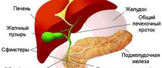
Psittacosis meningitis is one of the atypical forms of acute psittacosis and is rare. It begins acutely with an increase in body temperature and the appearance of symptoms of intoxication. Over the next 2-4 days, meningeal symptoms appear (severe headache, stiff neck, positive Kernig, Brudzinsky, etc. symptoms). There are no changes in the lungs. Ornithosis without lung damage begins acutely with an increase in body temperature (usually above 39 °C) and the appearance of signs of general intoxication. Patients complain of headache, loss of appetite, stool retention, and sometimes aching pain throughout the body. By the end of the first week, an enlargement of the liver and spleen is determined.
In addition, acute psittacosis can occur without any clinical manifestations in an inapparent (lat. inapparens) - asymptomatic - form. This form of the disease is more often observed in young people with good body reactivity. In this case, the infection does not develop, but immunity is formed in the same way as during the normal course of the disease.
Chronic psittacosis
Found in the following forms:
- Chronic ornithosis pneumonia develops if there is no timely adequate therapy in patients with an acute form of ornithosis. The course of the disease is sluggish and long-term, remission periodically alternates with exacerbations. Chronic psittacosis pneumonia is characterized by asthenia, an increase in temperature to subfebrile levels, symptoms of spastic bronchitis, and chronic intoxication. The disease can last more than three years.
- Chronic ornithosis (does not affect the lungs) is manifested by prolonged low-grade fever, vegetative-vascular disorders, enlarged liver spleen, asthenia, and symptoms of chronic intoxication.
Brief description of the disease
Psittacosis is an acute infectious disease that manifests itself as damage to the central nervous system and lungs, fever, intoxication, and an increase in the size of the liver and spleen.
The causative agent of the infection, chlamydia Chlamydia psittaci, enters the human body from the external environment, where it can persist for up to 2-3 weeks and develops intracellularly. Sources of ornithosis infection in humans are wild and domestic birds: ducks, turkeys, parrots, canaries, budgies and city pigeons. Most often, treatment for psittacosis is required by people who are constantly in contact with birds, in particular, workers in meat processing plants and poultry factories. Psittacosis mainly occurs in the cold season, and isolated cases of infection are usually detected. Familial outbreaks and epidemics are extremely rare. The pathogen enters the body through inhalation of dust, which contains tiny particles of bird feces, and, consequently, chlamydia. In addition, psittacosis in humans can occur after accidental inhalation of fluff particles from sick individuals. Patients with psittacosis are not dangerous to others, and therefore they do not require urgent hospitalization in infectious diseases departments of clinics or isolation from other family members.
Diagnostics
The diagnosis is usually made based on the study of symptoms and epidemiological history (presence of close contact with birds or a group case of disease). To confirm ornithosis infection, the following tests are performed:
- sputum microscopy;
- serological tests (ELISA, RTGA, RIF and RSK);
- bioassay on chicken embryos;
- analysis of bronchial biopsy specimens;
- clinical blood test.
To identify the lesion, the following studies are carried out:
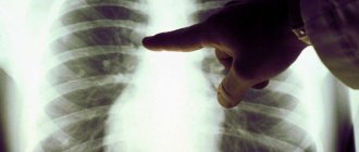
- X-ray of the lungs;
- bronchoscopy with biopsy;
- spinal puncture with collection of cerebrospinal fluid for analysis (if a meningeal form is suspected);
- intradermal allergy test.
To exclude an erroneous diagnosis, differential diagnosis is carried out with the following diseases:
- pneumonia of bacterial and viral origin;
- Q fever;
- tuberculosis;
- brucellosis;
- histoplasmosis;
- coccidioidosis;
- aspergillosis;
- nocardiosis;
- Infectious mononucleosis;
- legionellosis.
Diagnostic measures
Symptoms of psittacosis are nonspecific. They largely coincide with the signs of other pathologies. That is why diagnosing the disease causes some difficulties for infectious disease doctors. It includes collecting complaints and medical history, examining the patient and studying clinical symptoms, and conducting laboratory and instrumental examinations.
Epidemiological history is important in making a diagnosis. It is necessary to find out the most important thing - whether there was close contact with birds. Physical examination includes palpation, percussion, and auscultation of the chest. These methods allow us to identify signs of pneumonia in patients:
- Dullness of percussion sound indicates a decrease in the airiness of the lung tissue and the presence of inflammatory-congestive infiltration of the lung;
- Crepitation is a respiratory noise formed as a result of the simultaneous dissolution of a large number of alveoli and caused by the presence of sputum in the small bronchi;
- Pleural friction noise occurs when adhesions form in the pleural cavity, they stretch and rub against each other;
- Fine-bubble whistling and buzzing wheezing is a sign of excess accumulation of sputum and mucus in the respiratory tract;
- Increased vocal tremors indicate the presence of inflammation in the lungs, which lose their softness and airiness, become dense and well conductive to sound;
- Weakened vesicular breathing, hard inhalation and exhalation.
Instrumental and laboratory research methods:
Chlamydophila psittaci
Microscopy of sputum and epithelial scraping - the biomaterial is fixed and stained, and then the preparation is studied under a light microscope; only a bacteriologist is involved in deciphering the results,
- Serology - staging the complement binding reaction and hemagglutination using the method of paired sera and confirming the diagnosis with a fourfold increase in antibody titer,
- PCR - determination of a specific fragment of the chlamydia DNA molecule in the test sample,
- Immunogram: in the acute period - immunoglobulins M, during reinfection - immunoglobulins A, chronicity of the process - immunoglobulins G,
- A diagnostic allergy ornithine test in the early stages of infection is carried out by intradermal injection of the allergen,
- Histological examination of bronchial biopsy obtained during bronchoscopy,
- Infection of chicken embryos, white mice and tissue culture, followed by microscopic identification of the pathogen,
- Hemogram - increased ESR, leukopenia, lymphocytosis, aneosinophilia,
- X-ray of the lungs - a homogeneous shadow on the pulmonary field, increased pulmonary pattern, expansion of the lung root on the affected side.
How to treat ornithosis?
Patients with psittacosis, depending on the severity of the pathology, are recommended bed or semi-bed rest, as well as good nutrition with the inclusion of additional sources of vitamins in the diet. Persons with severe forms and complications are hospitalized in the hospital.
Treatment of psittacosis is antibacterial. Patients are prescribed tetracyclines - Vibramycin, Doxycycline, Tetracycline; macrolides - Azithromycin, Clarithromycin, Erythromycin; fluoroquinolones - “Ciprofloxacin”, “Ofloxacin”. The drugs are taken for 7-10 days until the temperature normalizes. Chlamydia is resistant to penicillins and sulfa drugs.
Symptomatic treatment of pneumonia consists of the use of bronchodilators, mucolytics, expectorants and antitussives, antipyretics, and oxygen therapy.
Pathogenetic treatment includes detoxification by oral or parenteral administration of saline solutions. To strengthen the body's defenses, immunomodulating and immunostimulating agents are used. Multivitamin complexes, antioxidants, herbal adaptogens and metabolic substances help the body to quickly rehabilitate after illness. Taking prebiotics or probiotics is necessary to restore intestinal microflora. In severe cases, NSAIDs, antihistamines, glucocorticosteroids, vascular and neuroprotective agents are used.
Treatment
Patients must be hospitalized and discharged only after complete elimination of signs of illness, but not earlier than 4 weeks after the onset of the disease. Patients are prescribed:
- Antibiotics. Usually these are tetracycline drugs, since Chl. psittaci is most susceptible to them. Medicines in this group include: Doxycycline, Doxal, Unidox Solutab, Monoclin, etc. In severe situations, antibiotics are administered intramuscularly or intravenously. They are taken throughout the febrile period, as well as another 5–7 days after its completion.
When pregnant women are affected by psittacosis, the use of tetracyclines is unacceptable, since they cause the development of deafness in the fetus. In such cases, taking erythromycin is indicated.
- Bronchodilators (Euphylline, Theophylline, Salbutamool). These are drugs that dilate the bronchi and thereby relieve their spasm.
- Diuretics (Furosemide, ethacrynic acid, Mannitol). The use of these medications is indicated mainly for psittacosis meningitis.
- Vitamin complexes.
Patients are also shown:
- Oxygen therapy with humidified oxygen, that is, inhalation of a concentrated oxygen-air mixture. The procedures are carried out for 45–60 minutes, 4 to 6 times a day during the entire febrile period.
- Exercise therapy. Basically, breathing exercises and exercises are prescribed for patients with lung diseases.
In severe cases and chronic forms of pathology, vaccine therapy is used. The method involves the introduction of ornithosis allergen diluted with saline intradermally in gradually increasing doses.
After discharge, the patient must be registered with a dispensary for at least 6 months, during which he must regularly visit the doctor and undergo the necessary tests. This is necessary in order to timely diagnose a relapse of the disease, if it occurs.
Drugs used to treat psittacosis - gallery
Doxycycline is a tetracycline antibiotic
Unidox Solutab is a tetracycline antibiotic
Eufillin is a popular bronchodilator
Furosemide is an available diuretic
Prevention
General preventive measures include strict veterinary supervision and the control of psittacosis among birds. As for personal prevention of this disease, this includes limiting direct contact with wild and domestic birds, with their physiological secretions, as well as the use of personal protective equipment during work.
A vaccine against psittacosis has not yet been created, but developments in this direction are actively underway, since this infection poses a serious occupational hazard for workers in poultry factories and farms. It is also worth noting the fact that immunity after an illness is short-lived, so you can get sick again.
Epidemiology
Psittacosis is a zooanthroponotic infection with natural focality and occupational morbidity.
Sources of infection are domestic and wild birds, patients or virus carriers. The respiratory mechanism of infection in psittacosis is the main one. Men and women are equally susceptible to the disease. A higher incidence among women is associated with their greater employment in poultry farms, poultry processing enterprises and at home. In groups professionally associated with poultry, psittacosis is registered in age groups from 16 to 40 years.
Forecast
In most cases, psittacosis has a favorable prognosis. Another problem with such an infectious zoonosis is the high probability of recurrence of the disease. In about ¼ of patients they occur after 14-30 days or after 4-6 months.
With timely initiation of antibiotic therapy, complications arise only in rare cases. The most dangerous of these can be conditions such as acute heart failure or pulmonary embolism.
After suffering ornithosis, the patient develops only unstable immunity, and repeated cases of infection with Chlamydophila psittaci remain possible.
Emphysema Tuberculosis Pulmonary sarcoidosis Lung cancer Pneumonia Main causes of shortness of breath
Treatment prognosis
In general, the prognosis is favorable if the patient seeks medical help in a timely manner and completes the full course of treatment. In such cases, the mortality rate from psittacosis is less than 1%. However, Chl. psittaci are able to persist for a long time in internal organs, and when favorable conditions are created for their reproduction, cause relapses of the disease.
Full recovery occurs after 1–1.5 months, and in severe cases after 2–2.5.