Hydronephrosis is a disease of congenital or acquired nature, in which the calyces and pelvis of the kidney significantly expand. Hydronephrosis of the kidney is a consequence of a violation of the outflow of urine, which leads to stagnation, poisoning of the entire body, and in advanced cases, death of the kidney.
The incidence of this disease in men and women depends on age. Women under 60 years of age are more likely to suffer, which may be due to pregnancy and gynecological diseases. After 60 years, men are at risk due to benign prostate hyperplasia.
At CELT you can get a consultation with a urologist.
- Initial consultation – 2,700
- Repeated consultation – 1,800
Make an appointment
Causes of hydronephrosis
Laparoscopic hydronephrosis repair
- Cost: 140,000 - 200,000 rubles.
- Duration: 1-3 hours
- Hospitalization: 3-4 days in hospital
More details
It is more correct to call the disease hydronephrotic transformation, since when the outflow of urine is disrupted, changes occur not only in the collecting system, but also in the parenchyma, ureter, vessels, bladder and the entire excretory system as a whole. The right and left kidneys are affected with approximately the same frequency; a bilateral process occurs only in 5-9% of cases.
The main cause of the pathology is difficulty in the outflow of urine from the kidney due to narrowing of the urinary tract. Narrowing can occur at any level, from the pelvis to the external opening of the urethra. This narrowing can be congenital or acquired.
The following congenital causes occur:
- anomaly in the location of the renal artery or its branches compressing the ureter;
- internal ureteral valves;
- underdevelopment of the ureter;
- spiral arrangement of the ureter when it covers the inferior vena cava;
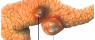
- ureterocele – saccular dilatation of the ureter;
- atresia of the urethra or complete or partial fusion;
- pouch-like protrusions of the ureter or diverticula.
Acquired causes are the result of other diseases or injuries of the urinary system:
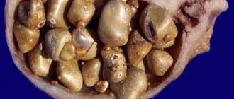
- nephrolithiasis or urolithiasis, in which a stone mechanically blocks the ureter;
- long-term chronic inflammation, in which the wall of the ureter changes and ureteral stricture develops;
- tumors of the urinary organs – benign and malignant;
- in women – chronic inflammation and tumors of the pelvic organs;
- in men – growing prostate adenoma;
- metastasis of tumors of various origins into the retroperitoneal space;
- injuries to the abdominal and pelvic organs;
- diseases of the spinal cord associated with impaired innervation of the kidneys and bladder.
That is, the cause of hydronephrosis is always the appearance of an anatomical obstruction in the path of urine movement.
Symptoms of pathology
Symptoms of hydronephrosis are initially scant. They depend on the reasons that caused this pathology. In pathology caused by stone disease, pain occurs in the lower back or lower pelvis.
The pain increases with increased urine pressure, and is sharp and does not go away after taking an analgesic.
The development of inflammation increases the number of symptoms. Frequent headaches, attacks of nausea with vomiting, and fainting appear. The patient's temperature rises, blood pressure increases, and pulse quickens.
When both kidneys are affected, intestinal bloating is observed and signs of the so-called “acute abdomen” appear. The skin becomes pale, sweating increases, and the urine takes on the smell of acetone. In the acute course of the disease, patients may experience confusion, swelling of the face and legs. Many people at this stage experience shortness of breath and breathing problems. No urine is produced.
Hydronephrotic transformation
This is essentially hydronephrosis. Regardless of which kidney is affected, the disease is severe. Due to the expansion of the pelvis, compression of the vessels occurs, which impairs the functionality of the kidneys.
More often, one of these organs is affected, equally the left and right. Transformation of both kidneys is rare.
Stages of disease development
There are several stages of the disease:
- Initial . It is observed when only the pelvis is enlarged (a stage called pyeloectasia). Hypertrophy of the pelvis tissue occurs due to the body's attempts to compensate for the increased pressure. There are no symptoms of pathology, and there are no noticeable changes inside the organ. It can only be diagnosed by chance.
- The second stage is characterized by even greater stretching of the renal pelvis and thinning of its tissues. The functions of the organ are reduced by 40%, and the healthy person increases his work, trying to do the work of the sick person. Symptoms of hydronephrosis appear, but not yet very obvious.
- The third stage, called thermal, is characterized by an atrophic process within the cortical medulla. Nephrons replace connective cells. Kidney activity decreases sharply. Sometimes the kidney stops working completely. The organ resembles a bladder, which consists of connective tissue and is filled with fluid. Inside this bubble there can be up to five liters of this liquid. Moreover, it does not represent urine.
What happens to the kidney when the outflow of urine is disrupted?
Urography
- Cost: 5,000 rub.
- Duration: 45 minutes
More details
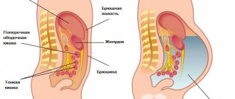
The blood is constantly filtered through the kidneys, forming urine, and this work stops only in critical conditions with an abnormal drop in blood pressure. If the produced urine has nowhere to go, the kidneys become enlarged and stretched. An increase in kidney size is one of the standard signs of hydronephrosis. All parts of this organ are stretched, but especially the elastic pelvis and calyx. In extreme cases, the pelvis can even rupture, spilling its entire contents into the abdominal cavity.
In stagnant urine, bacteria find a nutrient medium, and complications begin in the form of infection and suppuration. Salts of acids contained in urine, with prolonged stagnation of urine, can precipitate and form stones, which further worsens the situation.
In an enlarged kidney, the nephrons or structural units of the kidney tissue are affected. This leads to a decrease in the filtration function of the kidney. In urine tests, urea and creatinine levels increase, and electrolyte imbalance increases. The acid-base balance or pH of urine and blood changes. Such changes in homeostasis affect the entire body, and severe intoxication can develop.
Knowing the severe consequences of hydronephrosis, CELT doctors strive to eliminate the obstruction to the outflow of urine as early as possible in order to preserve kidney function.
Folk remedies
Treatment with folk remedies involves the use of various herbs and medicinal preparations that improve kidney function and alleviate the condition of patients with hydronephrosis. For this purpose the following is used:
- Pumpkin, namely the stalks. To prepare the medicine, the stalks are crushed, 500 ml of boiled water is poured in and infused in a water bath for 20 minutes. After this, the infusion must be removed, wrapped in a warm towel and left for about 2 hours. Take 4 times a day, half a glass per dose.
- 150 gr. birch leaves, 50 gr. nettle leaves, 50 gr. Adonis herbs, 50 gr. oat grains, 50 gr. bearberry and 50 gr. horsetail.
- In equal proportions take black currant leaves, raspberry leaves, calamus roots, kidney tea, string grass, chamomile flowers.
- Herbs for hydronephrosis are used in the form of preparations, which are recommended to be used for no longer than 3-4 months. It is necessary to change fees, after each course, waiting approximately 2 weeks. Plant infusions are taken on an empty stomach, approximately half an hour before meals.
- Chopped parsley root, 1 tbsp. l., pour 100 ml of boiling water. The product is infused all night. Carefully drain the liquid in the morning and drink 1 tbsp. l. on an empty stomach during the same day. If it is not possible to purchase plant roots, you can also use seeds. However, they give a less pronounced positive result. In the same way, you can prepare infusions for hydronephrosis from caraway seeds.
Manifestations of hydronephrosis
The disease occurs in two forms - acute and chronic. With a unilateral process, the disease is a little easier, but bilateral hydronephrosis often poses a direct threat to life.
Acute unilateral hydronephrosis is similar in course to renal colic: a rapid increase in lumbar pain, its paroxysmal nature, increased temperature, frequent urge to urinate, decreased amount of urine discharge, nausea and vomiting. The pain is very strong, radiating to the thigh, perineum, groin, genitals. There may be traces of blood in the urine that are visible to the eye. The prognosis for an acute process is sometimes better than for a chronic one, because acute pain and vivid symptoms of intoxication force one to consult a doctor immediately, which guarantees timely qualified assistance.
Chronic hydronephrosis occurs differently. If inflammation does not accompany the expansion of the pelvis for a long time, then such a process is not detected. Symptoms are nonspecific and occur in many other diseases:
- discomfort in the lumbar region;
- dull lumbar pain;
- increased discomfort after drinking large amounts of liquid and physical activity;
- constant or chronic fatigue;
- arterial hypertension;
- an admixture of blood in the urine, detectable only during laboratory examination.
A sign that may suggest that a person has hydronephrosis is the habit of sleeping on his stomach. In this position, urine drains better from the diseased kidney.
If you suspect hydronephrosis, you must consult a urologist.
Nutrition and diet
With hydronephrosis, the process of natural removal of decay products from the body is inhibited. To reduce the intoxication of the body with harmful substances, the patient must follow a special diet.
The diet for hydronephrosis is as follows:
- Limited consumption of animal protein;
- Exclusion of certain products;
- Consuming the required amount of plant foods.
For hydronephrosis, it is recommended to consume no more than 0.5 grams of protein per 1 kilogram of the sick person’s weight. So, if a patient weighs 70 kilograms, he can eat 35 grams of protein per day. You should replenish your protein intake with lean fish or meat, and low-fat dairy products. In this case, you need to choose natural (not frozen, not processed) products.
Foods that should be removed from the diet for hydronephrosis are:
- any types of meat or fish with high fat content;
- jellied meat and other types of rich broths from meat or fish;
- dishes prepared by frying, smoking, drying;
- industrial and home preservation (salted, pickled, pickled vegetables or fruits);
- sweets, cakes, pastries and other products with a lot of sugar;
- food products with a high salt content (chips, crackers, salty straws);
- any products that have an unnatural smell, color or taste.
- carbonated drinks, alcohol.
Products that are allowed for hydronephrosis are:
- meat (chicken, turkey, veal);
- offal (liver);
- fish (cod, pike perch, pike);
- dairy and fermented milk products (cottage cheese, kefir, milk, sour cream, yogurt).
It is also recommended to include at least 600 grams of fruits and vegetables in your daily diet. Plant products should be consumed mainly in raw form.
The most useful cultures for hydronephrosis are:
- watermelon;
- grape;
- apples;
- apricots;
- plums;
- pumpkin;
- beet;
- potato;
- cauliflower.
Diagnosis of hydronephrosis
In a conversation with the patient, after collecting anamnesis, the doctor determines the diagnostic search algorithm. Upon examination, almost nothing specific can be detected, except that in advanced cases an enlarged kidney can be felt.
It is mandatory to study urine and blood in the laboratory. Diagnosis includes mandatory instrumental examination - kidney ultrasound and radiography. Ultrasound reliably reveals signs of hydronephrosis; if necessary, ultrasound examination of renal vessels is prescribed.
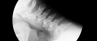
X-rays are performed with intravenous contrast. During the study, kidney function is assessed, it is seen how the contrast agent passes through the calyces and pelvis, where it accumulates, how long it takes to completely filter the blood (excretory urography). In some cases, contrast is injected from below through the ureter, which allows visualization of the level of narrowing (retrograde ureteropyelography). MRI and radioisotope studies are also used as diagnostics. Endoscopic ureteroscopy (examination using an endoscope, which is inserted through the urethra and bladder into the ureter) allows you to clarify all the details and see the narrowing with your own eyes before surgery.
How to confirm the diagnosis?
The above complaints in combination with examination data will form a picture of the disease.
Ultrasound
Ultrasound of the kidneys will allow you to assess the size of the kidney, the thickness of the parenchyma, the size of the CL itself, and the condition of the ureters. Also, using ultrasound, you can determine the condition of the second kidney, in order to form ideas about the prognosis of the disease and the possibility of compensation for renal function.
CT
The most accurate method will allow you to see the cause of hydronephrosis and the degree of pathological changes in the ureter and the kidney.
Other possible examination methods
Survey urography is carried out as a method of possible detection of stones and pronounced changes in the urinary system.
Excretory urography is a routine method, in the absence of intolerance to iodine-containing radiocontrast drugs in patients, it will reveal renal function and the level of obstruction.
Along with instrumental methods, laboratory studies are carried out:
- general blood test - if there is an increase in the level of leukocytes, an increase in ESR, changes in the leukocyte formula (increased levels of band forms, toxic granularity of neutrophils), the degree of the inflammatory process can be determined and adequate treatment can be prescribed;
- general urinalysis - the presence of leukocytes, protein, red blood cells will also indicate the activity of inflammation in the kidney;
- biochemical blood test - pay attention to the level of creatinine and urea, potassium and sodium - their significant increase is observed with bilateral hydronephrosis;
- bacteriological urine culture - prescribed simultaneously with determining the sensitivity of the cultured bacteria to antibiotics for the purpose of further targeted treatment;
- Urinalysis according to Nechiporenko will also indicate the activity of the inflammatory process.
Our doctors
Khromov Danil Vladimirovich
Urologist, Candidate of Medical Sciences, doctor of the highest category
35 years of experience
Make an appointment
Mukhin Vitaly Borisovich
Urologist, Head of the Department of Urology, Candidate of Medical Sciences
34 years of experience
Make an appointment
Perepechay Dmitry Leonidovich
Urologist, Candidate of Medical Sciences, doctor of the highest category
40 years of experience
Make an appointment
Kochetov Sergey Anatolievich
Urologist, Candidate of Medical Sciences, doctor of the highest category
34 years of experience
Make an appointment
Degrees of development of left-sided flow
There are several stages of development of the disease; at the initial stage it is difficult to recognize.
Later, a list of characteristic signs appears that help to recognize and classify hydronephrosis.
First stage
At the initial stage of development, the pathological process is difficult to recognize, since the outflow of urine is not impaired. The kidney does not function fully, the ureteral duct is narrowed, but not completely blocked. A healthy kidney compensates for the work of the affected organ.
Second phase
At stage 2 of development, the pathological process is clearly expressed, but it is not possible to notice its development without carrying out characteristic diagnostic procedures.
But you can suspect the presence of hydronephrosis if you evaluate the characteristic symptoms, compare them and carry out a number of diagnostic procedures that can help make a diagnosis.
At the second stage, the outflow of urine is completely disrupted, the kidney cannot perform its functions, and various complications arise. This is what makes the disease dangerous.
Treatment of hydronephrosis
If the course is asymptomatic and the degree of hydronephrosis is insignificant, a wait-and-see approach is chosen, and the patient is monitored over time. Conservative treatment of hydronephrosis is used in cases of inflammation in preparation for surgery.
The main treatment method for this pathology is surgery. Tumors, metastases, stones are removed, congenital malformations are eliminated.
Practice shows that the maximum number of narrowings occurs in the ureter. It is enough to eliminate the narrowing in this place, and the normal outflow of urine is restored, and the person recovers.
CELT urologists have been performing ureteroplasty using laparoscopic access for more than 15 years. This is the most gentle reconstructive surgery that leads to a complete recovery. We have accumulated vast practical experience that allows us to help a huge number of patients.
The operation is performed through punctures, without large incisions. The image is projected onto a large monitor, where all the details are visible. The narrowed, scarred ureter is dissected. A flap is cut out from the tissue of the pelvis, which is sewn into the place of narrowing, and plastic surgery is performed.
Endoscopic ureteroplasty causes minimal damage to the patient. During the operation, a small amount of tissue is dissected, therefore, there are much fewer possible postoperative complications than with open access. The risk of infection is reduced to almost zero. The rehabilitation period lasts no more than a month, most of which the patient spends at home. You only need to spend 2-4 days in the hospital.
CELT urologists try to help all patients who have a similar problem.
Prevention
It is difficult to prevent the congenital form of the disease. To do this, you need to plan your pregnancy and undergo a medical examination while carrying the baby. The expectant mother needs to avoid contact with teratogenic substances, not drink alcohol and not smoke. To prevent acquired hydronephrosis, it is necessary to:
- promptly treat diseases of the urethra, bladder and ureters;
- dress warmly in cold weather;
- Healthy food;
- drink more clean water;
- exclude injuries;
- observe the rules of intimate hygiene;
- do not engage in casual sex;
- empty your bladder in a timely manner;
- do not be stressed;
- to refuse from bad habits.
Our services
The administration of CELT JSC regularly updates the price list posted on the clinic’s website. However, in order to avoid possible misunderstandings, we ask you to clarify the cost of services by phone: +7
| Service name | Price in rubles |
| Ultrasound of the kidneys and adrenal glands | 2 700 |
| Urography intravenous | 6 000 |
| Kidney CT | 10 000 |
All services
Make an appointment through the application or by calling +7 +7 We work every day:
- Monday—Friday: 8.00—20.00
- Saturday: 8.00–18.00
- Sunday is a day off
The nearest metro and MCC stations to the clinic:
- Highway of Enthusiasts or Perovo
- Partisan
- Enthusiast Highway
Driving directions
Forecast
The outcome of the disease can give various indicators. The prognosis depends on the course of the operation and possible complications. After surgical intervention, half of the patients experience complications that lead to repeated operations, systematic hospitalization and continuous therapy. If hydronephrosis has developed only on the left or only on the right, then the prognosis is relatively favorable, since even with a severe form of the disease, one kidney can be removed, thereby saving the entire body from infection and further diseases.
In case of dual diagnosis, surgical intervention is not always effective. And although double hydronephrosis is too rare, the patient may not survive without transplantation of at least one kidney, since in most cases there are all indications for the removal of both kidneys, which is incompatible with life. If, nevertheless, both kidneys can be saved, then every second patient is diagnosed with renal failure. As with other diseases, the prognosis depends on the period and stage. The sooner the operation is completed, the greater the chances for a full, healthy life.
In 90% of cases, children under three years of age lead a normal life after surgery. Good results are determined in 80% of children aged 3 to 15 years. Adults recover completely in fifty percent of cases.
Pyelonephritis Urolithiasis Prostate adenoma - what is it? Symptoms and treatment Glomerulonephritis Hepatitis C: first signs and treatment regimen Diabetes mellitus
Possible complications
The most dangerous complication of hydronephrosis can be considered its transition to a higher stage. It is difficult to stop the ongoing process of enlargement of the pelvis area. After diagnosis, surgery is usually immediately prescribed to restore the urine output.
With significant stagnation of urine, infection often occurs, and pyelonephritis and cystitis can develop. Before surgery, it is necessary to cure inflammation.
The worst outcome of the disease can be the loss of a kidney; if the second organ is healthy, the entire load will be transferred to it.
If the second kidney is pathological, continuous hemodialysis will be required. Kidney transplantation is a rare and unlikely operation.