Diagnosisforeign body of the eye"is well known to almost everyone. It is unlikely that there is at least one person who has never in his life experienced unpleasant sensations caused by a small insect, a speck of dust, an eyelash getting into the eye, that is, what is called a “foreign body”.
Foreign bodies can be superficial, i.e. located on the surface of the eye or intraocular - penetrating into the eye cavity and damaging its membranes.
Most superficial foreign bodies are removed from the eyes as a result of intense blinking and increased tear production. If this does not happen, then it is necessary to seek specialized medical help as soon as possible.
Metal particles getting into the eye are especially dangerous. Often they are so small that they do not cause significant discomfort and the victim does not seek medical help.
However, after a few days, he begins to notice that his vision has noticeably deteriorated. This is due to the oxidation of the metal. The most dangerous in this regard are copper particles, the oxides of which have a toxic effect on the cornea, lens and retina. Therefore, it is so important to promptly contact an ophthalmologist in case of a foreign body in the eyes.
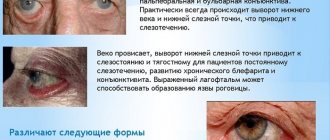
Foreign body of the conjunctiva
Depending on the force of the flight, foreign bodies that fall on the conjunctiva remain on its surface or are embedded in its tissue, causing not only discomfort in the patient, but also serious complications that, without adequate treatment, can lead to irreversible loss of vision.
Usually these are small particles:
- land,
- grains of sand
- coal particles,
- stone,
- metal,
- hairs of some caterpillars,
- hard hairs of cereal plants,
- burdock, etc.
A foreign body violates the integrity of the conjunctival epithelium. When introduced into the conjunctiva, an infiltrate appears around the foreign body. Caterpillar hairs embedded in the conjunctival tissue after a few days cause the development of granulations that resemble tuberculous lesions of the conjunctiva. Granulations consist of lymphocytes, epithelioid and giant cells.
Symptoms
A foreign body of the conjunctiva causes irritation of the eye - photophobia, blepharospasm, pain, foreign body sensation. With focal illumination or biomicroscopy, a foreign body lying on the conjunctiva or embedded in its tissue is determined. Conjunctival injection of the eye is expressed to varying degrees. Thanks to reflex blinking movements and increased tear production, a foreign body can move and most often linger on the inner surface of the eyelid in a groove running along the edge of the eyelid. It is therefore necessary to turn out the upper eyelid and carefully examine its mucous membrane, as well as the transitional fold. When hairs from cereal plants and burdocks penetrate into the conjunctiva, severe irritation of the eye occurs. When the eyelid is everted, a limited inflammatory focus is found on the conjunctiva with the development of papillae, in the center of which there is a hair. Often this hair causes desquamation of the epithelium of the corresponding area of the cornea.
Diagnostics
The diagnosis is not difficult and is made on the basis of anamnesis, the phenomenon of eye irritation and the detection of a foreign body when examining the conjunctiva, incl. using a slit lamp.
Foreign body of the cornea
A foreign body entering the eye, depending on its structure, the presence of sharp edges or teeth, as well as the speed of flight, either remains on the surface of the cornea or is embedded into its tissue to different depths. Metal particles are usually embedded deep into the corneal tissue. Foreign bodies violate the integrity of the epithelium of the cornea, thereby creating conditions for the possible development of infection (see Keratitis). After several hours of residence in the corneal tissue, a thin rim of infiltration is almost always visible around the foreign body. The vessels of the eye react to the introduction of a foreign body with a pericorneal injection.
Signs of a corneal foreign body
Complaints of photophobia, lacrimation, blepharospasm, pain in the eye, feeling of a “grain of sand in the eye.”
Conjunctival or mixed injection of the eye is observed.
The cornea contains a foreign body of various sizes and locations. It can be on the surface of the cornea or in its tissue (superficial, middle, deep layers).
A foreign body located in the deep layers can penetrate one end into the anterior chamber.
Foreign bodies can also be multiple and of varying depth.
If a superficial foreign body has not been removed for any reason, it may gradually be rejected by demarcating inflammation.
A chemically inactive foreign body located in the middle or deep layers of the cornea can encyst or give rise to purulent keratitis.
Diagnostics
The diagnosis is made based on the detection of a foreign body in the cornea, which usually looks like a small gray, yellowish or dark dot. To determine the nature and depth of a foreign body, focal illumination and biomicroscopy are used. If there are many foreign bodies in the cornea located at different depths, it is determined by radiography and gonioscopy whether there are foreign bodies that have penetrated into the anterior chamber.
Symptoms
Symptoms indicating the presence of foreign bodies in the eye range from mild discomfort to severe, high-intensity pain. First of all, there is a dependence of the degree of pain on the location of the foreign particle and the nature of the damage.
Superficial eye damage often manifests itself as:
- discomfort;
- pain in the eye area;
- lacrimation;
- redness of the protein membrane;
- burning sensations;
- scratches on the eyes;
- hypersensitivity to light;
- problems associated with opening the eyelids;
- swelling of the soft eye tissues;
- the presence of a foggy cloud in the field of view;
- bleeding of vessels located close to the surface.
When foreign bodies penetrate into the inner parts of the eyes, these symptoms appear stronger and more often.
Occasionally, when a small sharp foreign object is ingested, symptoms are almost completely absent or their number and degree of manifestation are minimal.
In a situation where the patient does not complain of various types of pain, but suspects that a foreign particle has entered the eye, he should immediately go to see a doctor. He will prescribe the appropriate diagnostic procedures, since sometimes the location of a given object is quite difficult to determine during examination without ophthalmological equipment.
First aid
Independent removal of a foreign body is possible only if it is freely located on the surface of the eyeball (without “intrusion”). This can be done by rinsing with clean water or using a scarf or tampon. After this, it is recommended to instill antibacterial drops (albucid, chloramphenicol, etc.). In all other cases (invasion into the mucous membrane or cornea, inability to establish the position, etc.), you must consult an ophthalmologist. If severe pain is present, the victim must be bandaged (on both eyes to prevent synchronous movement of the eyeballs) and taken to a specialized ophthalmological institution.
How to help a child?
Due to their age, children are not always able to react in time and protect themselves from environmental influences. Therefore, there are often cases when, having played on the playground, a child did not have time to dodge the sand flying at him or rubbed his eyes with dirty hands. The result is a speck in the eye, redness and sharp pain. The parent should not panic; they need to quickly and effectively help the baby if debris gets into the eye.
We will describe in detail how to get it:
- It is necessary to wash your hands thoroughly with soap.
- Calm the baby and prohibit rubbing his eyes.
- Take the eyelashes of the upper eyelid with your thumb and forefinger, lift them and bend them slightly.
- Using a cotton swab, carefully pick up the fragment that gets on the mucous membrane and remove it.
- Rinse the eye with saline solution and, if available, apply Levomycetin or Sodium Sulfacyl eye drops.
The child should be monitored for the next couple of days. If the redness does not go away and intensifies, discomfort is felt and the baby constantly rubs the eye, you should urgently consult an ophthalmologist.
Removal of a foreign body from the eye by a specialist
Scales, metal shavings, glass pieces, etc. can only be removed by specialists! As a rule, the procedure is performed under local drip anesthesia (Inocaine drops). In this case, the doctor uses a magnifying device - a slit lamp, and can turn the upper eyelid inside out for examination.
When removing inclusions from the conjunctiva, problems generally do not arise. When removing foreign particles from the cornea, insulin needles or special ophthalmic instruments are used. In this case, an epithelial defect is formed, which requires the use of antibacterial eye drops and keratoprotectors (Korneregel). The patient may be issued a sick leave certificate.
Penetrating injuries may require hospitalization of the patient. Read more about foreign body inside the eye
Treatment
Therapy for eye damage is aimed at eliminating the provoking factor, relieving pain and signs of inflammation, healing and fusion of corneal tissue.
For pain relief for up to 2 days, Lidocaine drops, Novocaine solution, Inocaine are used. To relieve pain and inflammation, medications from the NSAID group are used in the form of eye drops (Indomethacin, Indocollir, Broxinac, Diklo-F, Diclofenac).
In case of severe pain, it is possible to take additional painkillers (Analgin, Nimesulide, Ketoprofen, Nurofen).
When a bacterial infection occurs, antibacterial ointments and drops are used (Tobrex, Sulfacyl sodium, Levomycetin, Floxal).
For the healing of corneal tissue, the medications of choice are Korneregel and Solcoseryl.
Before using all medications, the eyes must be rinsed with an aqueous solution of Furacilin or fresh chamomile infusion.
Our clinic prices
Below we provide prices for services to remove superficial foreign bodies of the eye. In case of penetrating wounds, it is necessary to carry out full-fledged surgical operations (including vitreoretinal), the price of which is determined individually after consultation with a surgeon and a comprehensive examination.
| Name of procedure | Price (RUB, 1 eye) |
| Removal of a foreign body from the eyelids and surrounding tissues | 600 |
| Removal of a conjunctival foreign body | 1 200 |
| Removal of corneal foreign body | 1 500 |
| SEE THE FULL LIST OF CLINIC PRICES | |
Our advantages
The MGK clinic has all the necessary equipment and professional ophthalmologists to quickly solve problems that arise when a foreign body gets into the eye. Timely contact with professionals will allow you to avoid the development of serious complications and preserve your vision. Don't hesitate!
You can find out the price of a specific diagnostic or treatment procedure by calling the MGK hotline and Moscow number 8 (499) 322-36-36 or online, you must use the appropriate form on the website.
Mironova Irina Sergeevna
Why is it dangerous
If a foreign object enters the eye area, it can cause injury of both a toxic and mechanical nature. Inflammatory processes also often appear, manifested in the form of:
- conjunctivitis;
- blepharitis;
- uveitis;
- keratitis.
Sometimes hemorrhages and secondary complications are observed. All these factors often lead to retinal detachment, glaucoma, cataracts and other pathologies of the visual system.
The degree of danger from a foreign object depends on various factors. The main ones are:
- object shape;
- compound;
- the amount of substance entering the eye;
- location;
- duration of stay in the eye.
The safest case is when the foreign object enters the conjunctival sac and does not have sharp ends. Otherwise, penetration into the sclerotic membrane or corneal tissue may occur.