“The results of an ultrasound of the mammary glands revealed a formation suspicious for oncology,” “the results of mammography do not clearly exclude the presence of a malignant formation,” “a biopsy showed the presence of cancerous changes in the mammary gland and an extensive examination is required.” These are just a few of the worst words a woman can hear from her doctor.
The mammary glands consist of three main types of tissue - adipose, connective and glandular. Breast cancer (BC) is a malignant tumor that develops from glandular tissue cells. Contrary to popular belief, breast cancer affects both women and men, but it is approximately 100 times more common in women.
- How does breast cancer occur?
- Types of Breast Cancer
- Causes and risk factors
- Symptoms of breast cancer
- Self-diagnosis of breast cancer
- Diagnostics
- Stages of breast cancer
- Breast cancer treatment
- Prognosis for breast cancer
Signs of breast cancer
Uncharacteristic symptoms of a malignant tumor, the manifestation of which is fundamentally important to contact a specialist as soon as possible for examination and diagnosis:
- Rapid weight loss. Determined by comparing body weight at different times. If weight loss is stable over a period of more than 3 months with the presence of normal physical activity, without food restrictions and all kinds of diets (Attention! The mechanism of weight loss is purely individual for each organism);
- Stable and persistent rashes on the surface of the skin of the chest. Determined by characteristics. Rashes of various etiologies (allergic reaction, parasitic, infectious-bacteriological) or peeling, a rash that is distinguished by its undetectable cause, is characterized by: a rapid increase in the area of the rash;
- itching;
- certain stages of rash development (onset, formation itself, healing);
- growth or decline of their volumes;
Carcinoma in almost all cases is combined with the formation of nodular, dense formations. But sometimes there are forms combined with neoplasms in the mammary gland, which are considered cancerous until the moment of malignancy (transformation into a cancerous lump). Forms also arise that do not reveal themselves as a malignant tumor over a long period.
There are also mastitis, nodular, diffuse mastopathy, fibrotumors, characterized by focal or expanding violations of the integrity of the mammary gland epithelium.
Also in medical practice, other malignant formations of the gland are observed, similarly manifested at different stages and periods:
- During breastfeeding: Galactocele or milk cyst. It occurs as a result of blockage of the milk ducts and is a complication of acute mastitis. Retained milk collects in the duct, a jelly-like (fluctuating) increase in volume appears;
- Microtrauma of the chest. This means an insect bite, scratch or other. Occurs when the baby is uncomfortable when putting the baby to the breast, or when germs get into the wound. This is accompanied by the appearance of a hematoma (bruise);
- Wen , a benign formation of the subdermal layer of the connective epithelium. This situation is characterized by a slight risk of malignancy (malignant degeneration).
Malignant breast formations
These formations have a very complex classification according to the localization and metabolism of cells - more than 10 types. In addition, they are divided into sarcomas, adenocarcinomas and carcinomas. According to biochemical parameters, they are divided into hormone-dependent, invasive and estrogen-dependent, primary and secondary tumors.
Malignant breast tumors often also develop from the ducts. The body cannot control the uncontrolled growth and division of cells, and the cells, growing, begin to penetrate into neighboring areas. Breast cancer is a disease of older women. If under the age of 30 breast cancer occurs only in every 400 women, then after 50 years it occurs in every 38.
Forms of malignant tumors:
- Nodular - a compaction in the form of a painless node that does not have clear boundaries.
- Erysipelas is a very aggressive breast tumor that quickly metastasizes.
- Edema - there is no specific compaction, but the skin thickens diffusely, it is hyperemic, and has dense edges.
- Mastitis-like - symptoms resemble mastitis; differential diagnosis is required.
- Diffuse - has the form of a diffuse infiltrate that affects glandular tissue in different organs. Also an aggressive form of cancer.
- Hidden - with it, the reaction occurs from the lymph nodes, which hypertrophy, metastases appear in them, and signs in the gland itself are delayed.
Clinical forms of cancer:
- Nodular cancer is the most common. It is usually localized in the upper outer quadrants of the mammary gland. Quickly grows into the underlying tissues. Its risk of occurrence increases with age. The nodes in this cancer are dense and do not have clear boundaries. With rapid growth, the manifestation of this type of cancer is characterized by the change of nodes to tubercles, which grow on the surface of the skin and open in the form of bleeding ulcers. Over time, the tumor disintegrates and a foul odor appears. Necrosis may extend to the bones. Patients suffer from pain. Secondary infection and death easily occur.
- Diffuse cancer is less common, but has an unfavorable prognosis. The gland turns out to be completely riddled with this tumor, it increases in size, turns red, swells and hurts. It has no clear boundaries. Diffuse cancer has several types. The edematous form is characterized by skin in the form of a lemon peel. The mastitis form leads to necrosis. Armored - the gland is reduced in size, the nipple is retracted, the breast is severely deformed. The gland tissue and subcutaneous fat are completely damaged. There is a scattering of pinkish nodular infiltrates over the entire surface of the skin of the chest. This form of cancer metastasizes quickly.
- Nipple cancer, or Paget's disease, is the third form of breast tumors. This is intraductal carcinoma of the breast. It can also occur in men. Externally, nipple cancer is similar to eczema in the area of the nipple and areola. Its first signs are in the form of scales on or around the nipple. The nipple gradually begins to retract, and the dense infiltrate around it increases and grows into the underlying tissue. The skin in this part of the gland becomes inflamed and the color becomes crimson. Weeping ulcers form and periodically become crusty. The tumor grows slowly, metastases in the lymph nodes can occur in the absence of treatment. Treatment is only a mastectomy followed by radiation and chemotherapy, as well as hormonal treatment. The patient should be constantly monitored by a mammologist, because the tumor is prone to frequent recurrence.
- Breast sarcoma is also a malignant neoplasm of the gland. It develops from connective tissue, this differs from breast cancer, which more often occurs when the epithelium grows. Sarcoma is an aggressive tumor with rapid growth, germination and metastasis. On palpation it is lumpy, dense, the skin over it is thinned, hyperemic, the venous network on the chest is strengthened. Mastectomy for sarcoma is only extended - with complete removal of all axillary and subclavian lymph nodes. Next, radiation and chemotherapy are prescribed.
Causes of breast cancer
A number of root causes are identified that in one way or another help the formation of breast cancer. However, almost all of these factors have one common root - an increase in estrogen activity, or a hereditary predisposition.
The main causes of the disease include:
- Gender;
- Genetic feature (presence of episodes of pathology in close relatives on the female side);
- The onset of the first critical days before the age of 12 , or the onset of menopause after the age of 55, the presence of menstrual flow for more than 40 years (this fact indicates excessive activity of female hormones);
- Absence of conceiving and bearing a child throughout life or its occurrence after 35 years
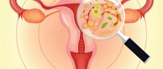
- Pathological formations in the female genitourinary area: uterine cavity, appendages, salivary glands);
- All kinds of deviations in heredity;
- The influence of radio waves: radiation treatment of diseases, close proximity to radioactive areas, frequent fluorographic examinations, occupational hazards and others;
- Other ailments in the chest: benign formations, matopathy, manifested in nodular tumors;
- The influence of carcinogens (compounds of chemical origin that promote the growth of negative tumors), certain pathogenic viruses (these factors are not fully studied);
- The patient is taller than average;
- Physical inactivity;
- Bad habits: drinking alcohol and smoking;
- Taking hormone-modulating contraceptives for a long period and in large dosages;
- Gaining excess weight after menopause.
A variety of causes contribute to the occurrence and growth of breast carcinoma in different forms. For example, if a patient is above average height and has an impressive figure, then this fact is not a reason to think that she will definitely get cancer. Danger in general involves the confluence of many factors.
Often carcinoma formations are different in their structure. They are formed from different types of cells, which multiply and grow at different rates, and respond in a unique way to methods of getting rid of them.
Therefore, it is very difficult to assume a picture of the development of the disease process. In some cases, all the signs clearly appear, and in some cases, when the tumor increases gradually, the symptoms are very vague.
Interpretation of mammography results
| Assessing what you see: is there a neoplasm? | Conclusions and recommendations |
| Negative | Everything is normal, next mammogram as planned (for women over 40 years old) |
| The neoplasm is benign | Continue mammography as planned |
| The neoplasm is more likely benign | Carry out a repeat mammogram in six months |
| The tumor is suspicious | A biopsy may be needed |
| The likelihood that the tumor is cancerous is high | Biopsy is required |
| Everything points to the presence of a cancerous tumor | A biopsy will confirm the doctor’s assumptions |
Thus, it is based on the results of ultrasound and mammography that a conclusion is made about the need for an additional examination - a biopsy.
Diagnosis of the disease
There are different diagnostic methods for diagnosing carcinoma.
These methods include physical examinations, which are divided into:
- screening
- additional examination.
Once the symptoms of cancer are identified, they are separated. The final stage of diagnosis is consultation with a specialist.
As an example, some standard examination techniques that are used in medical practice to diagnose carcinoma:
- Initial registration of the patient , taking tests and studying the patient’s medical card, the usual methods of examining the breast (surface examination, palpation, percussion, auscultation, measuring body temperature);
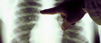
- X-ray examination (mammography and chest radiography);
- Ultrasound of the chest cavity in various variations with auxiliary examination (liver);
- Scincigraphy is a method of radioisotope visual diagnosis of cancerous branches in bone tissue; it is also known to use this method for other medical purposes.
Other methods of diagnosis are based on symptoms and depend on the technical equipment of the medical institution. Laboratory studies of biofluids (blood test without changes, stabilized test, blood plasma, etc.) for constituent, biochemical, and other indicators are additional in nature, for the most part, for the purpose of accurately determining the patient’s condition.
Chemotherapy
Comprehensive treatment for breast cancer includes chemotherapy. For this purpose, cytostatics are used - drugs that have an inhibitory effect on cancer cells. The preferred route of drug administration is intravenous infusion and oral administration. There are two types of chemotherapy for breast cancer:
- Adjuvant (neoadjuvant). Refers to a preventive treatment method. Adjuvant chemotherapy is given after surgery to target remaining tumor sites. Neoadjuvant chemotherapy is prescribed before surgery to determine the sensitivity of the tumor to therapy.
- Therapeutic. The goal of this type of chemotherapy is to reduce the size of the cancer lesion in the mammary gland. Therefore, it is carried out before surgery. The positive effect of treatment allows you to reduce the volume of surgical intervention.
The duration of chemotherapy for breast cancer depends on the individual reaction of the body, the stage of the tumor and its structure. The duration is determined by the attending physician and can be several months.
A significant disadvantage of chemotherapy is the large number of side effects. Common symptoms include:
- Hair loss
- Feeling nauseous
- Vomit
- Lack of appetite, resulting in weight loss
- Menstrual irregularities
- Drowsiness, weakness, sudden loss of strength
- Headache
- Unstable chair
- Decreased immunity
- Increased body temperature
The drugs used in chemotherapy are:
- alkylating agents (Cyclophosphamide, Ifosfamide, Melphalan, Sarcolysin, Thiophosphamide),
- anthracyclines (Doxorubicin, Farmorubicin, Mitoxantrone),
- antimetabolites (5-Fluorouracil, Ftorafur, Gemcitabine, Xeloda, Methotrexate),
- vinca alkaloids (Vincristine, Vinblastine, Navelbine),
- taxanes (Taxol, Taxotere, Intaxel),
- platinum drugs (Cisplatin, Oxaliplatin)
Self-examination
A breast self-examination for cancer does not last long, only half an hour. It should be done at least twice a month.
In some cases, the tumor may not be felt, and taking this into account, it is recommended to keep notes, noting your own sensations and all indicators based on the results of each independent examination.
It is best to examine the mammary glands on days 5-7 of the menstrual cycle, preferably on the same days.
Inspection
Visual examination should be carried out in a well-lit room, with a mirror available. You need to undress to the waist, and standing right in front of the mirror, calm your breathing rhythms.
Then you need to track the following parameters:
- How visible is the symmetry in the positions of the right and left breasts;
- The degree of enlargement of one breast in relation to the other, whether one mammary gland has increased in size compared to the other (it should be borne in mind that normal volumes may vary slightly)
- Skin surface (the occurrence of any changes, localizations;
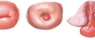
- Appearance of nipples;
- Other changes.
Probing
Palpation of the chest is carried out in a comfortable position (standing, sitting, or lying down).
Probing from a sitting or standing position can be considered effective.
You should feel each breast with your fingertips.
At the same time, there is no need to apply any pressure; it is best to feel the slightest changes in the content of the mammary glands.
Each mammary gland is palpated in turn. Starting from the nipple, gradually moving your fingers to the periphery. For comfort, you can palpate by looking at your reflection in the mirror, conditionally dividing the mammary gland into 4 components.
Points to pay attention to
Have there been any tightness in the chest:
- the presence of seals, nodes inside the gland;
- the presence of transformations, formations in the nipple.
If there are changes, you should definitely visit the following specialists:
- mammologist;
- gynecologist;
- oncologist;
- therapist (visual examination and referral to the right doctor).
With the help of self-examination, you can determine not only breast cancer, but also benign tumors and mastopathy. Situations where dubious formations are present do not yet indicate cancer. Only a specialist can say more definitely after conducting tests.
Is a biopsy dangerous? Does it hurt? What are the consequences?
With the skillful actions of a doctor, a breast biopsy is absolutely safe for a woman, who may feel slight discomfort during the procedure. It does not require special preparations, except that on the eve of diagnosis you should avoid taking blood thinning medications - by the way, you must tell your doctor about them.
The consequences of the puncture are also minimal: swelling and bruising may appear in the neck and chest area, which quickly disappear. To eliminate their appearance or minimize their visibility, you can put ice in your bra immediately after the biopsy. It is also easy to relieve pain if it appears after a biopsy: just take a painkiller. But the advantages of a biopsy are obvious: the doctor will be able to accurately determine the nature of the existing tumor and prescribe effective treatment.
Thus, the development of any disease, including such a terrible disease as breast cancer, can not only be cured, but also prevented. The main thing is to pay attention to warning signs in time, undergo all the necessary examinations and begin full treatment in the early stages of development. And do not forget that a person’s health largely depends on himself.
<
p style=”text-align: justify;”>
CLICK TO MAKE AN APPOINTMENT, TEST OR ULTRASOUND
If you find an error, please select a piece of text and press Ctrl+Enter
Medical examination
Diagnosis of malignant breast tumors begins with an examination by an oncologist or mammologist.
At the time of visual examination, the doctor:
- Will try to obtain complete information about the manifestations of the disease, possible root causes of its occurrence;
- Perform a visual examination and palpation (palpation) of the breast in two circumstances: lying down and standing with your arms along the body and with your arms raised.
Ultrasound
Now ultrasound has become an auxiliary method for detecting the disease, although it has a number of advantages, unlike radiography. For example, it makes it possible to study images from different angles, without harmful radiation.
The main reasons for using ultrasound diagnostics for tumors:
- Detection of growth after the tumor has been diagnosed using x-rays;
- The need to classify the nature of the fluid-filled cyst;
- Detection of breast diseases in young representatives of the fairer sex;
- General observation at the time of biopsy;
- Preventive need during pregnancy and lactation.
Mammography
Mammograms are in most cases carried out for screening, but they are advisable to use if cancer is suspected.
Therefore, they are often referred to as detection mammograms.
The examination clarifies the situation in the presence or absence of pathology, which is very acceptable for routine checks when no deviations are detected.
For other situations, a biopsy (removal of a piece of tissue for detailed examination under a microscope) may be necessary.
A biopsy may be needed in circumstances where mammography is negative but a breast mass is present. Such a detailed study is not carried out if an ultrasound reveals the existence of a cyst.
MRI
MRI mammography is the study of the chest cavity using magnetic resonance imaging.
| pros | Minuses |
|
|
Before analysis, all metal items should be removed. Do not hold any electronic device to avoid interference.
When a woman has any metal implants (pacemaker, prosthetic joints, etc.), the specialist should be informed - this fact is considered a contraindication to the examination.
The woman is placed in the apparatus in a lying position. The patient must remain motionless during the examination. The length of stay in the device is determined by the doctor.
Based on the results of the MRI examination, images are studied that show all the negative transformations in the chest.
PCR diagnostics using tumor markers
Tumor markers are individual substances that are present in the bloodstream during malignant formations. Any tumor is characterized by tumor markers identical to it.
CA 15-3 is a protein located on the passages of the mammary glands and secreting fragments. Its presence in the bloodstream is increased in 10% of patients with the initial stages of the disease and in 70% of patients with formations associated with metastases.
For analysis, material is taken from the intracubital vein.
Smoking is prohibited before blood collection. Reasons for implementing the analysis:
- detection of recurrence of compactions;
- monitoring the effectiveness of treatment;
- need for tumor classification;
- Detection of the size of the tumor: the greater the presence of tumor markers, the larger the lesion.
Targeted therapy
A breakthrough in modern breast cancer therapy lies in the development of a method that allows targeted action on the tumor. For this purpose, targeted drugs are used. Unlike chemotherapy, this method does not affect healthy cells. This allows you to minimize the occurrence of side effects. Targeted drugs are used as independent therapy, as well as in combination with other methods of treating breast cancer. The following targeted drugs are used for the treatment of breast malignancy:
- Estrogen receptor blockers. This group of drugs includes tamoxifen, toremifene, fulvestrant. They are used to reduce the level of estrogen receptors, thereby causing the death of cancer cells.
- Aromatase inhibitors. Used to reduce the level of the aromatase enzyme, which is actively involved in the production of estrogen. This action causes a decrease in its concentration, provoking the death of the tumor cell. The scope of use of drugs in this group is limited. They are prescribed to women during menopause, because it is not possible to suppress aromatase with medications when the ovaries are functioning. Aromatase inhibitors include exemestane, letrozole, and anastrozole.
- Inhibitors (blockers) of PARP protein. They belong to one of the most promising groups of targeted drugs. The point of application of PARP protein blockers is damaged DNA. This group of medications includes olaparib, iniparib, and veliparib.
- Selective growth factor blockers. Targeted drugs in this group include bevacizumab, panitumumab, cetuximab, trastuzumab. The action of the drugs is based on inhibiting the development of the vascular network around the tumor focus. This helps slow the growth of breast cancer.
Targeted therapy for breast cancer is prescribed for local action on the tumor site. However, some side effects may develop, including chills and increased body temperature. Symptoms are relieved by taking nonspecific anti-inflammatory drugs. The prescription of targeted therapy drugs is carried out exclusively by an experienced doctor after previous diagnosis.
Myths about breast cancer
Every year, more than a million episodes of breast cancer are diagnosed in women worldwide. This fact often becomes a reason for panic, a feeling of fear for one’s own life and a whole series of myths that do not correspond to reality.
Some of these misconceptions, in turn, become the basis for a categorical refusal to undergo an examination and conduct a completely safe diagnosis:
- Myth – None of my female relatives have had breast cancer, so I have nothing to fear. Of course, if there is no family history of breast cancer, the threat of its occurrence in relatives is minimal. However, today a large number of episodes of oncology are recorded when no one in the patient’s family had previously had cancer;
- Myth – My body is too young for cancer. Even a very young girl is at risk of developing carcinoma; in modern ecology, cancer can affect a woman of any age group;
- Myth – Cancer prevention. Unfortunately, the root causes of the formation of the neoplasm are not yet fully understood, and research is still being carried out. It should be mentioned that the panacea for hormone-dependent oncology is the preventive use of anti-estrogen medications. Prevention of other forms of tumors still involves timely detection of the onset of the pathological process;
- Myth – There are no prospects for mammography, because breast cancer is somehow fatal. Women in the so-called “risk group” should strictly carry out this analysis annually, because during this time no significant changes occur, and this fact contributes to the timely detection of pathology;
- Myth: A large dose of radiation during mammography, especially every year, is very harmful and will inevitably lead to cancer. The dose of radiation during this procedure is negligible and cannot have a serious negative impact on health, but it can save lives if the tumor is detected in time at an early stage, when treatment provides a high chance of recovery;
- Myth – I’m not bothered by pain, why should I get examined, I’m healthy . The initial stages of carcinoma occur without any manifestations. If the patient has benign tumors in the breast - cysts, fibrotumors, nodular type of mastopathy - they require constant monitoring by a mammologist. Strict consultation with a doctor is necessary for the following manifestations: Dark red, bloody, yellow discharge from the nipples, the slightest density - small, large, painful or painless, of any shape, if chest pain occurs not seven days before the critical days, but after 2 weeks before the onset of menstruation, intense pain in one or both mammary glands.
The main rules of palpation: how to identify a tumor
You need to palpate the mammary gland gently, without aggressive pressure, with the pads of three fingers pressed together. This is done while standing in front of a mirror, or lying on your back, but in any case - with the hand opposite the chest being examined. Many women prefer to palpate in the shower, as their fingers glide well over wet, soapy skin.
- If the “lying down” position is chosen, a cushion or pillow should be placed under the shoulder blade on the side of the breast being examined - an elevated chest will provide better palpation of the mammary gland;
- The free hand should be placed behind the head or extended along the body - it is recommended to use both positions alternately;
- When palpating, you can use circular (fingers move in a circle from the outside to the nipple and vice versa), longitudinal (top to bottom and bottom to top) movements of the fingers, but the same for each examination;
- You should feel your breasts slowly, taking each section of the gland and armpit in turn, so as not to miss any lumps that appear;
- It is necessary to examine the nipple: with movements reminiscent of the process of expressing milk, you need to make sure that there is no discharge or pain.
When doing self-diagnosis, you must understand that breast cancer develops gradually in women , so the first signs may not appear immediately.
Stages of breast cancer
The formation of breast cancer occurs in 4 stages:
- Zero. Carcinoma of the mammary ducts (the neoplasm forms inside the mammary ducts, without affecting nearby organs), invasive lobular carcinoma (structured by cells developing lobules).
- First. The volume of the negative cavity is less than 2 cm. The lymph nodes are not affected.
- Second. The volume of the negative cavity is up to 5 cm, embedded in the fat layer, can involve the lymph nodes, or remain within the gland. The possibility of complete cure at this stage is 75-90%.
- Third. The volume of the malignant cavity is more than 5 cm, manifests itself on the surface of the skin of the chest, lymph nodes, and chest.
- Fourth. The cancer expands and crosses the borders of the chest, growing in the bone tissue, liver cavity, lungs and brain. At this stage, cancer is unlikely to be cured.
Breast cancer treatment
Treatment options for breast cancer:
- surgical;
- chemotherapy;
- hormone therapy;
- immunostimulation;
- radiation treatment.
Treatment is usually carried out in association with additional methods.
Surgery
Surgery is often the main way to get rid of breast cancer. Modern surgeons strive to remove small amounts of breast tissue using auxiliary methods: drug treatment and laser therapy.
Types of surgical procedures to get rid of a tumor:
- Complete mastectomy. Complete removal of the breast, along with the fat layer and nearby lymph nodes. This method of surgery is the most radical ;
- Complete resection. Removal of a section of the breast along with the subcutaneous fat layer and lymph nodes. Modern surgeons mainly use this option because, unlike a complete mastectomy, resection increases the chances of survival. With this option, auxiliary therapy is used: chemo-radiation treatment;
- Quadrantectomy – removal of the tumor itself and adjacent tissues in a location of 2–3 cm, as well as lymph nodes located in the immediate vicinity. This operation can be performed only at the initial stage of the disease. The cavity that needs to be removed is necessarily sent for a biopsy;
- Lumpectomy is a minimalist operation in which only the tumor and lymph nodes need to be removed. The conditions for performing this surgery are similar to those for quadrantectomy.
The extent of surgical removal is determined by the doctor himself, influenced by the type, area of damage, location and volume of carcinoma.
Breast examination - mammography
The basic principle of mammography is an examination using X-rays, and therefore it is considered less preferable than ultrasound. The disadvantages of mammography include the fact that it produces a static image and the inability to see the condition of the armpits. However, it is mammography that makes it possible to diagnose intraductal formations, and therefore is prescribed as a preventive procedure once every 1-2 years to all women over 40 years of age. Modern X-ray equipment, designed specifically for mammography, is guaranteed to reduce the level of radiation to a minimum and, unlike ultrasound, makes it possible to study all the details of a tumor if it occurs.
Both ultrasound and mammography do not require special preparation, are painless and allow the specialist to get a clear picture of the condition of the patient’s mammary glands. The examination results are recorded in a special protocol and become the basis for further actions by the gynecologist and oncologist.
About consultation with gynecologist, oncologist / Consultation with gynecologist, oncologist
Our clinic sees gynecologists - oncologists of the highest and first certification categories. All doctors have certificates confirming their qualifications, issued in St. Petersburg and Moscow. The cost of an initial appointment with a gynecologist or oncologist is 1000 rubles, a consultation based on test results or ultrasound is 500 rubles.
Gynecologist appointment
Oncologist appointment
You can make an appointment with a gynecologist or oncologist without an insurance policy, registration in St. Petersburg or Russian citizenship. We have doctors who speak English.
You can apply to us without having an insurance policy, registration in St. Petersburg and Russian citizenship. ATTENTION! IN THE CLINIC IS A DOCTOR SPEAKING IN ENGLISH LANGUAGE!
Hormone therapy
The main focus of hormonal treatment is to block the effect of female sex hormones (estrogens) on the tumor.
Such methods are used only in situations with seals that have a certain reaction to hormones. Methods include:
- surgery to remove appendages;
- drug blockade;
- taking antiestrogenic medications;
- use of androgens (male hormones);
- taking drugs that inhibit aromatase enzymes;
- use of progestins.
Chemotherapy
Chemotherapy (chemistry) is a drug treatment for breast cancer that uses cytostatics. These drugs eliminate cancer cells and inhibit their reproduction.
Cytostatics are medications that have many side effects.
Therefore, in each individual case they are prescribed strictly in accordance with generally accepted regulations and taking into account the characteristics of the disease. Cytostatics most often used for malignant breast tumors:
- Adriblastin;
- Methotrexate;
- 5-fluorouracil;
- Paclitaxel;
- Cyclophosphamide;
- Docetaxel;
- Xeloda.
Associations of medications often used in the treatment of breast cancer:
- CMF (Cyclophosphamide, Fluorouracil, Methotrexate);
- CAF (Cyclophosphamide, Fluorouracil, Adriablastin);
- FAC (Fluorouracil, Cyclophosphamide, Adriablastin).
Radiation therapy
Effective short irradiation sessions are carried out.
Directions for presurgical radiation therapy for breast tumors:
- Complete elimination of malignant tissue along the tumor borders to prevent relapses.
- Transformation of formation from an inoperable form to an operable one.
Postoperative
The main focus of radiation therapy in the postoperative period is to prevent relapses.
Areas exposed to irradiation postoperatively :
- Actually the tumor itself;
- lymph nodes that could not be removed at the time of surgery;
- lymph nodes close in location, for prevention.
At the time of operation
Radiation treatment can be done right at the time of surgery, when the surgeon strives to preserve breast tissue.
This is acceptable at the following stages of the disease:
- T1-2;
- N0-1;
- M0.
Independent
Indications for the use of gamma therapy without surgery:
- the impossibility of getting rid of the tumor surgically;
- contraindications to surgery;
- patient's refusal to undergo surgery.
Interstitial
The radiation source is as close as possible to the localization of the pathology. Intracavitary radiation treatment is used simultaneously with external beam treatment (the source is located at a distance), often for nodular types of cancer.
Direction of therapy : to bring the maximum increased dose of radiation closer to the oncological formation, with the goal of its complete elimination.
Targeted therapy
Targeted therapy is essentially the activity of monoclonal antibodies that adhere to certain receptors on the membrane of the cancer cell.
These proteins are prototypes of real human antibodies that reproduce B lymphocytes. But B lymphocytes do not produce antibodies against molecular receptors located on the membrane of cancer cells.
For example, targeted treatments can disrupt the function of negative proteins (like HER2) that help carcinoma cells grow.
In cases where laboratory tests have detected the presence of a significant excess of HER2 protein in the breast tumor, the patient is prescribed trastuzumab (Herceptin®) or lapatinib (Tykerb®).
Monoclonal antibody (MAB) drugs have become the ultra-nanotechnological drugs of our time.
Targeted treatment can be used both in radical therapy in association with other methods of combating breast cancer (adjuvant regimen), and for the treatment of carcinoma with branches (therapeutic regimen).
Metastases
In breast cancer, the appearance of metastatic lesions occurs in the early stages. The spread of metastases occurs through lymphogenous and hematogenous routes. A feature of breast malignancy is the long-term persistence of metastases in a latent state. In accordance with the route of spread, the following possible localization of metastases is distinguished:
- Regional lymph nodes. The spread of the tumor occurs through the lymphogenous route. In this case, the thoracic, axillary, periosternal, sub- and supraclavicular lymph nodes are affected.
- Brain. Tumor cells reach the organ through the blood. Metastatic brain damage is characterized by headache, weakness in the limbs, and blurred vision. The development of mental disorders and seizures is possible.
- Lungs. Cancer cells reach the lungs by hematogenous route. Metastases cause cough with or without sputum. The progression of the oncological process inhibits the functioning of the lung tissue, which is accompanied by shortness of breath with minor physical exertion, and subsequently at rest.
- Liver. Metastases enter the organ through the bloodstream. Such a lesion is characterized by the appearance of abdominal pain of varying severity, heaviness and bloating. As the tumor develops, jaundice develops, a sharp weight loss occurs, and fluid accumulation in the abdominal cavity is possible.
- Bones and vertebrae. The hematogenous route of metastasis affects the bone structure. Bone metastases are characterized by pain that intensifies after a few weeks. The pain syndrome is relieved by taking potent analgesics. When the metastatic lesion is localized in the vertebrae, back pain and numbness of the limbs appear. Due to damage to the nerve roots, urinary and fecal incontinence may develop.
Forecast
Still, the prognosis for recovery with such a serious illness is a little more optimistic than with ordinary cancer, which is not resistant to hormonal treatment.
Some circumstances are important for a positive prognosis:
- diagnosing the disease at the initial stage;
- provision of biological material for the presence of ER, PR, HER2 receptors;
- dependence on the time of surgery;
- the woman’s age and general body strength;
- dependence on the effectiveness of prescribed medications.
In order to achieve a positive prognosis, the treatment of carcinoma must be given the same pace as the development of the disease. Patients whose pathology was identified at the initial stage have a chance of full recovery.
In today's realities, many of the world's minds in medicine are busy with the problem of curing cancer, or rather with a close study of the root cause and nature of the appearance of malignant cells.
Most scientists have not yet determined exactly what the main reason is for the situation when a completely healthy mammary cell begins to acquire pathological properties, forming oncology, which is capable of deceiving the body by synthesizing pseudohormones.
If we celebrate some victories on this topic, we can talk about the creation of experimental medications that can inhibit this process.
Nutrition for breast cancer
Nutrition for breast cancer should consist of healthy foods rich in vitamins. Non-dietary meats, animal fats, fried, salted and smoked products, and canned food should be excluded. Sea fish and poultry meat should be included in the diet. The patient should be provided with a sufficient amount of vegetables and fruits. The diet includes whole grains and legumes. To maintain skeletal health, it is recommended to take 2 grams of calcium every day. You should not take dietary supplements containing phytoestrogens. Women with hormone-dependent forms of cancer are not recommended to include soy and soy products in their menu. You should limit sugar and give up alcohol.
Disease prevention
It is no secret that excess levels of female sex hormones in the blood cause breast cancer.
The level of estrogen decreases during pregnancy and breastfeeding.
Independent and unregulated use of hormonal contraceptives also leads to disruption of the normal levels of hormones in the blood and causes tumors.
During the pre-menopausal period and during the onset of menopause, the presence of hormones in the blood should be monitored in order to recognize and prevent the formation of the disease.
Bearing, childbirth and breastfeeding are the best prevention of breast tumors and help defeat cancer.
Causes of tumors in women
The exact reasons are still unknown today, but a number of provoking factors have been identified that can serve as a trigger:
- genetic predisposition - with it the risk of cancer doubles in descendants;
- old age - from 55 to 65 years;
- early menarche;
- late menopause - after 55 years;
- prolonged menopausal syndrome;
- absence of pregnancy and childbirth before age 30;
- the woman did not breastfeed;
- frequent abortions;
- lack of sex life;
- late childbirth and pregnancy - after 35 years;
- inflammatory diseases of the ovaries;
- infertility;
- any tumors and cysts of the ovaries;
- endocrinopathies - diabetes, disorders of the thyroid gland, adrenal glands, pituitary tumors;
- long-term use OK;
- any trauma to the mammary glands;
- radiation;
- obesity;
- physical inactivity;
- stress;
- tight underwear;
- hypovitaminosis A, E, D, C;
- smoking and alcohol - drinking even small portions of alcohol, but regularly, increases the risk of breast cancer, CSA by 50%;
- insolation, topless sunbathing;
- bad ecology;
- hepatitis;
- chronic inflammatory diseases of the genital area.