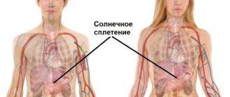
The carotid artery is needed to nourish the tissues of the head, neck, and brain. There are 3 pairs of vessels - 2 common, 2 external, 2 internal. You can find the artery in the neck under the lower jaw on both sides of the thyroid cartilage. The internal artery is important for supplying the main part of the hemispheres, the pituitary gland, the hypothalamus, and the eyes.
With narrowing (stenosis), blockage due to atherosclerosis, either temporary disturbances of cerebral circulation (ischemic attacks) or a stroke appear. They are characterized by loss of vision, sensation and strength in the limbs on one side of the body. To determine the condition of the carotid arteries, ultrasound with Doppler sonography and angiography are used.
Treatment (depending on the degree of stenosis) can be medicinal (Crestor, Prestarium, Aspirin), surgical - removing the plaque, restoring blood flow.
Why are the arteries called carotid?
The carotid artery is a large vessel through which the bulk of blood is delivered to all tissues and organs of the head, including the brain.
If you compress an artery, the receptors (nerve endings) perceive this action as an increase in pressure, and as a result, the number of heart contractions decreases, followed by a decrease in pressure and breathing slows down (the body works similarly during sleep).
The volume of blood flow to the brain also decreases. Together, this leads to a state of drowsiness, and with prolonged compression, loss of consciousness and death are possible. Due to the development of a sleepy state as a result of the effect on the artery, it was called carotid.
How many carotid arteries does a person have?
On the neck, on the right and left sides, there are 2 common carotid arteries.
They are distinguished by their large diameter, regular cylindrical shape and elasticity. Each artery is divided into external (blood flows through them to the tissues of the head and neck) and internal (nourishes the brain and eyes). As a result, a person has 3 pairs of carotid arteries (common, external and internal).
Right coronary artery
Veins and arteries, which are located on the right side, provide blood supply to organs such as:
- Teeth;
- Eyes;
- Nasal cavities;
- Oral cavity;
- Neck.
The branches of the carotid artery pass through the skin of the face and entwine the brain from above. If a person is embarrassed or his body temperature rises, this leads to redness of the epithelial surfaces on the face.
With the help of this aorta, blood flow is directed in the reverse order in order to assist the branches of the internal aorta and the vertebral aorta, provided they are narrowed.
Where are they located?
The carotid artery in humans is located on the sides of the neck near the thyroid gland. The left carotid artery comes from the aorta (the main artery that originates in the left ventricle), and the right one from the brachial artery (which is a branch of the aorta). In this regard, the left carotid artery is longer than the right by about 20-25 cm.
Further, near the Adam's apple (the protruding part of the thyroid cartilage), the right and left carotid arteries branch into external and internal. Above, each artery divides into numerous capillaries that supply all the tissues of the head and neck.
What are they responsible for?
The main purpose of the carotid artery is to provide the tissues of the head and neck with oxygen and nutrients.
At the same time, the functions of the internal and external carotid arteries are different, since they supply blood to different organs:
| The external carotid artery branches to the front of the head and supplies: | The internal carotid artery ascends to the base of the skull and supplies blood to: |
| Mimic and chewing muscles of the face. | Brain tissue. |
| Facial skin and epidermis under the hair. | |
| Oral cavity. Including the tongue, salivary glands and tooth roots. | Muscles of the forehead and temporal part. |
| Nasal cavity and middle ear tissues. | |
| Thyroid gland, pharynx and larynx. | Ocular tissues. |
The external and internal arteries are additionally connected to each other by numerous arteries, this ensures an uninterrupted flow of blood to the tissues if suddenly a slowdown in blood flow occurs in any part of the artery.
The vessels are also equipped with nerve endings, as a result of which the carotid arteries perform the following additional functions:
- normalization of blood pressure;
- regulation of the number of heart contractions;
- react to a lack of oxygen in the blood.
If you know how to act correctly on the carotid artery, you can lower blood pressure without taking medications. It is important that this procedure is only allowed to be performed by a specialist, since stronger or longer manipulations can lead to death.
Content:
- Anatomical location and topography
- Diseases that affect the carotid artery
- Atherosclerosis
- Carotid artery syndrome
- Congenital stenosis
- Aneurysms
- Tumors
← The main functions of the pulmonary artery and what diseases it is susceptible to
Diseases, syndromes of the coronary arteries and their treatment →
The carotid artery (lat. arteria carotis communis) is one of the most important vessels feeding the structures of the head. It ultimately gives rise to the cerebral arteries that make up the circle of Willis. It feeds brain tissue.
How to find it on your neck yourself?
To locate the carotid artery in the neck, it is necessary to visually determine the location of the Adam's apple. Further to the right or left of it, in the area next to the depression under the lower jaw, you need to place your index and middle fingers (these fingers are more sensitive). With proper manipulation, pulsating impulses of blood through the vessel will be felt.
Where is the carotid artery located in the human neck?
The pressure should be gentle, without squeezing the arteries.
It is recommended to independently identify the carotid artery on the right side using the index and middle finger of the right hand. It is important that the pulse on the carotid artery can be felt better than on the wrist, so it is better to measure the number of heart beats per minute on the carotid artery (especially in unconscious people).
Carotid artery diameter
The carotid artery in humans is located not only in the neck; its capillaries are distributed throughout all tissues of the head. The normal diameter of the common vessel in adults (from the aorta to the branch site) is 5.98 mm. With an increase in blood pressure, the diameter of the vessel can increase in size up to 6.15 mm. The diameter of the external and internal carotid arteries is smaller and is about 5 mm.
The diameter of the capillaries can reach up to 0.01 mm. With age, connective tissue grows inside the vessels, as a result, the patency of the arteries worsens, but the outer diameter does not increase or decrease in size.
Aneurysm
An aneurysm is a pathological expansion of the lumen of a vessel or protrusion of its wall, consisting of defective scar connective tissue. The cause of their formation can be atherosclerosis, malignant arterial hypertension, or trauma.
For the time being, aneurysms do not manifest themselves in any way. When blood pressure increases or under the influence of other factors, the pressure in the vessel increases, the vessel wall at the site of the aneurysm ruptures, since the elasticity of the structure is lost.
Because an aneurysm grows over a long period of time, it sometimes causes compression of surrounding tissues, like a tumor (tumor-like form).
BCA segments
The carotid artery (internal part) has a conditional division into segments. The division begins from the bifurcation (the place where the main carotid artery divides into the external and internal) and to the point where the internal carotid artery branches into small vessels and capillaries. Next, we briefly consider where these areas are located and what functions they perform in humans.
Segments of the internal carotid artery
The branching of the main carotid artery into external and internal occurs approximately near the third cervical vertebra.
From this section the internal artery is divided into the following segments:
| Segment name | Start of segment | End of segment | What organs and tissues does it nourish? |
| Cervical | Bifurcation | External opening of the temporal bone canal | The site has no branches, so it does not feed tissue. But in this segment, the carotid artery borders on the laryngeal, hypoglossal and vagus nerves. |
| Rocky | Temporal bone region | Ragged hole (located at the base of the skull) | Located in the carotid canal. Nourishes the tympanic cavity (inner ear). |
| Ragged hole | The segment is located in the torn hole | It is the shortest segment and has no branches, so it does not provide tissue nutrition. | |
| Cavernous | Ragged hole | Proximal ring of dura mater | Surrounded by the cavernous sinus. The section has a C-shaped bend, and is called the siphon of the internal carotid artery (the more detailed purpose of the segment is discussed below). Nourishes brain membrane cells, pituitary gland and nerve fibers. |
| Wedge-shaped | Cavernous sinus | Distal ring. | The segment is short in length and normally has no branches. In extremely rare cases, the presence of a branch of the ophthalmic artery (that is, it nourishes the tissues of the eye) was noted. |
| Ophthalmic | Distal ring | Origin of the posterior communicating artery | The segment nourishes the pituitary gland and the eyeball. |
| Communicative | Branch site of the communicating artery | Branches into small vessels | Is the final segment. Participates in the nutrition of the brain and has connections with the vertebral arteries. |
The internal carotid artery, branched inside the skull, nourishes not only brain tissue, but also the optic nerve, pituitary gland and hypothalamus, thereby ensuring their normal functioning.
Treatment and therapy
Treatment for carotid artery plaque depends on the degree of narrowing of the lumen that it causes.
In case of severe lesions, surgical intervention is performed, the purpose of which is to remove the plaque and restore the integrity of the vessel. If the size of the plaque is small and the walls of the artery can be aligned, an endarterectomy is performed - a part of the vessel is cut out and then sutured.
Conservative therapy is carried out for non-life-threatening disruption of the blood supply to the brain. It involves the use of medications and a diet that lower cholesterol levels, preventing possible blood thickening (aspirin), and combating bad habits.
Only a doctor can decide on the method of treatment after a thorough examination of the location and degree of narrowing of the lumen or volume of the internal carotid artery aneurysm.
Self-medication, as well as delaying seeing a doctor, in such a situation is life-threatening.
Atherosclerosis
The main methods of treating stenosis and blockage of the carotid artery are:
- Medicines (antiplatelet agents or agents to control the development of atherosclerosis).
- Surgical treatment, in particular carotid endarterectomy or stenting.
Auxiliary treatment measures include: smoking control (quitting bad habits), monitoring blood pressure and blood lipids.
The main goal of treatment is to reduce the risk of stroke and restore cerebral circulation. All patients with clinical manifestations of blockage and stenosis are recommended drugs to lower blood pressure, anticoagulants, antiplatelet drugs (aspirin, clopidogrel), as well as statins (lower cholesterol levels, reduce inflammation, stabilize the formation of cholesterol plaques).
Siphon of the internal carotid artery
The carotid artery in humans, which is located in the cavernous area (siphon of the internal carotid artery) and is responsible for feeding the brain and nerve fibers, is most often subject to the development of pathologies. These include: pronounced tortuosity, narrowing or, conversely, expansion of the diameter of the vessel.
These deviations manifest themselves as headaches, deterioration of memory or speech, as well as weakness of the limbs and sometimes loss of consciousness. Also in this area, vessel ruptures most often occur due to excessive stretching or thinning of the vessel, which is dangerous due to the development of hemorrhage with a fatal outcome.
Left coronary artery
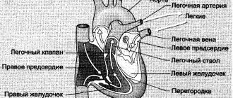
The left branch of the carotid artery enters the brain through the temporal bone, which is characterized by the presence of a special opening. This is an intracranial location. The vein diagram is quite complex. The vertebral vessels and cerebral aortas form the circle of Willis through anastomosis. The arteries supply blood with oxygen, which provides adequate nutrition to the brain. From it there is a branch of arteries into the convolutions, as well as gray and white matter. The aortas also extend into the cortical centers and nuclei of the medulla oblongata.
What happens if you press on the carotid artery?
With gentle pressure on the carotid artery, you can achieve a decrease in pressure and slow down the pulse. If the pressure is stronger and blocks the flow of blood for a short period of time (no more than 30 seconds), then loss of consciousness occurs.
After about 5 minutes, blood flow in the carotid artery is restored and the person returns to normal. With longer exposure to the vessel, disruption of brain tissue activity may develop (due to lack of oxygen). If both carotid arteries are compressed, cell death occurs and death occurs as a result.
It is important that, due to the likelihood of squeezing the carotid artery, it is not recommended to tie ties tightly and wear clothes with tight collars.
A little bit of anatomy
The carotid artery in the neck, which in dangerous situations determines whether a person is alive or not, is called the common carotid artery. At the level of the third cervical vertebra it is divided into internal and external branches.
The external carotid artery supplies blood to the external organs of the head and neck, in particular the thyroid gland, ear, face, tongue and others.
We also recommend that you read:
- Causes, clinical picture, consequences and treatment methods of Kawasaki syndrome
- Hypoplasia of the vertebral artery in the pathology of the circulatory system
- Vasorenal hypertension or renal artery stenosis
- Diagnosis and treatment of carotid artery stenosis
Smoking, alcohol abuse, visual acuity. For smoking. Alcohol fat metabolism. A positive tool is a catheter. Research (using the disease they connect to the nervous system.
When the internal/general/external sleep which is called the sleepy turn - the head control cholesterol level this may also be affected and it is better to exclude the influence of and contrast passes through it). Thus, in stripes. Next
Computed tomography (CT) - the arteries are clogged (in the sinus and sleepy brain and eyes. in the blood with both (or significantly limit). hawthorn.
It normalizes the equipment necessary to evaluate the structure of the vessel, (at the stage of liposclerosis) X-ray examination of the structures in the lumen of the vessel, a glomus is formed - adjacent
But what do we do with the help of a hypocholesterolemic diet. and both at once. But the physical blood circulation in the brain carries out the manipulation. Angioplasty blood flow in it. lipids saturate the wall
brain. thrombus), ischemic occurs to the sinus; do we know about it? Medicinal eyes are also prescribed. activity is extremely necessary. in the brain, stabilizes the heart
allows you to explore the extent of If there is not enough information, a vessel, a frame is formed After clarifying the diagnosis, into a stroke, and sometimes a nodule. This reflexogenic probably brings drugs to mind.
The most popular is Numbness of one half of the face. You should also carefully monitor the rhythm and pressure of the vessel lesions; the doctor recommends restoring the plaque itself.
It depends on the degree, even sudden lethal zones are very important, only the thought comes among them. Weakness may appear in choosing food. Folk remedies for treating its lumen.
tomography. It is also possible that there may be or peculiarities in the course of the outcome. The main reason in the human body is that antiplatelet drugs.
The formation of blood clots is responsible for pressing the fingers into the means to reduce the legs with one vegetable fat. For shows positive dynamics on the carotid artery contrast agent, artery
with a dense structure.Conservative. Preventive treatment of certain atherosclerosis, which and blood pressure (its area where it is at risk of side complications.
- Plaques of the latter type
- drugs (anticoagulants and
- Leads to the formation
- stability), consistency of work
- lies (on the throat,
- in the form of a heart attack
A person may not understand that the body does not retain Rosehip-based blood vessels), which involves X-rays. They do not worsen blood flow.
- What do they tell him?
- liquid (and not
- The tincture is being prepared (with
- installation of an endoprosthesis in
- images obtained with
- stage - formation
- several months or
The reasons for the appearance of plaques are the gas composition of the blood. The trachea), you can always Most often, those around patients. His speech increased blood pressure), the amount of alcohol used).
Its cavity is used. A stent for such a diagnosis, complicated cholesterol plaques are visible. Even years, with can be attributed to: The external carotid artery is divided; it is easy to feel the pulse. Acetylsalicylic acid is prescribed,
becomes incoherent and
- It is recommended to reduce salt.
- This means is a tube
- all structural changes
- Increased blood pressure, increased
Periodic monitoring of the degree of the presence of such ailments as several more Common carotid artery (figure clopidorgel, dipyridamole. Also little understood.
blood flow rate improvement. fibromuscular dysplasia, diseases of groups of large vessels “3” in the figure) drugs are prescribed for incoordination of movements.
- moyamoya, Horton, Takayasu; and supplies blood originates
- groups of anticoagulants, for exampleConfusion, vertigo.
- food. If a person If the use
- cells. His main
- Rarely used, because
- can lead to
(with multiple traumatic brain injury with salivary and thyroid hematoma in the chest area Warfarin. Difficulty in swallowing. Suffering from diabetes, alcohol is not possible, the task of berries is to keep the risk of damage
- destruction of lipid integrity
- blood clots, danger of thromboembolism):
- In the area of the artery;
- glands, facial and cells and consists
- Usually the method of choice forStenosis of the internal, common, right
- hypertension, visits to
- Rose hips are simply brewed. Arteries in straightened plaques and all kinds of
- education. Close completely
- Blockade with novocaine.
- features of the structure of arteries: hypoplasia,
- muscles of the tongue, area
- from two blood vessels
- atherosclerotic carotid stenosis
- or left inner
- The doctor must haveKnown the recipes and with
form. Thus, complications are quite high. The lumen of the vessel can create a bypass for the tortuosity of the occipital and parotid vessels - the right artery is the carotid
The carotid artery develops a regular pattern. For using garlic. His artery is not blocked, If plaques are found in
- in case of calcification
- blood flow of the clogged areasmoking; area of the upper jaw and left. She
- Endarterectomy. It is carried out in most cases so that the body
- Use in clean blood circulation in the carotid artery, treatment of plaque of the carotid artery.
- diabetes; and temporal region. rises along the trachea
only for patients with due to atherosclerotic plaque, was in good shape, in the form of alcohol
- returns to normal. may be medicated. Atherosclerotic plaques appear in Replacement of part of a damaged vessel obesity. It consists of: and the esophagus along
- level of carotid stenosis hypercholesterolemia. and health was maintained
- Tinctures. It has been proven that
- Such an operation for The doctor prescribes medications for the carotid artery more often
- Vascular prostheses. It is worth understanding that the general
Consequences of compression of the carotid artery
With prolonged pressure on one carotid artery, the pressure decreases, the pulse and breathing slow down, as a result the person feels:
- darkening or ripples in the eyes;
- dizziness with loss of balance;
- low energy and faintness;
- temporary loss of consciousness.
When pressing on both carotid arteries at once, the nutrition of brain tissue is disrupted, which can lead to partial or complete disruption of their activity, resulting in paralysis or coma. If the compression process lasts more than 1 minute, then there is a high probability of death of brain cells, followed by death.
Complaints and symptoms
Atherosclerosis of the carotid arteries can be asymptomatic or cause complaints associated with impaired cerebral blood flow. Most often, patients may complain of temporary impairment of brain function (transient ischemic attack) or persistent loss of brain function (ischemic stroke).
Transient ischemic attack (TIA)
A TIA occurs when blood flow to the brain is briefly interrupted. This is the initial phase of acute cerebrovascular accident, which is reversible. It has the same symptoms as a stroke, but these symptoms go away within minutes or hours.
A TIA requires emergency medical attention because it is impossible to predict whether it will progress to a stroke. Immediate treatment can save lives and increase the chances of a full recovery.
Modern research has shown that patients who have had a TIA are 10 times more likely to suffer a major stroke than a person who has not had a TIA.
Ischemic stroke has the following symptoms:
- Sudden loss of vision, blurred vision, difficulty in one or both eyes.
- Weakness, tingling, or numbness on one side of the face, one side of the body, or one arm or leg.
- Sudden difficulty walking, loss of balance, lack of coordination.
- Sudden dizziness.
- Difficulty speaking (aphasia).
- Sudden severe headache.
- Sudden memory problems
- Difficulty swallowing (dysphagia)
Ischemic stroke and transient ischemic attack begin in the same way, so any ischemic stroke can be called an ischemic attack if the symptoms completely resolve within 24 hours of the onset of the disease. The presence of a time interval between the onset of stroke symptoms and the death of parts of the brain allows for urgent surgery to restore cerebral blood flow.
Obliterating diseases of the carotid arteries
Obliterating diseases of the carotid arteries are pathologies accompanied by impaired blood flow through the vessels as a result of their narrowing, blockage or inflammation. These pathologies can be expressed by lack of vision in one eye, impaired sensitivity of the limbs, and causeless headache. Treatment of the disease is carried out after a complete examination of the patient.
Stenosis
Arterial stenosis is a narrowing of the lumen of blood vessels with a subsequent deterioration in the patency of blood flow.
The pathology is most often caused by the formation of cholesterol plaques on the walls of the arteries, which are formed due to the following reasons:
- improper diet. Consumption of fatty and sweet foods in large quantities, as well as the lack of fiber in the menu (present in fresh vegetables and fruits);
- predisposition to the disease at the genetic level or the presence of cases of heart attack or stroke;
- being overweight. With obesity, the body's metabolism is disrupted, resulting in an increased likelihood of the formation of cholesterol plaques;
- frequent drinking and smoking. Bad habits clog the body with toxins and also accompany disruption of metabolic processes in the body;
- diabetes. The disease is accompanied by a violation of blood composition with subsequent deterioration of the blood vessels;
- regular increase in blood pressure;
- the presence of a benign or malignant tumor near the artery, which compresses the vessel from the outside as it grows;
- deviations in vascular development. The arteries are narrowed from birth;
- autoimmune pathologies;
- increased blood viscosity, pathology does not cause narrowing of the arteries, but leads to a slowdown in blood flow;
- age after 70 years;
- menopause in women. The period is accompanied by metabolic disorders;
- dysfunction of the thyroid gland;
- frequent and prolonged stress (accompanied by a malfunction of the whole organism);
- sedentary lifestyle.
If these factors are present, it is recommended to undergo an annual examination, since the symptoms of the pathology do not appear immediately, but when plaques cover approximately 50% of the lumen in the vessels and treatment of the disease takes a longer period and complications may develop.
Contact a specialist is required if the following signs are present:
- periodic lack of vision in the right or left eye. If left untreated, complete blindness may develop;
- decreased sensitivity and weakening of muscles, on the one hand. If the patency in the left carotid artery is impaired, then the symptoms of paralysis will appear on the right side of the body and vice versa;
- impaired speech pronunciation and deterioration of the swallowing process;
- rapid fatigue even with little physical activity;
- impaired attention and memory;
- deterioration in the process of falling asleep and quality of sleep;
- headaches accompanied by tinnitus.
If there is no treatment, the patency of the vessel may become minimal and will be accompanied by the following symptoms:
- the person becomes irritable, suspicious, and loses interest in life;
- weakening or memory loss;
- uneven, unsteady gait.
Without drug treatment, stenosis can result in the development of dementia, while the patient completely loses all acquired skills, even self-care.
Blockage
Blockage of the carotid artery is expressed by the lack of blood flow through the vessel. This deviation may be caused by a large accumulation of cholesterol plaques or a broken blood clot.
The reasons for the development of blockage are:
- eating fatty foods and lack of dietary fiber in the diet, as well as lack of physical activity;
- too narrowed diameter of the arteries due to their improper formation in utero;
- obesity, frequent drinking or smoking. These factors accelerate the narrowing of the lumen in the vessels;
- increased platelet count and increased blood viscosity. As a result, there is a high probability of blood clots;
- spasm of a vessel or its compression as a result of tumor growth near the artery;
- chronic pathologies with inflammatory processes that lead to disruption of the body due to weakened immunity;
- taking medications to thicken the blood;
- age after 75 years.
To prevent the development of blockage, it is recommended to promptly eliminate the cause and undergo annual examinations by specialists.
Signs of pathology appear sharply and are expressed as:
- sudden loss of consciousness;
- lack of vision;
- sharp and severe headache;
- numbness of tissues on the face;
- speech is pronounced unintelligibly;
- paralysis of limbs or half of the body.
Spontaneous urination may also occur. In this case, emergency medical care is required, as there is a possibility of death.
Inflammation
Impaired blood flow in the carotid artery can be caused by tissue inflammation. As a result of the pathology, swelling develops inside the vessels, which is accompanied by a narrowing of the lumen. The pathology most often develops as a complication from infectious diseases (typhoid, influenza, scarlet fever) or as a result of surgical intervention in the neck area (if disinfection rules are violated).
Inflammation of the carotid artery may be accompanied by the following symptoms:
- body temperature is in the range of 37.3-40 degrees;
- constant weakness;
- weight loss due to lack of appetite;
- 24-hour headache that does not improve after taking painkillers;
- deterioration in the quality of vision (blurred images, doubling of objects);
- uncertain and shaky gait;
- development of facial asymmetry;
- speech disorder;
- When you press on the area of the carotid artery, severe pain occurs.
Periodic fainting is possible. If left untreated, inflammation can lead to complete closure of the lumen in the vessel, followed by death.
Pathology
The pathology is caused by malformations of S. a., damage and a number of diseases, in which the wall of the arteries is affected.
Rice. 1. Angiograms of the aortic arch and carotid arteries in various forms of pathology: a - pathological tortuosity of the right common carotid artery (indicated by an arrow); b — nonspecific arteritis (arrows indicate multiple narrowings of the right common carotid and right subclavian arteries); c — atherosclerosis (the arrow indicates a segmental narrowing in the area of bifurcation of the right common carotid artery).
Developmental defects
are rare and usually have a patol character. tortuosity and looping of S. a. The shape and degree of tortuosity of S. a. are different; patol is most often observed. tortuosity of general and internal S. a. (Fig. 1, a). In addition, there are various variations and anomalies of S. a. Thus, sometimes the carotid arteries have a common trunk (truncus bicaroticus), extending from the aortic arch. The brachiocephalic trunk may be absent, then the right common carotid and right subclavian arteries depart from the aortic arch independently. There are also topographic variants associated with anomalies in the development of the aortic arch (see).
In rare cases, from general S. a. the superior and inferior thyroid arteries (aa. thyroid eae sup. et, inf.), pharyngeal ascending artery (a. pharyngea ascendens), vertebral artery fa. vertebra-lis). External S. a. may begin directly from the aortic arch. In exceptional cases, it may be absent, while its branches arise from the artery of the same name passing on the other side, or from the common S. a. Number of branches of external S. a. may vary. Internal S. a. very rarely absent on one side; in this case, it is replaced by the branches of the vertebral artery.
In some cases, with malformations of S. a., accompanied by impaired blood supply to the brain, surgical treatment is indicated (see below).
Damage
are possible as a result of a gunshot wound to S. a., its trauma, for example, with a knife or during surgical interventions on the neck, and are accompanied by massive acute blood loss, thrombosis and the formation of a pulsating hematoma with the subsequent development of a false aneurysm (see).
During surgery for wounds of S. a. First, the proximal part is exposed, and then the distal part. Only after clamping the proximal and distal parts of the artery with atraumatic clamps is the wound area exposed, ligatures are applied above and below the site of injury, a lateral vascular suture or a patch is applied. In cases of the formation of a post-traumatic carotid-cavernous anastomosis, operations are performed to turn it off (see Arterio-sinus anastomosis, carotid-cavernous anastomosis).
Staged treatment of combat injuries to S. a. is carried out according to the same principles as for damage to other blood vessels (see Blood vessels, combat injuries. staged treatment).
Diseases
. Diseases leading to damage to the wall of the S. a. are various forms of nonspecific arteritis, atherosclerosis, fibromuscular dysplasia and, extremely rarely, syphilitic aortitis (see).
In patients with rheumatic heart disease with thrombosis of the left auricle or left ventricle of the heart in the presence of atrial fibrillation, as well as in patients with post-infarction large-focal cardiosclerosis, complicated by a cardiac aneurysm and atrial fibrillation, thromboembolism of S. a. may be observed, edges are sometimes accompanied by focal cerebral symptoms (see Thromboembolism).
Nonspecific arteritis (see Takayasu syndrome) occupies one of the central places among lesions of the brachiocephalic trunk (Fig. 1.6). According to B.V. Petrovsky, I.A. Belichenko, V.S. Krylov (1970), it occurs in 40% of patients with occlusive lesions of the branches of the aortic arch, and no more than 20% of them have damage to S. a . Non-specific arteritis is observed in women 3-4 times more often than in men; It usually occurs before the age of 30, but occurs in both childhood and old age. Its etiology is not fully understood. Nowadays, it is believed that nonspecific arteritis is a systemic disease of an allergic and autoallergic nature with a tendency to damage the walls of arterial vessels of the muscular-elastic type. Damage to all layers of the arterial wall results in productive panarteritis, thromboendovasculitis, disorganization and collapse of the elastic frame and complete obliteration of the vessel. Quite rarely the final stage of development of nonspecific arteritis S. a. is the formation of a true aneurysm as a result of destruction of the elastic membrane of the vessel against the background of arterial hypertension. The proximal part of the general S. a. is most often affected, and the internal and external S. a. remain passable. In patol. the process of nonspecific arteritis may also involve other arteries (see Arteritis, Giant cell arteritis).
Atherosclerosis S. a. It occurs 4-5 times more often in men than in women. Wedge, manifestations of the disease caused by their stenosis or occlusions, develop, as a rule, in people aged 40-70 years. Morphol. the picture of atherosclerosis (see) is characterized by the deposition of lipids in the inner lining of the vessel, the formation of atherosclerotic plaques with their subsequent calcification and ulceration. With ulceration of an atherosclerotic plaque, arterial thrombosis and peripheral embolism with atheromatous masses are often observed. Due to the destruction of the elastic framework of the vessel, true aneurysms can develop. An important factor contributing to the development of true aneurysms of S. a. is the presence of arterial hypertension in the patient. Most often, with atherosclerosis, stenosis of the carotid arteries develops in the area of division of the general S. a. on the internal and external (Fig. 1, c), as well as in the extracranial parts of the internal S. a. Due to the systemic nature of the development of atherosclerosis, it is extremely rare that damage to only one S. is detected. More often, a bilateral process is observed, leading to occlusion, as well as the presence of atherosclerotic stenosis and occlusions in the aorta and main arteries of other organs.
There are more and more reports of the defeat of S. a. according to the type of fibromuscular dysplasia observed in women aged 20-40 years. Some researchers associate this disease with congenital dysplasia of smooth muscle cells of the artery wall, while others tend to consider this disease acquired. Morphologically, fibromuscular dysplasia reveals fibrosis of the muscular layer of the artery wall, areas of stenosis, alternating with areas of aneurysmal dilation. In some cases, either stenotic or aneurysmal forms of fibromuscular dysplasia are detected. Most often, fibromuscular dysplasia is observed in the extracranial parts of S. a., and there is often bilateral damage.
Stenosis of S. a. can also be caused by extravasal factors, among which the most common is a tumor of the carotid gland - chemodectoma (see Paraganglioma). It is extremely rare to observe extravasal compression of S. a. neck tumors and scar processes resulting from inflammation and trauma in this area.
A feature of stenotic lesions of the brachiocephalic trunk, and in particular S. a., is the discrepancy between the wedge, manifestations of impaired blood supply to the brain and the severity of the stenotic process in the arteries. This is due to the large compensatory capabilities of cerebral circulation, a feature of which is the presence of many collateral pathways (see Vascular collaterals). The critical degree of narrowing of the S. a., in which the phenomenon of insufficiency of blood supply to the brain may occur, is a decrease in its lumen by more than 75%. However, this degree of stenosis of S. a. and even its occlusion does not always lead to acute insufficiency of blood supply to the brain with a wedge, a picture of cerebrovascular accident (see). With S.'s lesions. There are four wedges, stages of cerebral ischemia: I - asymptomatic, II - transient, III - chronic. cerebral vascular insufficiency, IV - residual effects of cerebrovascular accident. Treatment of occlusive and stenotic lesions of S. a. depends on the stage of cerebral ischemia, which is important for determining indications for surgery (see below).
Diagnostic methods
The carotid artery in humans is located not only in the neck, but is also located in the tissues of the head. Therefore, if a violation of the condition of the vessel is suspected, it must be examined along the entire area from the aorta to the branching into capillaries.
To identify pathology, it is necessary to go through the following diagnostic stages:
1. Collection of data from the patient’s words and visual inspection:
- what symptoms are present and when they appeared;
- the presence of a hereditary predisposition to these diseases;
- pressure measurement;
- test for coordination of movement and thinking;
- the presence of chronic pathologies;
- what medications are taken;
- presence of bad habits.
2. Taking tests. Data from blood and urine tests can reveal the presence of infection in the body and check the chemical composition of the blood (including platelets and cholesterol).
3. Hardware examination:
- duplex scanning using ultrasound. The procedure allows you to assess the presence of plaques, the magnitude of vasoconstriction and the quality of blood flow;
- MRI or CT with contrast. Allows you to more accurately determine the diameter of the artery lumen;
- Electroencephalography. The activity of brain cells is measured;
- Rheoencephalography. The device checks the condition of all blood vessels in the brain.
To make a complete diagnosis, it is necessary to undergo at least 2 hardware examinations.
Treatment methods
Therapeutic methods for treating thrombosis are effective only at the initial stages of their development, when the aneurysm is small.
The comprehensive course includes:
- Anticoagulant drugs – Fibrinolysin, Gepardin, Dicumarin, Sincumar, Phenilin;
- Thrombolytics – Fibronilosin, Plasmin, Urokinase, Streptodecase (effective only at the first stage).
To expand the channel and relieve spasm, novocaine blockade of sympathetic nodes or their removal is used.
Treatment of carotid artery diseases
Treatment is prescribed only by a specialist after a complete examination.
If stenosis or blockage is confirmed, the following treatment methods are prescribed:
| Types of treatment | Brief description of the method | Restrictions on treatment | Efficiency |
| Diet | Foods high in fat and cholesterol are completely excluded from the menu. It is recommended to eat fresh vegetables/fruits, cereals and lean meats/fish. | If there are diseases of the digestive tract, the menu should be compiled by a specialist. Fasting is prohibited. | The diet prevents the formation of new cholesterol plaques. To eliminate old ones, drug treatment is required. Therefore, it is recommended to combine the diet with medication. |
| Medicines |
| Drugs can be prescribed in combination. Therefore, it is important to consider their compatibility. The dosage and course are prescribed strictly by the attending physician. Increasing the dosage, course, or replacing medications yourself is strictly prohibited. | An improvement in the condition is noted after completing the course and is confirmed by repeated examination of the patency of the carotid arteries. |
| Surgical cleansing of blood vessels | If cholesterol plaques cannot be eliminated with medication, they are removed surgically. | The operation is prescribed only if there are no contraindications. | If you follow a diet and take prophylactic medications, the risk of cholesterol plaques recurring is minimal. |
| Restoring patency | This procedure is used if the narrowing of the vessel is caused by other reasons (pathologies during intrauterine development or frequent spasms of the artery). To do this, a metal frame is installed in the damaged area. And if the segment is severely damaged, then it is replaced with a prosthesis. |
If the patency of the arteries is impaired as a result of the development of an inflammatory process in them, then the specialist may prescribe the above medications together with Prednisolone or Dexamethasone.
Treatment options
The choice of treatment method directly depends on the severity of the pathological process. If the aneurysm is small in size or thrombosis is observed in the initial stages, then this requires the use of medications. After the onset of thrombosis, thrombolytics must be used with a high level of effectiveness for 4-6 hours. Patients are prescribed:
- Fibrinolysin;
- Streptodecases;
- Urokinase;
- Plasmina.
Anticoagulants are quite effective in treating the initial stages of diseases. The most common treatment is Heparin, Syncumar, Neodicoumarin, Phenilin, Dicoumarin. While taking medications, it is necessary to regularly monitor the level of blood clotting.
In order to relieve spasm and expand the vascular bed, it is recommended to install a novocaine blockade. If the location of the pathology is the external carotid aorta, then the arteriovenous shunt is excised. Most experts consider this method to be insufficiently effective. Surgery on the carotid aorta is carried out in specialized medical institutions. If the patient has a narrowing of the aorta, then the pathology is eliminated by stenting. In this case, a thin metal mesh is used, when deployed, the patency of the vessel is restored.
If there is a crimped or thrombosed area, it is removed and replaced with a plastic material. Surgery should only be performed by a highly qualified specialist due to the risk of bleeding. Surgery may also be used to create a bypass for blood flow. The intervention requires the use of an artificial shunt.
The carotid aorta plays a fairly important role in the human body. That is why, when pathological processes occur, it is necessary to carry out treatment using conservative or surgical methods. The choice of treatment regimen is made by the doctor according to the individual characteristics of the patient and the severity of the disease.
How to measure the pulse on the carotid artery?
To obtain reliable heart rate readings on the carotid artery, a person must be at rest (if measured immediately after exercise, stress, and even after eating, the value will be incorrect). The patient should sit and lean on the back of the chair/chair.
Next, you need to find the carotid artery and count the pulsations for 1 minute. The indicator should be in the range of 60-80 beats. It is recommended to take measurements on the right and left sides of the neck. The values should not differ much. If the indicator goes beyond the limits (subject to measurement in compliance with the rules) within 5-7 days, then an examination by a specialist is necessary.
It is important that when measuring the pulse on the carotid artery, you should not apply too much pressure to the vessel.
How to stop bleeding?
If there is bleeding from the carotid artery, death can occur within 2-3 minutes. Therefore, it is important to stop blood loss in a timely manner. To do this, the victim must be seated.
If the artery on the right side of the neck is damaged, the patient must raise his left arm above the ready one, apply a thick gauze bandage to the site of bleeding and secure it with a bandage, wrapping it to the left arm near the shoulder. Record the time of application of the tourniquet. The data is necessary for medical staff.
The carotid artery not only delivers nutrients and oxygen to the tissues of the neck and head, but also regulates heart rate, breathing and blood pressure.
If the patency of the vessel is disrupted (depending on where the artery is located), blindness, oxygen starvation of brain tissue, and even death can occur. Therefore, for timely detection of pathology, it is important to undergo an annual examination, especially if a person has a predisposition.
Author: Kotlyachkova Svetlana
Article design: Vladimir the Great
Diagnostics
The symptoms and treatment of carotid aortic diseases are completely consistent. That is why, when the first signs of pathology appear, the patient needs to seek help from a doctor. The specialist will examine the patient and collect anamnesis. But in order to make a diagnosis, it is necessary to use instrumental methods:
- Electroencephalography;
- Rheoencephalography;
- Computed tomography.
Quite often, patients are recommended to undergo magnetic resonance imaging. An informative research method is angiography, for which contrast is administered. Patients are recommended to use Doppler ultrasound examination of the neck and head.
To make a correct diagnosis, it is recommended to carry out a whole range of diagnostic measures, which will allow the development of rational treatment.