Home » Heart disease or cardiac disease » Third degree atrioventricular block (complete block)
October 08, 2021 No comments
Third degree atrioventricular (AV) block, also called complete heart block, is a heart rhythm disorder resulting from a disturbance in the cardiac conduction system in which there is no conduction through the atrioventricular node, resulting in complete dissociation of the atria and ventricles. The ventricular exit mechanism can occur anywhere from the AV node to the Purkinje system.
Third degree AV block on the ECG is characterized by:
- At regular PP interval
- Regular RR interval
- No visible connection between P waves and QRS complexes
- More P waves than QRS complexes
Note that not all patients with atrioventricular dissociation have complete heart block. For example, patients with ventricular tachycardia have AV dissociation but not complete heart block; in this example, AV dissociation is due to the fact that the ventricular velocity was faster than the intrinsic sinus intensity. On electrocardiography (ECG), complete heart block is represented by QRS complexes, carried out on their own scale and completely independent of P waves.
Electrocardiogram of a patient with complete heart block
AV block occurs due to various pathological conditions that cause infiltration, fibrosis, or loss of communication in parts of the healthy conduction system. It can be either congenital or acquired.
Initial diagnosis of patients with complete heart block involves establishing symptoms, assessing vital signs, and looking for evidence of compromised peripheral perfusion. In particular, the results of the physiological examination of patients with third-degree AV block will be important in cases of bradycardia, which may be severe.
Treatment for third degree block is based on the level of the block. The first, and sometimes most important, treatment for heart block is to eliminate any potentially aggravating or irritating medications. Treatment of complete heart block is limited to patients with atrioventricular node conduction disorder.
Initial treatment efforts should focus on assessing the need for temporary stimulation and initiation of stimulation. Most patients whose block does not respond to treatment will require placement of a permanent pacemaker or implantable cardioverter defibrillator.
Classification of AV blocks
The division is carried out on three grounds.
Depending on the nature of the flow:
- Spicy. It is relatively rare and occurs as a result of severe external factors. Injuries, vomiting, sudden changes in body position, the course of somatic pathologies, all these are moments in the development of the process. The risks of cardiac arrest are maximum. Correction of the condition and stabilization of patients is carried out in a hospital setting, under the supervision of a group of doctors.
- Chronic form. Diagnosed in every second case of the total number of AV blocks. It is a lightweight option. Manifestations are minimal, and the likelihood of death is also not high. Restoration is carried out as planned. Treatment is medicinal or surgical, depending on the stage.
According to the degree of impairment of the functional activity of fibers:
- Complete AV block. There is no conduction of the electrical impulse from the sinus node to the atrioventricular node at all. The result is cardiac arrest and death. This is an emergency condition and can be treated in intensive care.
- Partial blockade of the atrioventricular node. It occurs more easily and accounts for the majority of clinical cases. But we must remember that progression may be abrupt, but this is relatively rare.
It is possible to subdivide the process according to the duration of the flow:
- Constant blockade. As the name suggests, it does not go away on its own.
- Transient (transient). The duration of the episode is from a couple of hours to several weeks and even months.
- Paroxysmal or paroxysmal. Duration about 2-3 hours.
Four degrees of severity
The generally accepted clinical classification is based on severity. Accordingly, there are 4 stages in the development of the process.
1st degree (mild)
It occurs against the background of other cardiac and extracardiac pathologies. Manifestations of the subjective plan are minimal or completely absent. At the level of diagnostic techniques, there are minor deviations in the ECG picture.
Recovery is possible within 6-12 months, but is not always required. Dynamic observation is indicated, and the use of medications as needed.
2nd degree (medium)
It is further divided into 2 types, depending on electrocardiographic data.
- 2nd degree AV block Mobitz 1 is characterized by a gradual prolongation of the PQ interval. Symptoms are also uncharacteristic. Minimal manifestations occur that are practically unnoticeable if you do not overload the body. Provocative tests are quite informative, but can pose a danger to health and even life. Treatment is identical, with more emphasis on medication.
- 2nd degree AV block Mobitz 2 is determined by the loss of ventricular complexes, which indicates incomplete contraction of cardiac structures. Because the symptoms are much brighter, it’s difficult not to notice them.
3rd degree (severe)
It is determined by pronounced deviations in the functioning of a muscular organ. Changes on the ECG are easy to detect, the manifestations are intense - an arrhythmia occurs, such as a slowdown in contractions.
Such signs do not bode well. Against the background of complex organic defects, weakening of hemodynamics, tissue ischemia occurs, and multiple organ failure is possible in the initial phase.
4th degree (terminal)
Determined by complete blockade, heart rate 30-50. As a compensatory mechanism, the ventricles begin to contract at their own rhythm, and separate areas of excitation arise.
All chambers work in their own way, which leads to fibrillation and ventricular extrasystole. The death of the patient is the most likely scenario.
Clinical classifications are used to identify a specific type of disease, stage, determine treatment tactics and diagnosis.
Mechanism of occurrence
Normally, electrical impulses pass through the myocardium at a certain speed and in a strictly established sequence. The signal path begins in the right atrial appendage - in the sinus node. From here, the excitation gradually spreads through the tissues of the atria and slows down briefly in the atrioventricular node. Next, the impulse spreads along the branches of the His bundle, which cover the right and left ventricles. The conducting system ends with small Purkinje fibers.
The problem occurs when the conduction of the impulse slows down or is completely blocked at a certain point. The cause may be functional and organic changes. In the first case, the impulse reaches cells that are in the refractory (inactive) phase - and its further passage is disrupted. The next signal can travel through the tissues without obstruction. With organic changes (for example, in the case of scar formation after a heart attack), the impulse will “stumble” over an obstacle, and the failure will become persistent.
If we talk about the pathophysiology of disorders, we should note the work of Na+ channels in cardiomyocytes. As long as these pathways are open, the impulse can penetrate the cells without obstacles. But, if the channels are inactivated, the signal transmission slows down or stops. This happens, for example, in the zone of myocardial ischemia - where the blood supply to the tissues stops.
Signs of heart block are nonspecific and are not always noticeable without special examination. The problem can be identified using an ECG. The film shows how the impulse travels through the heart muscle, whether there are obstacles to tissue excitation, and in which zone they are localized. Electrocardiography is the main method for making a diagnosis and prescribing treatment.
Causes of 1st degree AV block
These are mainly external factors. They can be eliminated by the patient himself, with rare exceptions.
- Intense physical activity, excessive activity. There is such a thing as a sports heart. Conduction disorders are the result of the development of cardiac structures. Such causes account for up to 10% of all clinical situations. But such a diagnosis can be made after long-term observation and exclusion of organic pathologies.
- Excess of medications. Cardiac glycosides, psychotropic drugs, calcium channel blockers, antispasmodics, muscle relaxants, narcotic analgesics, corticosteroids.
- Violation of the processes of inhibition of the nervous system. A relatively harmless factor. Usually it is part of a symptom complex of a disease.
Features in children
AVB 1 tbsp. occurs quite often in children. Usually a variant of the norm. It can develop during sleep and during rest in child athletes. This is explained by a relative increase in the tone of the parasympathetic nervous system.
The cause of the development of blockade in newborns can be congenital heart defects, degenerative myocardial diseases (Lewy and Lenegra syndromes), etc. Up to 90% of newborns without concomitant pathology do not have any signs of blockade. At grades 2-3, the most striking manifestation is Morgagni-Adams-Stokes attacks.
Causes of 2-3 degree blockade
Much more serious. Possible factors include:
- Myocarditis. Inflammatory pathology of the muscular layers of an organ of infectious or autoimmune (less commonly) origin. Occurs as a consequence in most cases.
Treatment in a hospital, the clinical picture is clear. A terrible complication - destruction of the ventricles is determined in every tenth case.
Especially without special antibacterial and supportive effects.
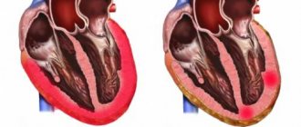
- Heart attack. Acute disturbance of trophism of cardiac structures. Occurs at any age, mainly in elderly patients. Also against the background of current ischemic heart disease, as a complication.
It ends with necrosis of cardiomyocytes (heart cells), replacement of active tissue with scar tissue. It is unable to contract and conduct a signal. Hence the AV block.
Depending on the extent, we can talk about the degree of severity. The more structures damaged, the more dangerous the consequences.
Complications of a major heart attack are described in this article, the symptoms of a pre-infarction condition are here, the causes and risk factors are here.
- Rheumatism. Autoimmune process affecting the myocardium. The treatment is long-term, lifelong maintenance therapy is the result.
It is possible to slow down the destruction and prevent relapses, but complete relief is unlikely.
The neglected phenomenon ends with damage to the His bundles and conduction disturbances.
- Ischemic disease. It is similar in nature to a heart attack, but the process does not reach a certain critical mass, since the blood supply still remains at an acceptable level. However, necrosis of the muscle layer will not take long to occur without treatment. This is the logical conclusion of IHD.
- Coronary insufficiency. As a result of atherosclerosis with narrowing or occlusion of the corresponding arteries supplying cardiac structures. Manifestations occur in later stages. Blockade is one of the organic disorders. Read more about coronary insufficiency here.
- Cardiomyopathy. The generic name of a group of processes. Arises as a consequence of severe somatic pathologies.
The essence lies in dystrophy of the muscular layer of the heart. Contractility decreases, the signal is transmitted through damaged tissues worse than in the normal position.
Weakened hemodynamics, ischemia, multiple organ failure as a consequence. Types of cardiomyopathy, causes and treatments are described in this article.
The presence of pathologies of the adrenal glands of the deficient type, the thyroid gland, and blood vessels, including the aorta, also affects.
The list goes on. There is an opinion about the participation of a hereditary factor in the process. Whether this is true or not is not entirely clear. In recent years, the role of the genetic component has been actively studied.
Why does the disorder develop?
There are many different conditions that can disrupt the conduction of electrical impulses through the myocardium.
We cannot always accurately determine the cause of this phenomenon. Often we can only guess why the blockade occurred and try to slow down the progression of the process. In clinical cardiology, it is customary to distinguish two groups of cardiac conduction and excitability disorders:
- Cardiac, that is, caused by pathological processes occurring in the heart muscle. This may be coronary heart disease (CHD) or previous myocardial infarction, inflammatory diseases, cardiomyopathies. Often the source of the problem is congenital and acquired heart valve defects. Failure of the conduction system can be caused by tissue trauma during surgery.
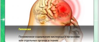
- Non-cardiac - the cause of such disorders lies outside the myocardium. Most often we have to deal with endocrine diseases - diabetes and thyroid pathology. Among the possible causes, it is also worth highlighting hypertension, chronic bronchitis, asthma and other conditions leading to the development of hypoxia. In women, failure is often recorded during pregnancy, with the onset of menopause.
It is important to understand: the occurrence of blockade is not always associated with organic changes in the myocardium or serious non-cardiac diseases. Heart failure can be a temporary phenomenon due to stress or physical activity. The nature of the disorders can be determined by examining the patient.
Symptoms depending on the degree
The clinical picture depends on the stage of the pathological process.
Stage 1:
Manifestations are completely or mostly absent. The patient feels normal, there are no deviations in his vital functions.
Functional defects can be detected only by electrocardiography results. Often this is an accidental finding, discovered during a preventive examination of a person.
Mild shortness of breath is possible during intense physical activity (work, running, exhausting sporting events).
Attention:
First degree atrioventricular block is clinically favorable. If detected early, there is a chance of complete recovery without consequences.
Stage 2:
- Chest pain of unknown origin. Occurs in most cases. This is a non-specific sign. The duration of the episode is no more than a few minutes.
- Shortness of breath due to intense physical activity. She is not in a calm state.
- Weakness, drowsiness, lack of performance. Apathy and reluctance to do anything are possible.
- Bradycardia. Change in heart rate downwards. It is not yet threatening.
Stage 3:
- Shortness of breath with little physical activity. Even with simple walking.
- Headache. Determined by ischemic disorders in cerebral structures. The duration varies from a few minutes to hours and even days. It is necessary to differentiate from migraine.
- Vertigo. Up to uncoordination of movements, inability to navigate in space.
- Arrhythmia of several types at once. The slowdown in cardiac activity is accompanied by a change in the time intervals between contractions.
- Fainting.
- Lowering blood pressure to critical levels.
- A sharp drop in heart rate.
All three symptoms presented above are part of the structure of the so-called Morgagni-Adams-Stokes syndrome. This is an emergency. It lasts a few minutes, but poses an enormous danger to life.
Injury, stroke, heart attack or cardiac arrest are possible. If this kind of manifestation occurs, surgical treatment is necessary, the essence of which is the implantation of a pacemaker.
Stage 4:
It is not always called. It is a variation of the previous one, but is characterized by even more severe symptoms. Massive organic disturbances are noted in all systems.
Death with 4th degree AV block becomes inevitable, it’s a matter of time. However, in order to trigger pathology in this way, you need to try and consciously ignore all the signals from your own body.
Epidemiology
There is no exact data on the prevalence of this condition, since it is often not accompanied by clinical manifestations. It is reliably known that the frequency of detection increases with age. This is explained by the development of age-related and ischemic changes in the myocardium. AVB develops more often in the presence of heart disease: about 5% have the first and 2% have the second degree.
First degree atrioventricular block can occur in 0.6-8% of children. It is detected in healthy young people as a normal variant. May develop during sleep or in trained individuals. About 1% of young people under 20 have AVB, and among athletes it is detected in 9% of cases.
Diagnostic methods
Management of persons with abnormalities in the conduction of cardiac structures is under the supervision of a cardiologist. If the process is complicated and dangerous, consult a specialized surgeon.
An approximate survey scheme includes the following elements:
- Questioning the patient about complaints, their age and duration. Objectification of symptoms and fixation.
- Anamnesis collection. Including family history, determination of lifestyle, presence of bad habits and others. Aimed at early identification of the source of the problem.
- Blood pressure measurement. Against the background of an advanced process, by the second stage, or even more so the third, jumps in blood pressure are likely. It is difficult to catch such a condition using routine techniques.
- 24-hour Holter monitoring. More informative way. Blood pressure and heart rate are assessed every half hour or more often, depending on the program. Can be performed repeatedly to improve accuracy.
- Electrocardiography. Allows you to identify functional disorders of the heart. Plays a key role in early diagnosis.
- EFI. Modified version of the previous survey. However, it is invasive. A special probe is inserted through the femoral artery. The activity of individual areas of cardiac structures is assessed. Quite a difficult study, but sometimes there are no alternatives to it.
- Echocardiography. In order to identify organic disorders. The classic variant resulting from a long course of blockade is cardiomyopathy of varying severity.
- Blood analysis. For hormones, general and biochemical. For a comprehensive assessment of the state of the body, in particular the endocrine system and metabolism in general.
As necessary, if previous methods do not provide answers to questions, CT, MRI, coronography, and radioisotope studies are used. The decision is made by a group of leading specialists.
Variants of deviations on the ECG
Among the characteristic features:
- Prolongation of the QT interval by more than 0.2 s. At the first stage this is a typical finding.
- PQ extension. Changes in the frequency of occurrence of ventricular complexes. The so-called Mobitz 1.
- Complete loss of contractions of the lower chambers of the heart. Or alternately, in a symmetrical order.
- Decreased heart rate (bradycardia) of varying severity. Depends on the stage of the pathological process.
AV block is noted specifically on the ECG, and the more severe the phase, the easier it is to diagnose the problem.
Case from practice
Patient R., 75 years old, turned to his local doctor with complaints of interruptions in his heart function. Upon examination and history taking, it turned out that the patient was also bothered by headaches, attacks of dizziness, and shortness of breath during exercise and at rest. Considers himself sick for 10 years. Suffered a myocardial infarction at the age of 67 years. She is seen by a cardiologist with a diagnosis of coronary artery disease. Post-infarction cardiosclerosis.
https://www.youtube.com/watch?v=m4sgcd4JLI0
After further examination, ECG revealed signs of second degree AV block, and echoCG revealed left ventricular hypertrophy. After consultation with a cardiologist, the patient was referred to the cardiac surgery center. A pacemaker was installed. The patient was discharged with improved condition. Observation by a cardiologist is recommended.
Treatment depending on the degree
The therapy is complex.
1st degree.
Long-term follow-up is indicated. Such tactics can be repeated for more than one year. If there is no progression, gradually the frequency of consultations with a cardiologist becomes less frequent.
Against the background of deterioration, medications from several pharmaceutical groups are prescribed:
- Antihypertensive drugs. Different types.
- Antiarrhythmic.
If there is an inflammatory infectious pathology, antibiotics, NSAIDs and corticosteroids are used. Treatment is strictly in a hospital.
Transient 1st degree AV block (transient) is the only dangerous case requiring therapy, provided it is of viral or bacterial origin.
2nd degree
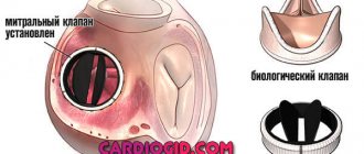
The same type of drugs are used. If the condition quickly worsens, there is no point in waiting. The installation of a pacemaker is shown.
Regardless of age. The only exception is older patients who may not be able to withstand surgery. The issue is resolved individually.
3-4 degrees
Implantation of an artificial pacemaker is mandatory. Once the terminal phase occurs, the chances of cure are minimal.
Throughout the entire period of therapy, lifestyle changes are indicated:
- Quitting bad habits.
- Diet (treatment table No. 3 and No. 10).
- Adequate sleep (8 hours).
- Walks, exercise therapy. The main thing is not to overwork. The duration is arbitrary.
- Avoiding stress.
Traditional recipes can be dangerous, so they are not used.
Doctor's advice: how to properly see a doctor if there is a blockage
After the diagnosis is established, the patient remains in outpatient treatment or is hospitalized in a hospital. Tactics are determined by the severity of the blockade. After achieving remission, the patient should not be left without specialist supervision. We recommend:
- If your condition is stable and there are no complaints, visit a cardiologist and do an ECG every 6 months.
- If the condition worsens, new complaints appear or existing disorders progress, make an appointment with a doctor as soon as possible.
If the doctor prescribes drug therapy, you should adhere to it and not violate the drug dosage schedule. Stopping the medication on your own is unacceptable - this leads to the development of complications.
If the patient has had a pacemaker installed, the monitoring tactics change. You should visit your doctor 3, 6 and 12 months after surgery to make sure that the device is working smoothly. The further observation schedule will depend on the patient’s condition.
Prognosis and possible complications
Typical consequences:
- Heart failure. Resuscitation in such a situation is minimally effective; as soon as it is restored, the rhythm will change again. A relapse is likely within a few days.
- Cardiogenic shock. Potentially fatal consequence. Moreover, death occurs in almost 100% of cases.
- Fainting and, as a result, injury may be incompatible with life.
- Heart attack or stroke. Acute malnutrition of cardiac structures and brain, respectively.
- Vascular dementia.
Forecasts depend on the stage of the pathological process:
| Stage 1. | Survival rate is close to 100%. There are risks only if there are infectious lesions. |
| Stage 2. | The probability of death is about 20-30% without therapy. With full treatment it is 2-4 times lower. |
| 3rd degree. | Mortality 40-60%. |
In the terminal phase, death is inevitable. Therapy is ineffective.
Radical surgery with the installation of a pacemaker significantly improves the prognosis.